-
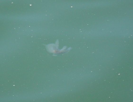
Melibe leonina swimming near surface, about 3 m from the bottom, in a harbor. Length about 8 cm Click the photo for a short video of this individual swimming
-
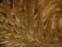
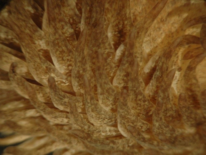
The cerata are flattened, widest above the base, and taper to a point. The hepatic diverticula cannot be readily seen within them if there is pigment present. The tips often take on the coloration of their anemone food. The mid-dorsal band, which is cerata-free but has light cororation on it, can be seen to the right.
-
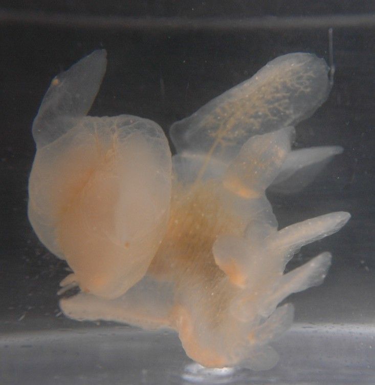
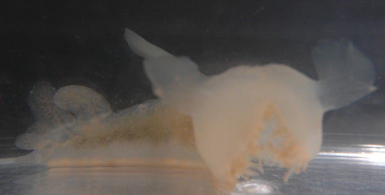
This is a top left view of a swimming individual, who has been swimming away but is making a strong turn to the left. The head and oral hood are visible to the right. The flaplike extensions of the oral hood are the rhinophores. The large dorsal cerata with an internal network of vessels (hepatic diverticula?) are visible at the top and right. The foot is facing down and away from view. The oral hood is closed in this view.
-
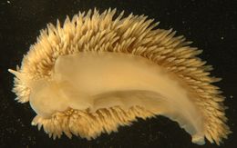
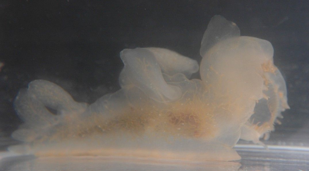
The foot tapers but is not drawn out into a long, sharp point.
-
In this nearly head-on view the open oral hood with its filiform tentacles can be seen.
-
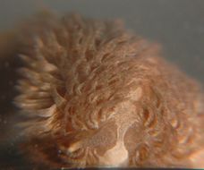
This head-on view shows the white triangle anterior to the rhinophores. It also shows the smooth, tapering rhinophore with a light-colored tip, and the fact that the rhinophore seems to have a pore in the end. Notice also the cerata-free band that runs mid-dorsally and has light coloration.
-
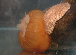
Melibe leonina about 10 cm long. Collected from eelgrass at Padilla Bay. The head and oral hood is to the right. Some of the tentacles from the opening in the oral hood can be seen. (Photo by: Dave Cowles July 2006)
-
This head-on view shows the rhinophores (the one on the right seems to have been injured and truncated), plus one of the two pedal tentacles extends to the right.
-
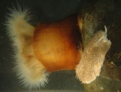
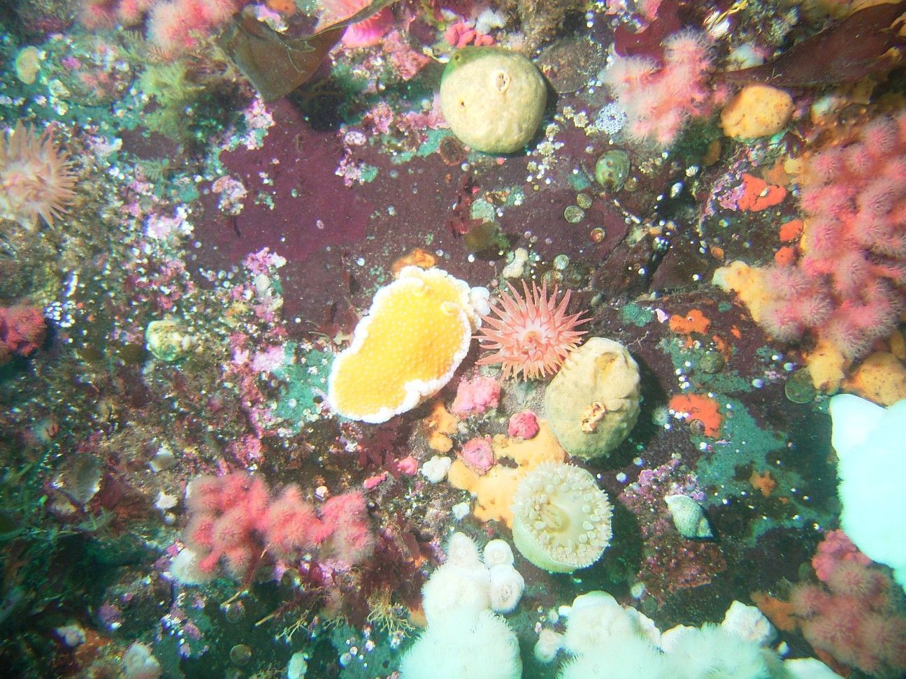
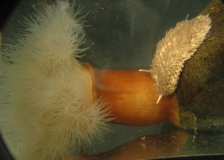
This larger view of the same individual as above shows pink patches of sea strawberry, Gersemia rubriformis, one of this species' preferred foods, around the nudibranch.The pink lumps beside the nudibranch may be Gersemia that the animal has already grazed on.The anemones present are Cribrinopsis fernaldi (pink striped tentacles, beside the nudibranch) and Metridium giganteum (white, bottom of picture)Photo by Jim Nestler, July 2005
-
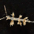
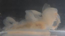
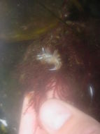
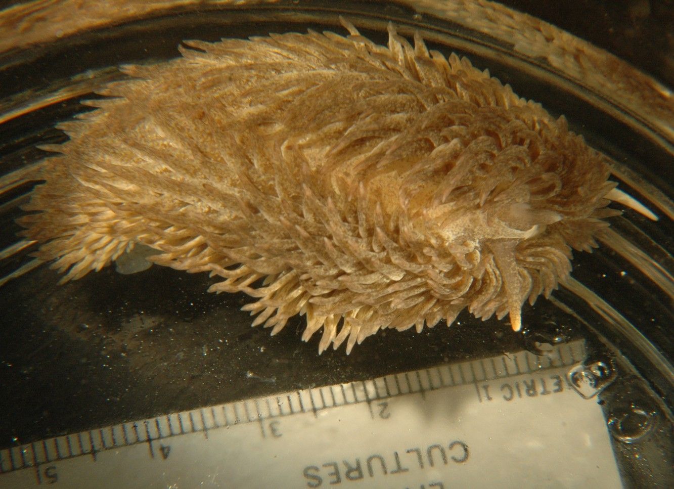
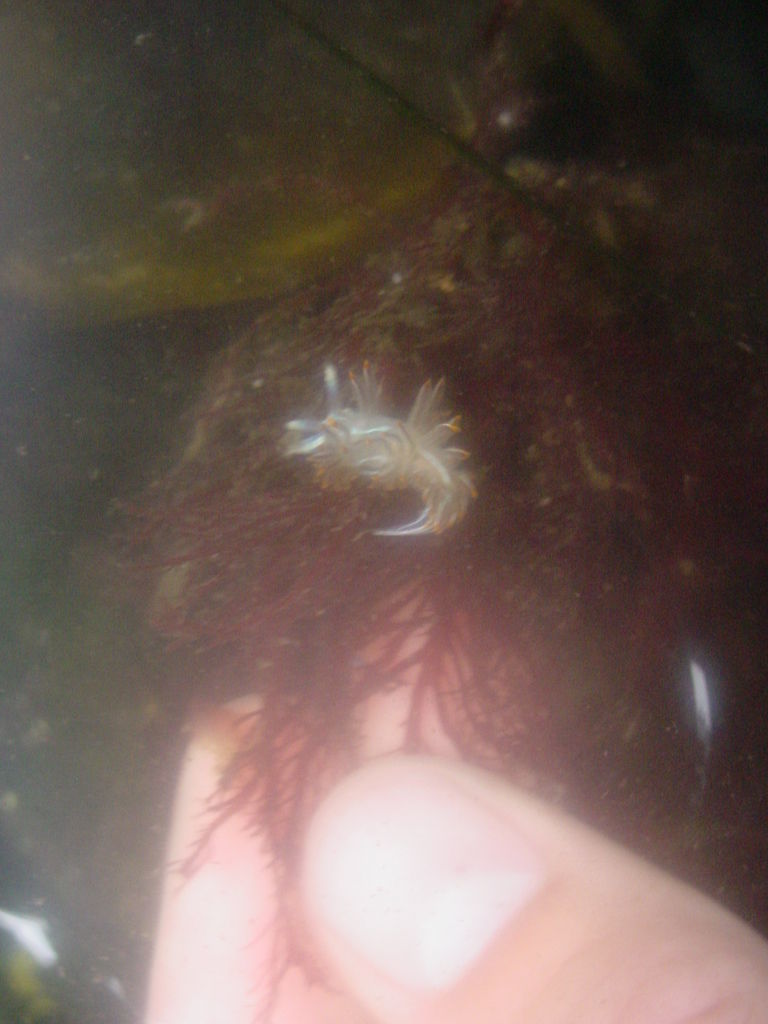
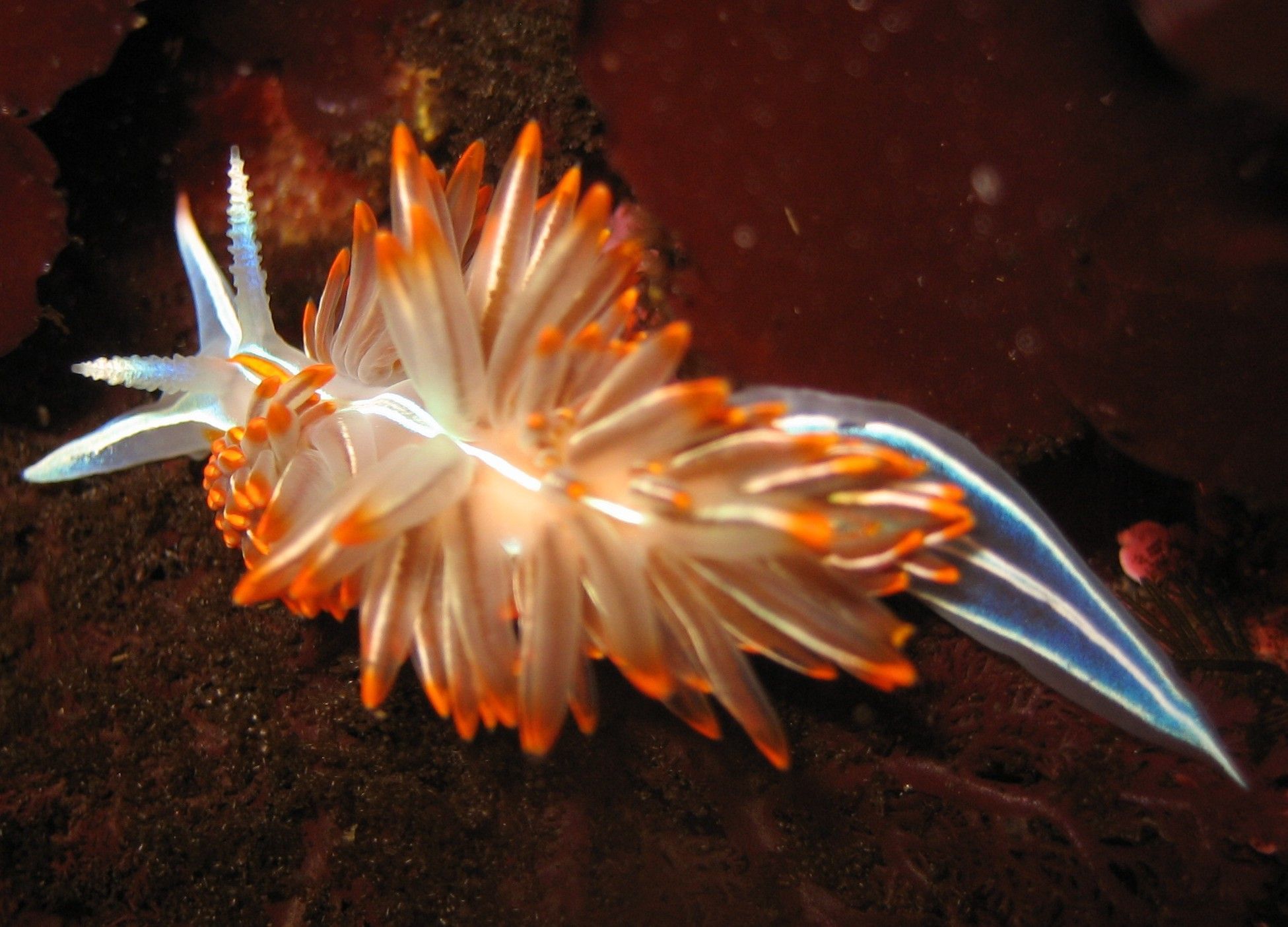
Aeolidia papillosa, approximately 4.5 cm long, found on a rock in Padilla Bay. This individual is crawling around the side of a dish. The rhinophores are visible in front with light tips, while one white pedal tentacle is visible on the extreme right. (Photo by: Dave Cowles, July 2008)
-
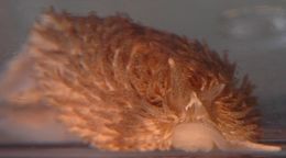
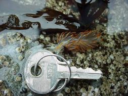
Tochuina tetraquetra, about 12 cm long. (Photo by: James Nestler subtidally off Northwest Island, July 2006)
-
-
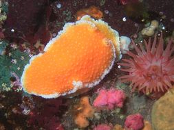
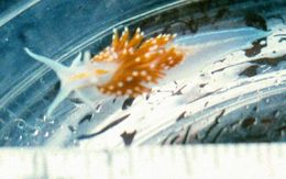
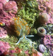
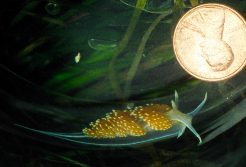
Hermissendra crassicornis: Beach 4, WA (Photo by: Robbie Wheeling, July, 2002)
-
-
-
-
-
-
-
-
-
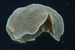
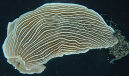
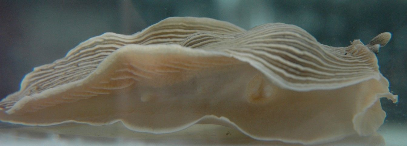
This species has a deep groove along the sides between the wide flap of the dorsum and the wide flap of the foot. Here the groove on the left side can be clearly seen as the animal turns.
-
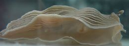
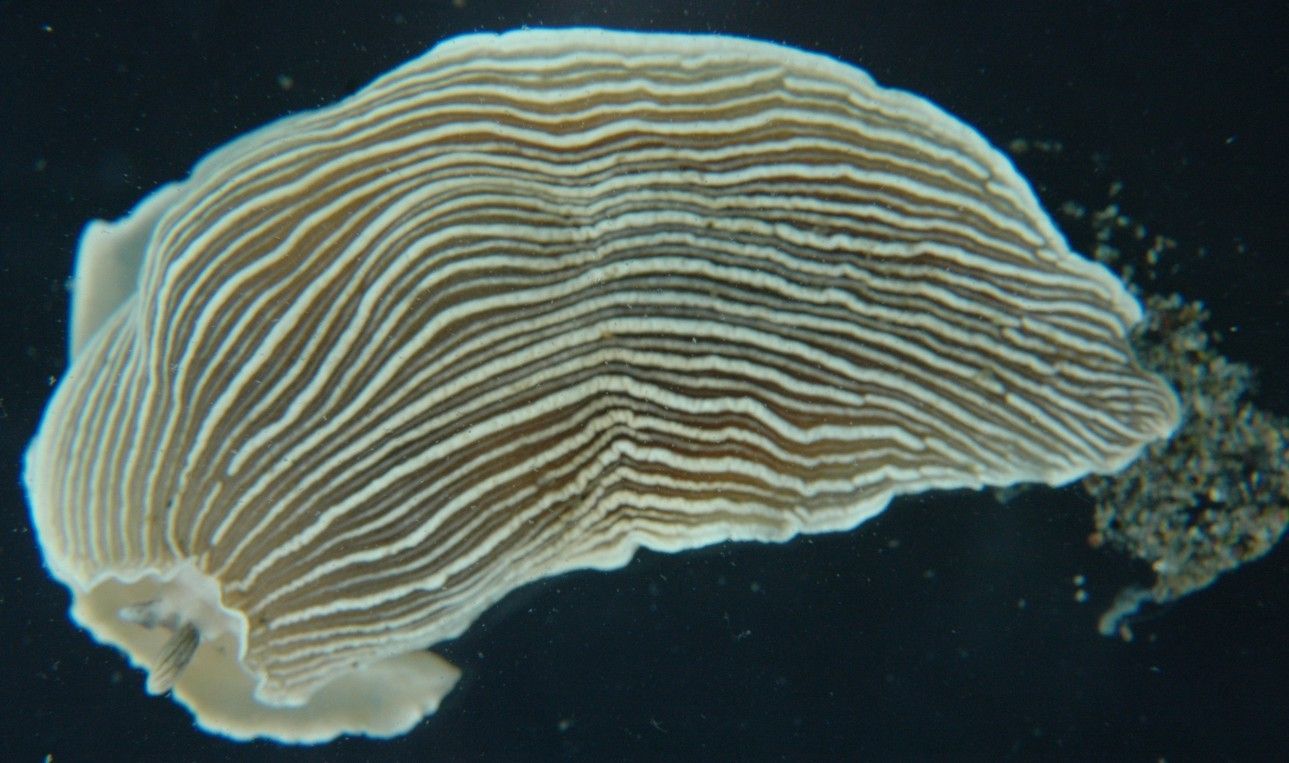
This species has its anus on a prominent papilla on the right side, in a groove between the flaps of the dorsum and the foot. The view above shows the groove on the right side of the animal, with the anus to the left (posterior) and the gonopore to the right (anterior)
-
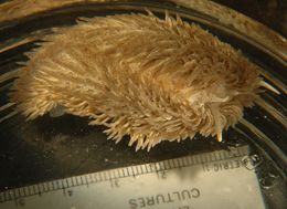
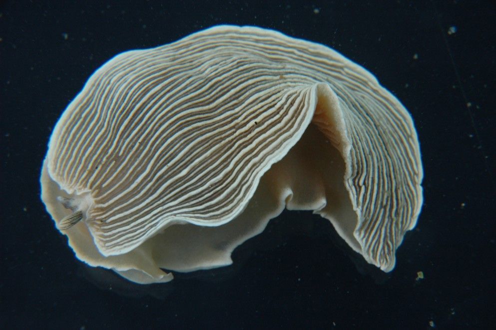
Armina californica, 6.5 cm long, from 6m depth in Burrows Bay. Photo taken at Shannon Point Marine Station (Photo by: Dave Cowles August 2006)