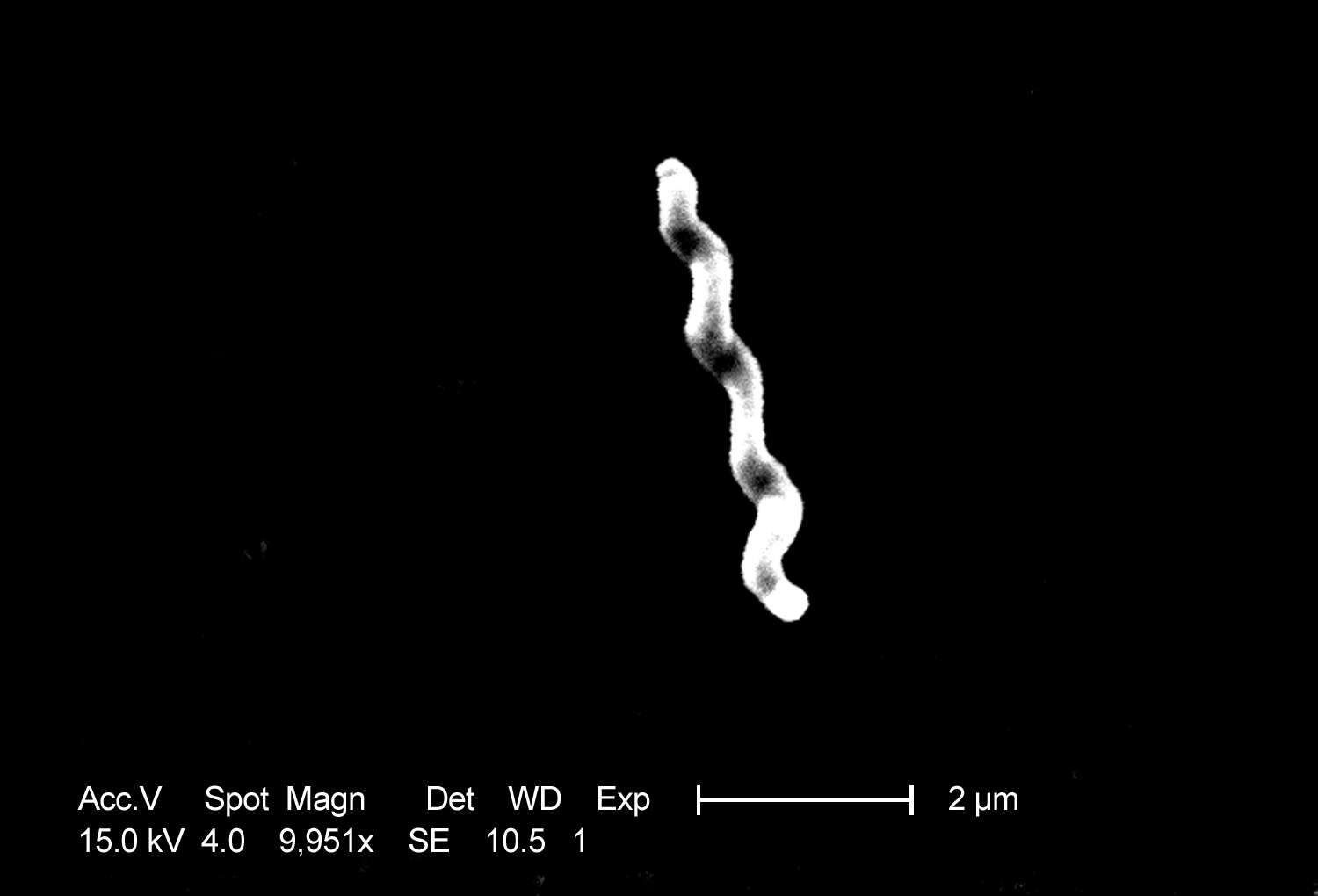
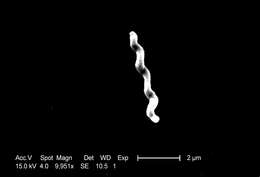
Description: English: This scanning electron micrograph depicts a grouping of Gram-negative Campylobacter fetus bacteria, magnified 4,976x. The “S-shaped” C. fetus bacterium, also known as C. fetus ssp. intestinalis or Vibrio fetus var. intestinalis, is an opportunistic human pathogen with a worldwide distribution pattern. 日本語: カンピロバクター・フェタスの電子顕微鏡写真. Date: 2004. Source: : This media comes from the Centers for Disease Control and Prevention's Public Health Image Library (PHIL), with identification number #5776. Note: Not all PHIL images are public domain; be sure to check copyright status and credit authors and content providers. العربية | Deutsch | English | македонски | slovenščina | +/−. Originally from en.wikipedia; description page is/was here. Author: Photo Credit: Janice Carr Content Providers(s): CDC/ Dr. Patricia Fields, Dr. Collette Fitzgerald. Permission(Reusing this file): PD-USGov-HHS-CDC English: None - This image is in the public domain and thus free of any copyright restrictions. As a matter of courtesy we request that the content provider be credited and notified in any public or private usage of this image.
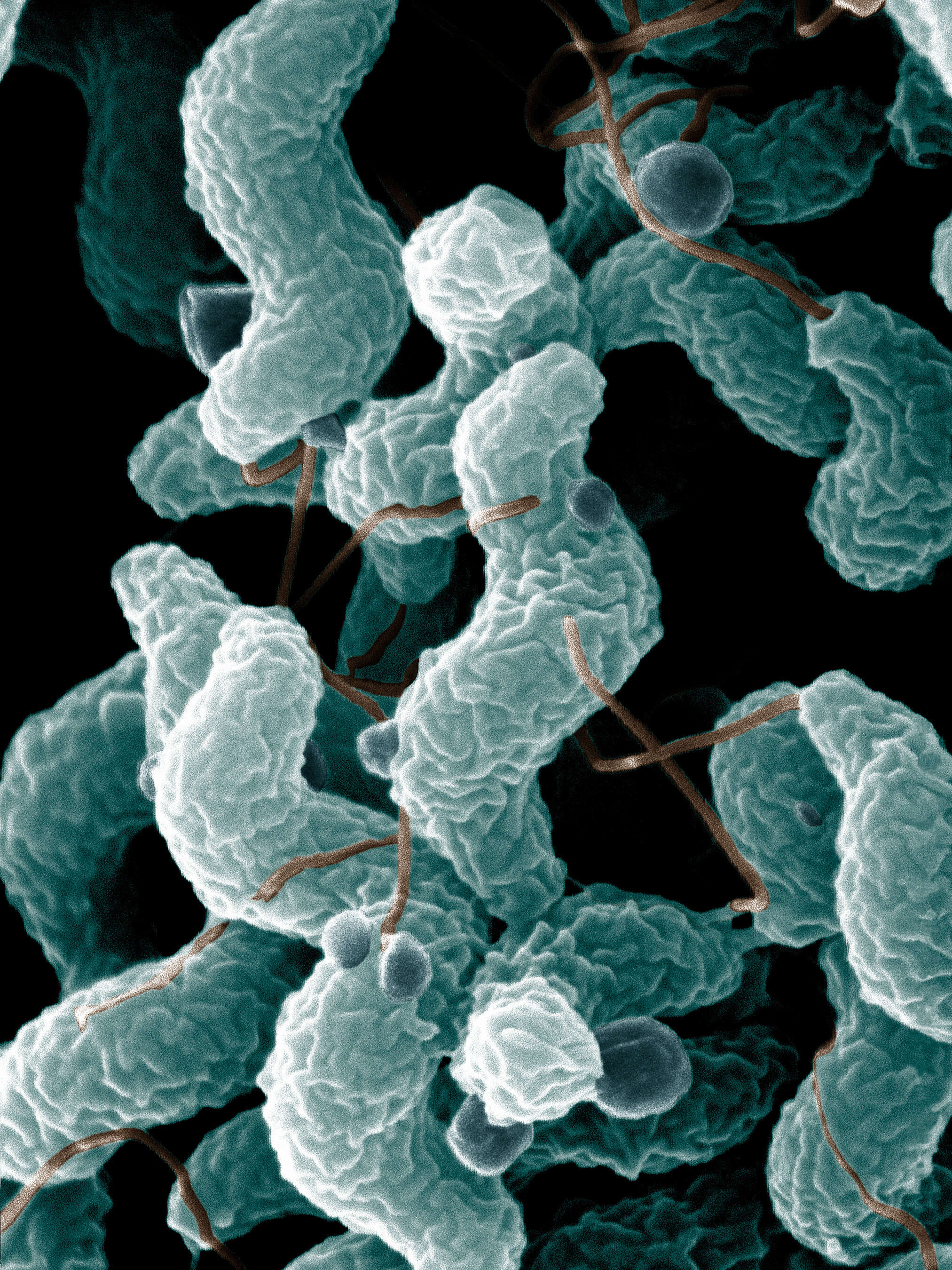
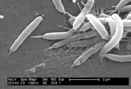
Description: English: Campylobacter bacteria are the number-one cause of bacterial food-related gastrointestinal illness in the United States. To learn more about this pathogen, ARS scientists are sequencing multiple Campylobacter genomes. This scanning electron microscope image shows the characteristic spiral, or corkscrew, shape of C. jejuni cells and related structures. Photo by De Wood; digital colorization by Chris Pooley. Date: 1/2/2008. Source: Agricultural Research Service (ARS) is the U.S. Department of Agriculture's chief scientific research agency. Author: De Wood, Pooley, USDA, ARS, EMU. 193 283 9 9 480 640 two species,campylobacter jejuni and cam. coli,are foodborne pathogens,small(0.2*1 micrometer)microaerophilic,helical,motile cells found in intestinal tract of humans. Licensing[edit] Public domainPublic domainfalsefalse. : This image is in the public domain because it contains materials that originally came from the Agricultural Research Service, the research agency of the United States Department of Agriculture. dansk | Deutsch | English | español | فارسی | français | italiano | македонски | മലയാളം | sicilianu | Türkçe | 中文(简体) | +/− :.
Description: Scanning electron micrograph of en:Helicobacter bilis bacteria (originally classified as Flexispira rappini, now deprecated). Obtained from the CDC Public Health Image Library. Image credit: CDC/Dr. Patricia Fields, Dr. Collette Fitzgerald (PHIL #5715), 2004. Source: This file is lacking source information. Please edit this file's description and provide a source. Author: This file is lacking author information.
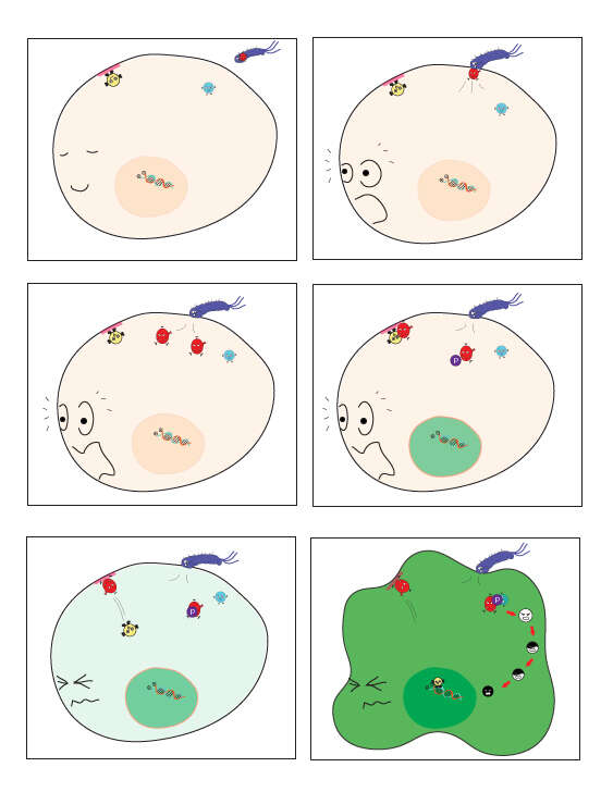
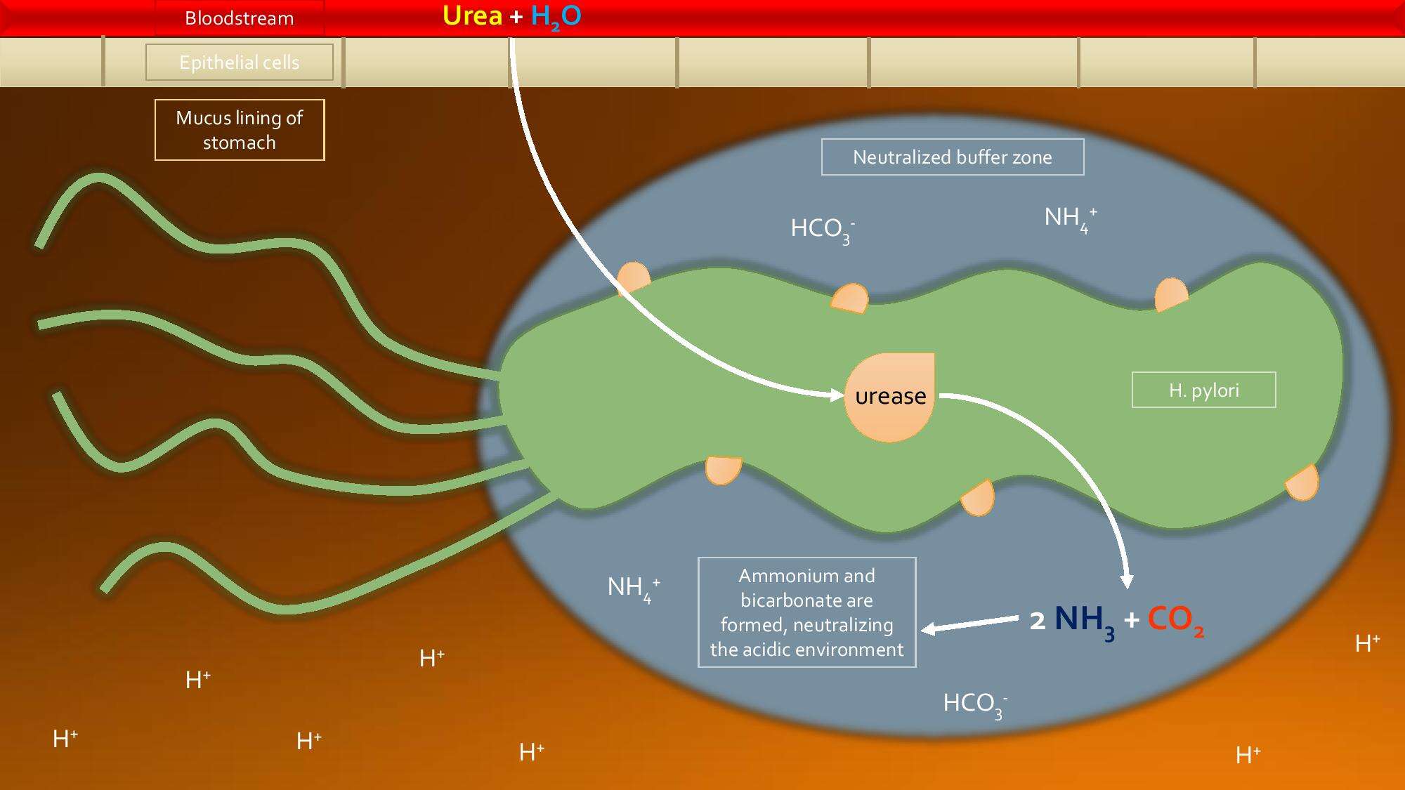
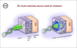
Description: A team of researchers from Boston University, Harvard Medical School and Massachusetts Institute of Technology have shown that the bacterium that causes human stomach ulcers uses a clever biochemical strategy to alter the physical properties of its environment, allowing it to move and survive and further colonize its host.Contact with stomach acid keeps the mucin lining the epithelial cell layer in a spongy gel-like state. This consistency is impermeable to the bacterium Heliobacter pylori. However, the bacterium releases urease which neutralizes the stomach acid. This causes the mucin to liquefy, and the bacterium can swim right through it. Read more about this research. Illustration Credit: Zina Deretsky, National Science Foundation Visit NSF’s Multimedia Gallery, at www.nsf.gov/news/mmg, for more images, and for video. Date: 11 August 2009, 11:50. Source: Ulcer-causing Bacterium (H.Pylori) Crossing Mucus Layer of Stomach. Author: National Science Foundation.
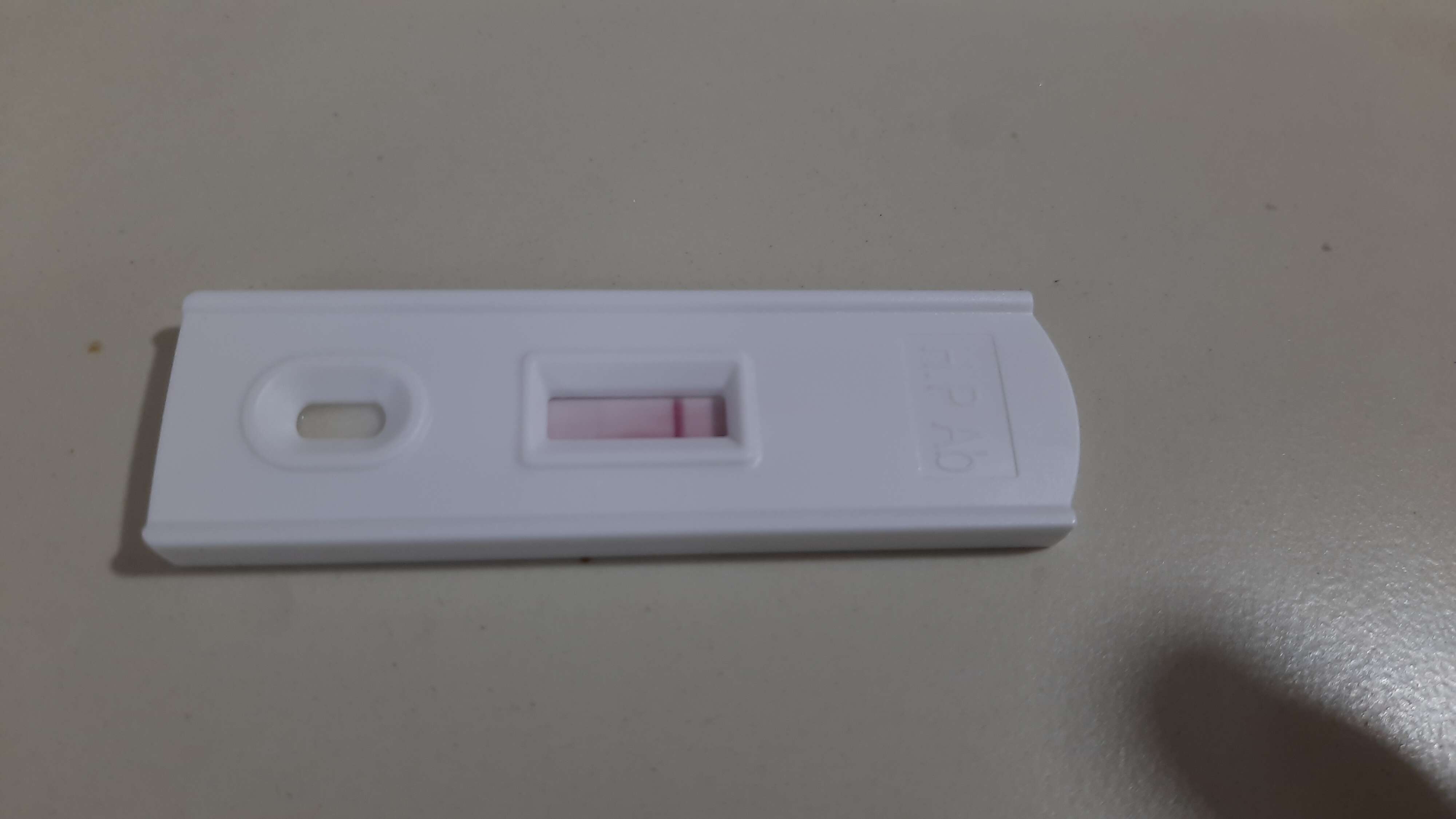
Description: English: Rapid urease test made in Poland. The inscription on the tests translates as: "rapid urease test / for detecting heliobacter pylori / positive / negative / manufacturer and distributor" Polski: Szybki test ureazowy. Date: 30 September 2008. Source: Own work. Author: Louve.pl.
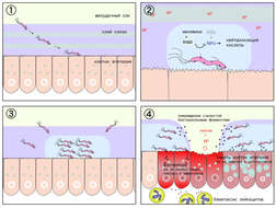
Description: English: H. pylori adheres to the gastric mucosal wall, injecting CagA into gastric lining cells. Date: 7 August 2015. Source: Own work. Author: WassermanLab.
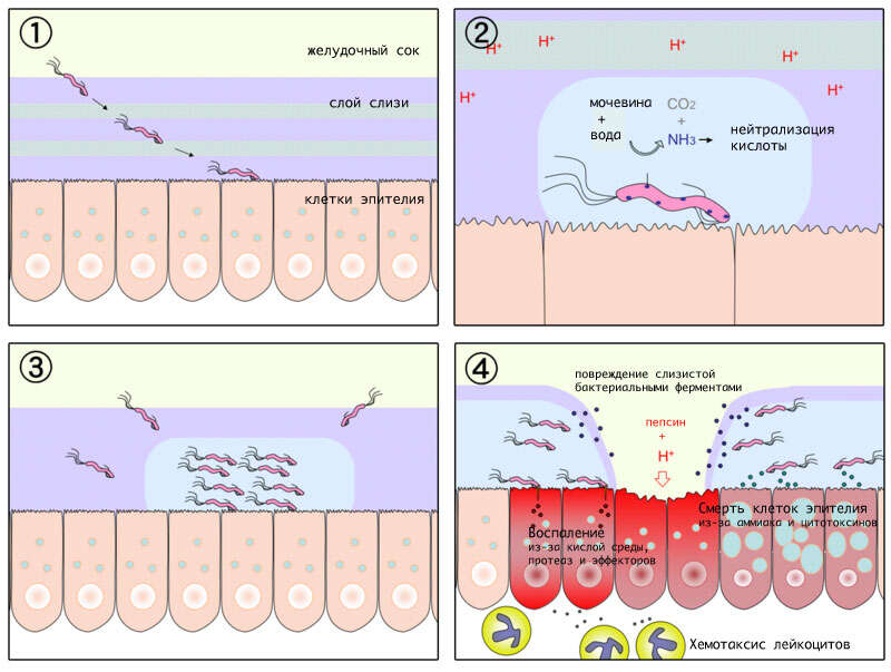
Summary.mw-parser-output table.commons-file-information-table,.mw-parser-output.fileinfotpl-type-information{border:1px solid #a2a9b1;background-color:#f8f9fa;padding:5px;font-size:95%;border-spacing:2px;box-sizing:border-box;margin:0;width:100%}.mw-parser-output table.commons-file-information-table>tbody>tr,.mw-parser-output.fileinfotpl-type-information>tbody>tr{vertical-align:top}.mw-parser-output table.commons-file-information-table>tbody>tr>td,.mw-parser-output table.commons-file-information-table>tbody>tr>th,.mw-parser-output.fileinfotpl-type-information>tbody>tr>td,.mw-parser-output.fileinfotpl-type-information>tbody>tr>th{padding:4px}.mw-parser-output.fileinfo-paramfield{background:#ccf;text-align:right;padding-right:0.4em;width:15%;font-weight:bold}.mw-parser-output.commons-file-information-table+table.commons-file-information-table,.mw-parser-output.commons-file-information-table+div.commons-file-information-table>table{border-top:0;padding-top:0;margin-top:-8px}@media only screen and (max-width:719px){.mw-parser-output table.commons-file-information-table,.mw-parser-output.commons-file-information-table.fileinfotpl-type-information{border-spacing:0;padding:0;word-break:break-word;width:100%!important}.mw-parser-output.commons-file-information-table>tbody,.mw-parser-output.fileinfotpl-type-information>tbody{display:block}.mw-parser-output.commons-file-information-table>tbody>tr>td,.mw-parser-output.commons-file-information-table>tbody>tr>th,.mw-parser-output.fileinfotpl-type-information>tbody>tr>td,.mw-parser-output.fileinfotpl-type-information>tbody>tr>th{padding:0.2em 0.4em;text-align:left;text-align:start}.mw-parser-output.commons-file-information-table>tbody>tr,.mw-parser-output.fileinfotpl-type-information>tbody>tr{display:flex;flex-direction:column}.mw-parser-output.commons-file-information-table+table.commons-file-information-table,.mw-parser-output.commons-file-information-table+div.commons-file-information-table>table{margin-top:-1px}.mw-parser-output.fileinfo-paramfield{box-sizing:border-box;flex:1 0 100%;width:100%}} Description: Diagram of gastric ulceration by H. pylori, with russian annotation. Date: 22 December 2007. Source: User:Y_tambe's file. Author: User:Vicki Doronina. Permission(Reusing this file): GDFL. Other versions: [edit] English: numberedEnglishJapanesePolishRussian.
Description: English: A combination of pantoprazole, metronidazole, amoxicillin, clarithromycin, and bismuth subsalicylate as used to treat H. pylori. Date: 8 January 2022. Source: Own work. Author: Doc James.
Description: العربية: صورة للملتويات البوابية بالمجهر الإلكتروني الماسح. English: Helicobacter pylori bacteria Polski: Fotografia H. pylori w mikroskopie elektronowym. Italiano: Vista al microscopio elettronico di una colonia di H. pylori staziata nel muco gastrico. Português: M.E. fotografia da H. pylori. Español: Micrografía de barrido de H. pylori. Русский: Электронно-микроскопическая фотография H. pylori. Svenska: Helicobacter pylori, sedd med elektronmikroskop. Català: Micrografia electrònica d' H. pylori. Українська: Електронна мікрофотографія H. pylori. Türkçe: EM'da çekilmiş H. pylori. नेपाल भाषा: एच पाइलोरीया इलेक्त्रोन माइक्रोस्कोपी किपा. Simple English: Helicobacter pylori under microscope. Date: February 7-9, 1994. Source: Helicobacter Pylori in Peptic Ulcer Disease National Institutes of Health Consensus Development Conference Statement February 7-9, 1994 http://consensus.nih.gov/1994/1994HelicobacterPyloriUlcer094html.htmhttp://consensus.nih.gov/IMAGES/Art/094.jpg. Author: Originally uploaded to the English Wikipedia by w:User:AxelBoldt. Permission(Reusing this file): PD-USGov-HHS-NIH.
Description: English: This scanning electron micrograph depicts a grouping of Gram-negative ”Flexispira rappini” bacteria, magnified 13,951x. Its name ”F. rappini” is considered provisional, for it was never formally proposed or accepted. Subsequently determined to be closely related to Helicobacter spp., it is referred to as Helicobacter sp. flexispira in the literature. Italiano: Helicobacter pylori. Colonia di Helicobacter pylori. Беларуская (тарашкевіца): Helicobacter pylori. Date: 2004. Source: : This media comes from the Centers for Disease Control and Prevention's Public Health Image Library (PHIL), with identification number #5715. Note: Not all PHIL images are public domain; be sure to check copyright status and credit authors and content providers. العربية | Deutsch | English | македонски | slovenščina | +/−. Author: Photo Credit: Janice Carr Content Providers(s): CDC/ Dr. Patricia Fields, Dr. Collette Fitzgerald. Permission(Reusing this file): PD-USGov-HHS-CDC English: None - This image is in the public domain and thus free of any copyright restrictions. As a matter of courtesy we request that the content provider be credited and notified in any public or private usage of this image.
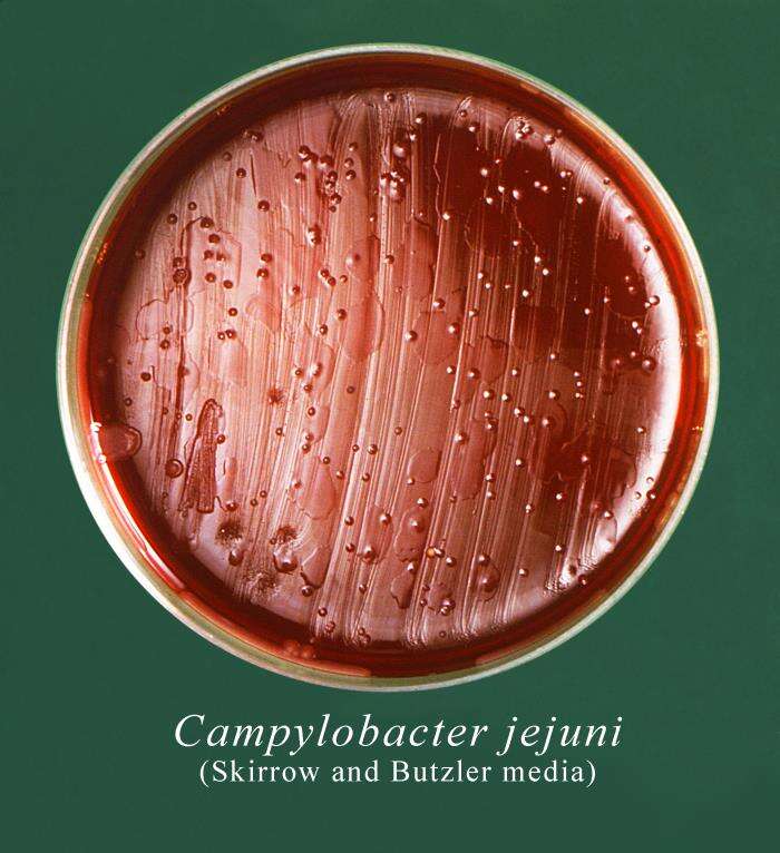
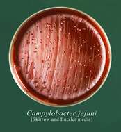
Description: English: This Petri dish culture plate contained Skirrow and Butzler growth medium, which had been inoculated with a culture of Campylobacter jejuni, formerly known as Campylobacter fetus subsp. jejuni, and had produced numerous, small round bacterial colonies. Campylobacteriosis is an infectious disease caused by bacteria of the genus Campylobacter. Most people who become ill with campylobacteriosis have diarrhea, cramping, abdominal pain, and fever within 2 to 5-days after exposure to the organism. Date: 1979. Source: https://phil.cdc.gov/Details.aspx?pid=15985. Author: CDC/ Sheila Mitchel.
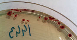
Description: English: Campylobacter jejuni colonies on CASA chromogenic medium, isolated from a canine stool sample. Date: 22 October 2014, 17:10:01. Source: Own work. Author: Stefan Walkowski.


