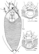
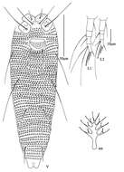
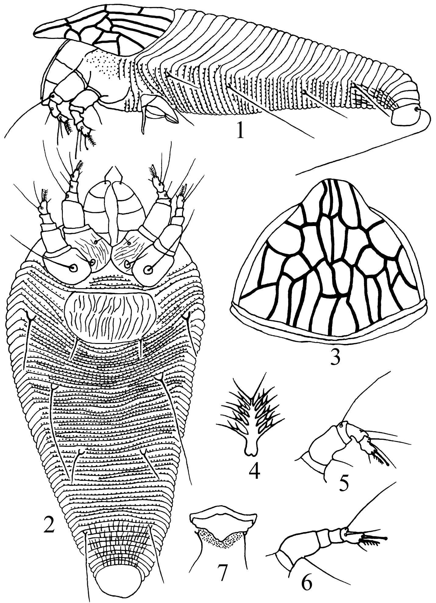
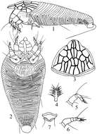
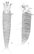
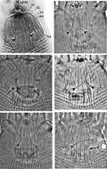
Figures 1–7.Kolacarus reticulatus sp. n. 1 lateral view of female 2 ventral view of female 3 anterior dorsal view of female 4 tarsal empodium 5 leg I 6 leg II 7 male genitalia
Parisa Lotfollahi, Enrico de Lillo, Karim Haddad Irani-Nejad
Zookeys
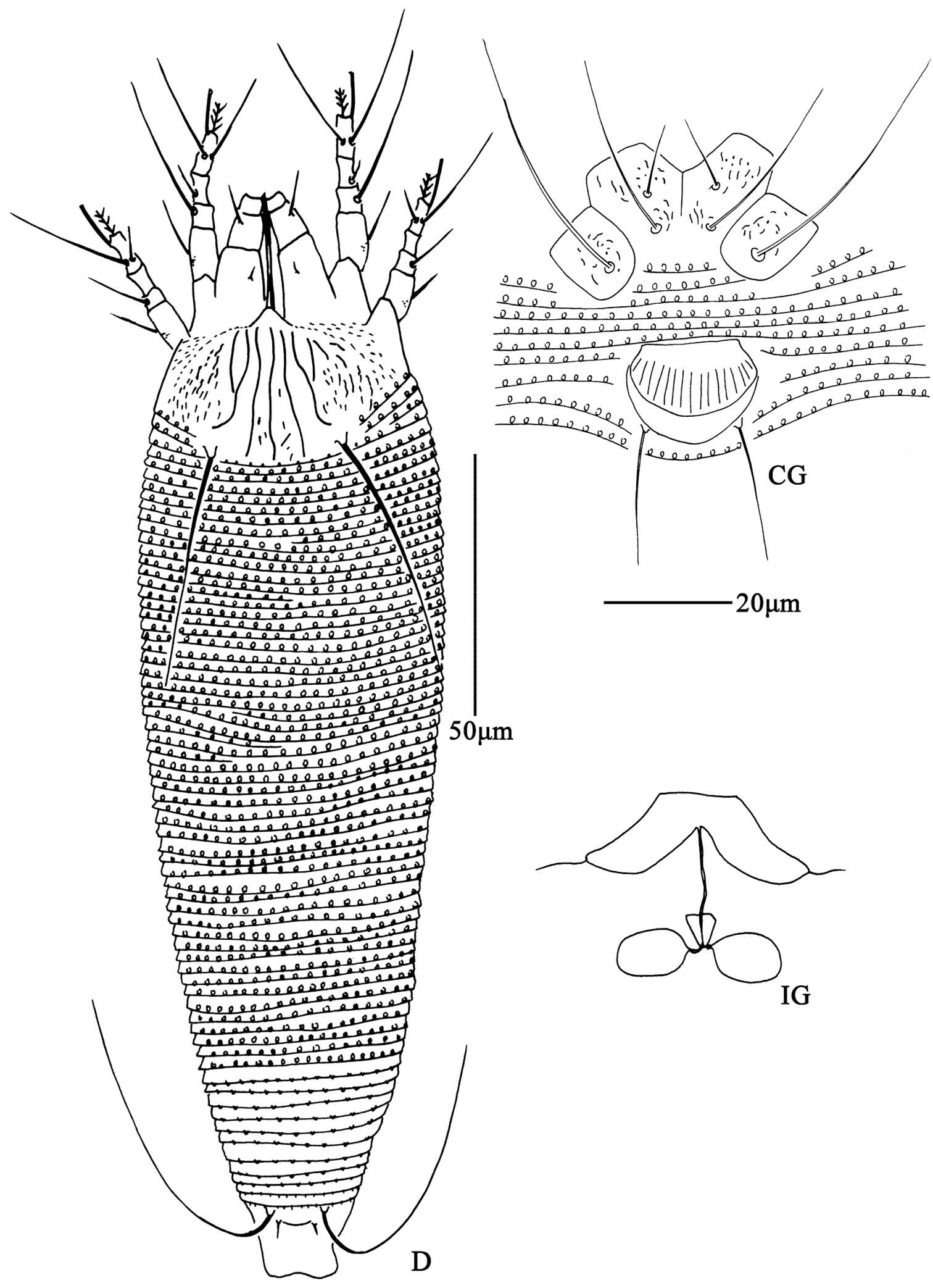
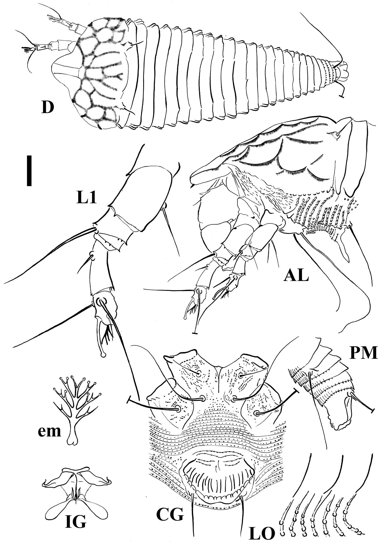
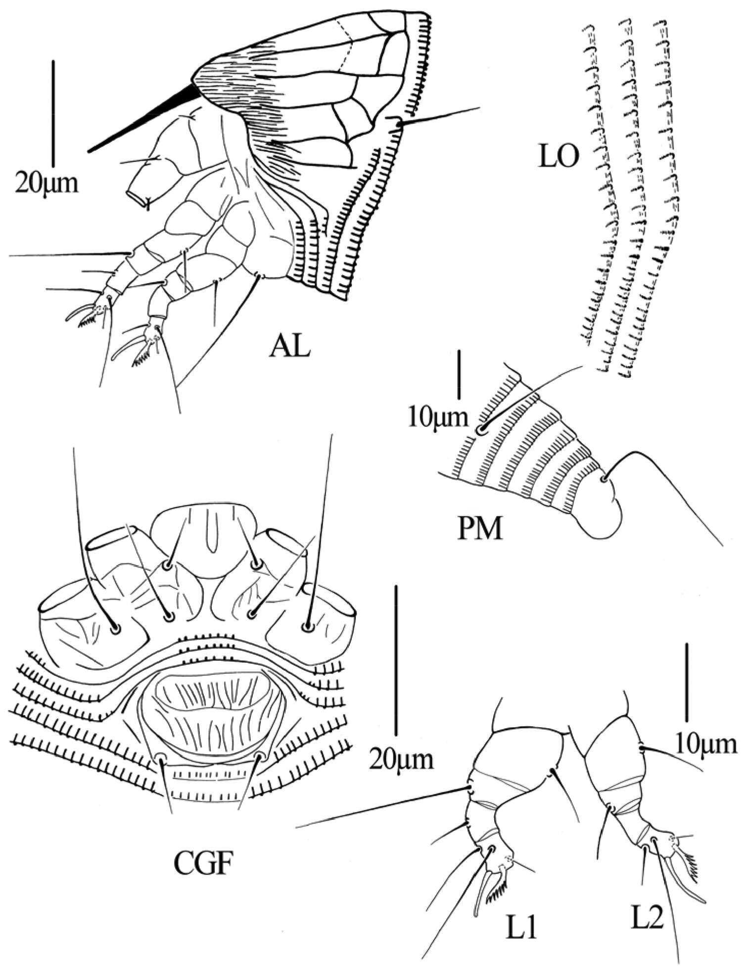
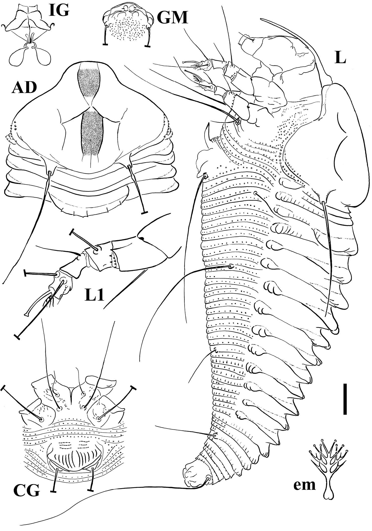
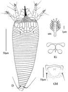
Figure 1.Schematic drawings of Shevtchenkella denticulata sp. n.: AL Lateral view of anterior body region CG Female coxigenital region D Dorsal view em Empodium IG Internal female genitalia LO Lateral view of annuli L1 Leg I PM Lateral view of posterior opisthosoma. Scale bar: 17.5 μm for D; 10 μm for AL, CG, IG, PM; 5 μm for LO, L1; 2.5 μm for em.
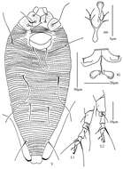
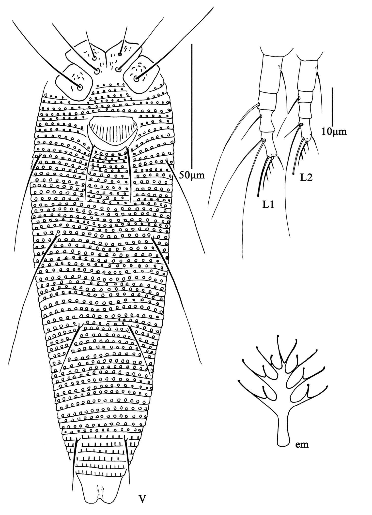
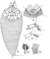
Figures 8–13.Gammaphytoptus schimae sp. n. 8 dorsal view of female 9 coxigenital area of female 10 leg I 11 leg II 12 tarsal empodium 13 lateral view of annuli.
Qiong Wang, Xiao Han, Xiao-Feng Xue, Xiao-Yue Hong
Zookeys
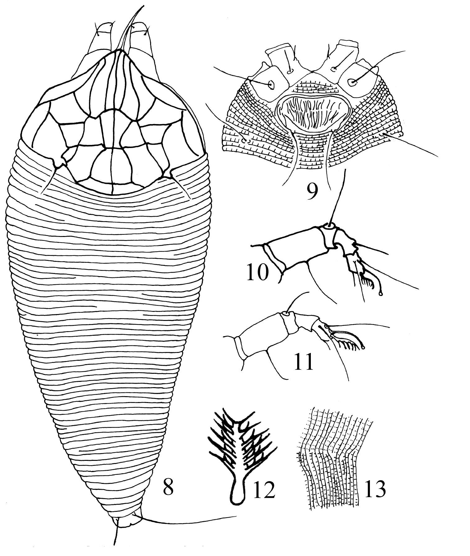
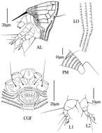
Figure 3.Gammaphytoptus striatilobus sp. n.: AL lateral view of anterior body region LO lateral view of annuli PM lateral view of posterior opisthosoma CGF female coxae and genitalia L1 leg I L2 leg II.
Parisa Lotfollahi, Enrico de Lillo, Karim Haddad Irani-Nejad
Zookeys
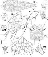
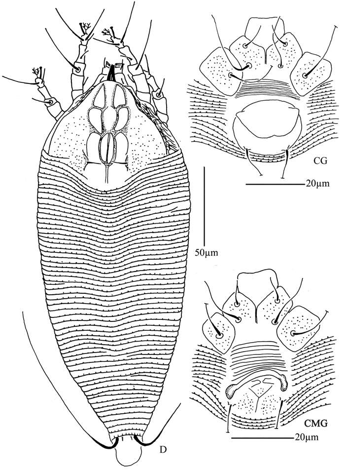
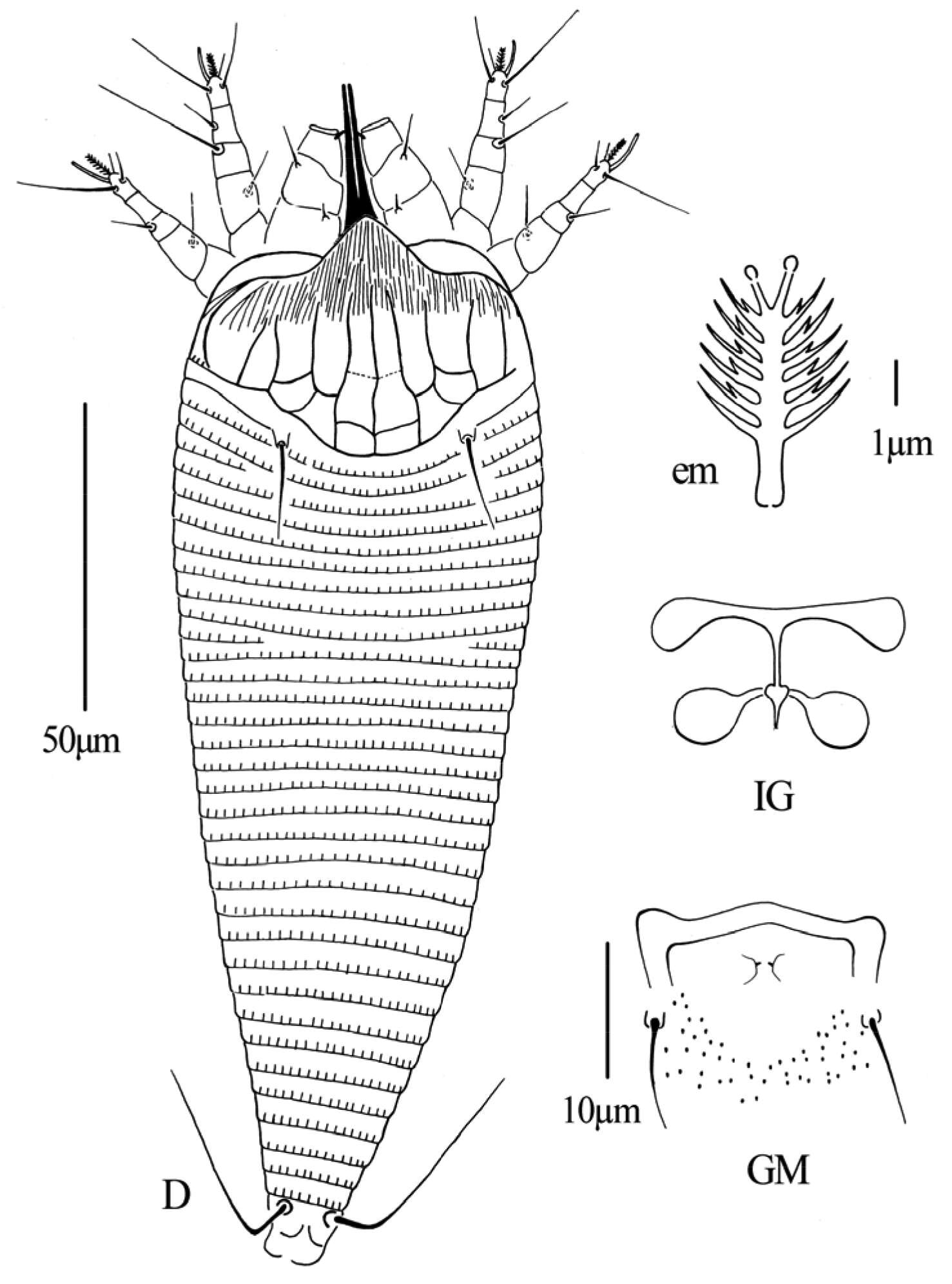
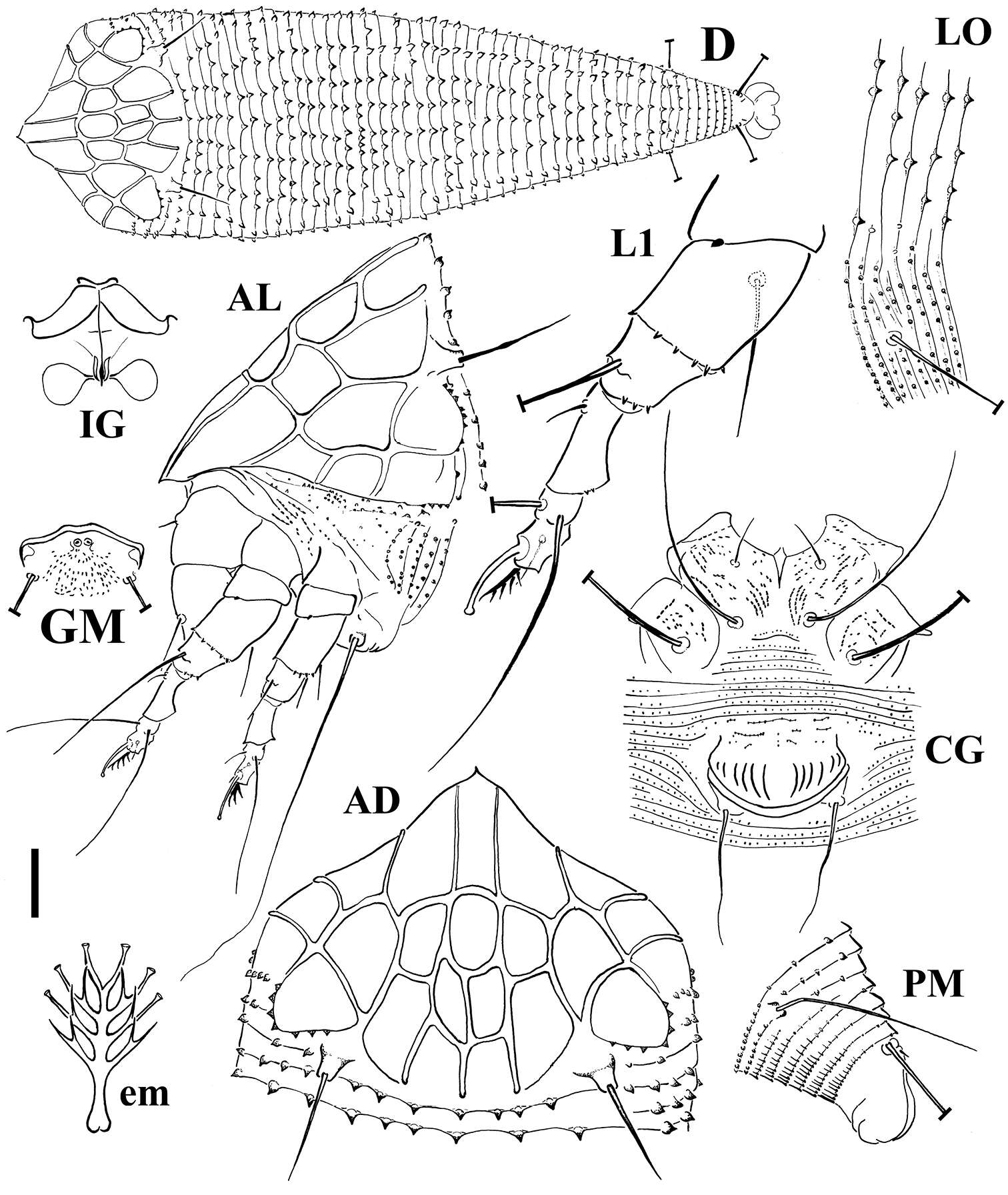
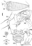
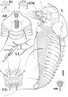
Figure 2.Schematic drawings of Echinacrus ruthenicus sp. n.: AD Dorsal view of anterior body region AL Lateral view of anterior body region CG Female coxigenital region D Dorsal view em Empodium GM Male genital region IG Internal female genitalia LO Lateral view of annuli L1 Leg I PM Lateral view of posterior opisthosoma. Scale bar: 20 μm for D; 10 μm for AD, AL, CG, IG, GM, PM; 5 μm for LO, L1; 2.5 μm for em.
Angsumarn Chandrapatya, Ploychompoo Konvipasruang, Carlos H. W. Flechtmann, Gilberto J. de Moraes
Zookeys
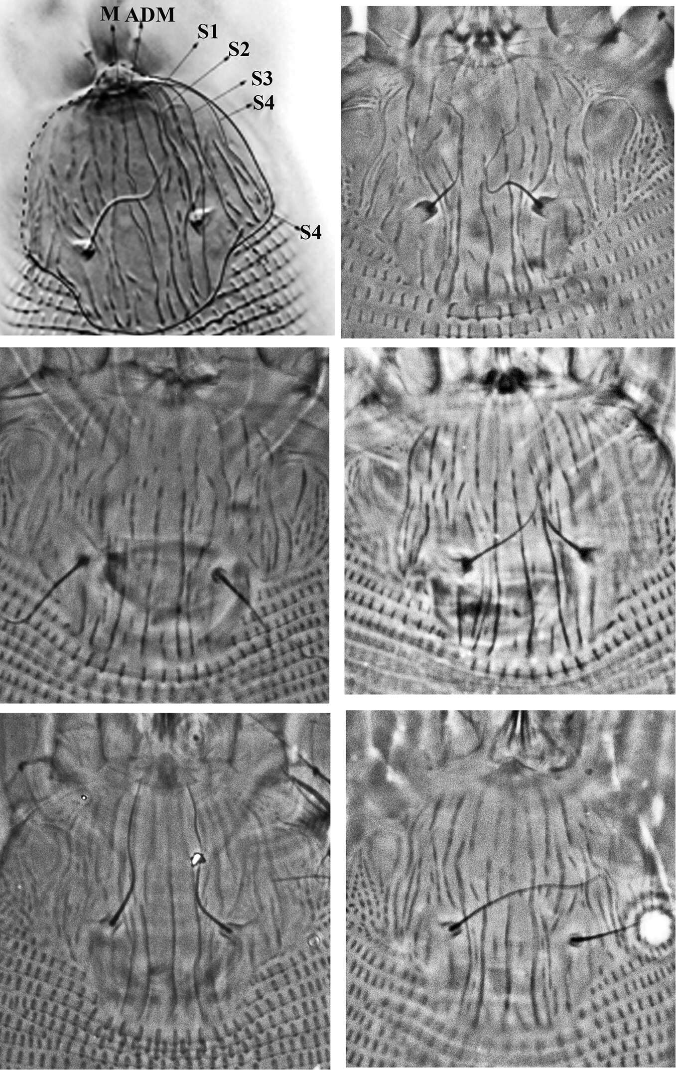
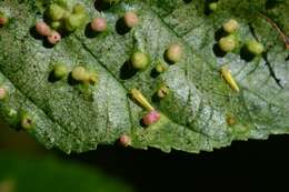
Figure 3.Variation of prodorsal shield sculpture of Colomerus novahebridensis Keifer. The top left figure highlights the prodorsal shield lines: from center to lateral margin, lines running from anterior to posterior margin are interpreted as median (M), admedian (ADM) and submedian lines (S1–S4). Specimens collected in Thailand.
Figure 3.Acaphyllisa tuberculumae sp. n.: A dorsal view of female B ventral view of female C dorsal view of female posterior part D ventral view of female posterior part E prodorsal shield F coxae and female genitalia.
Qiong Wang, Xiao Han, Xiao-Feng Xue, Xiao-Yue Hong
Zookeys
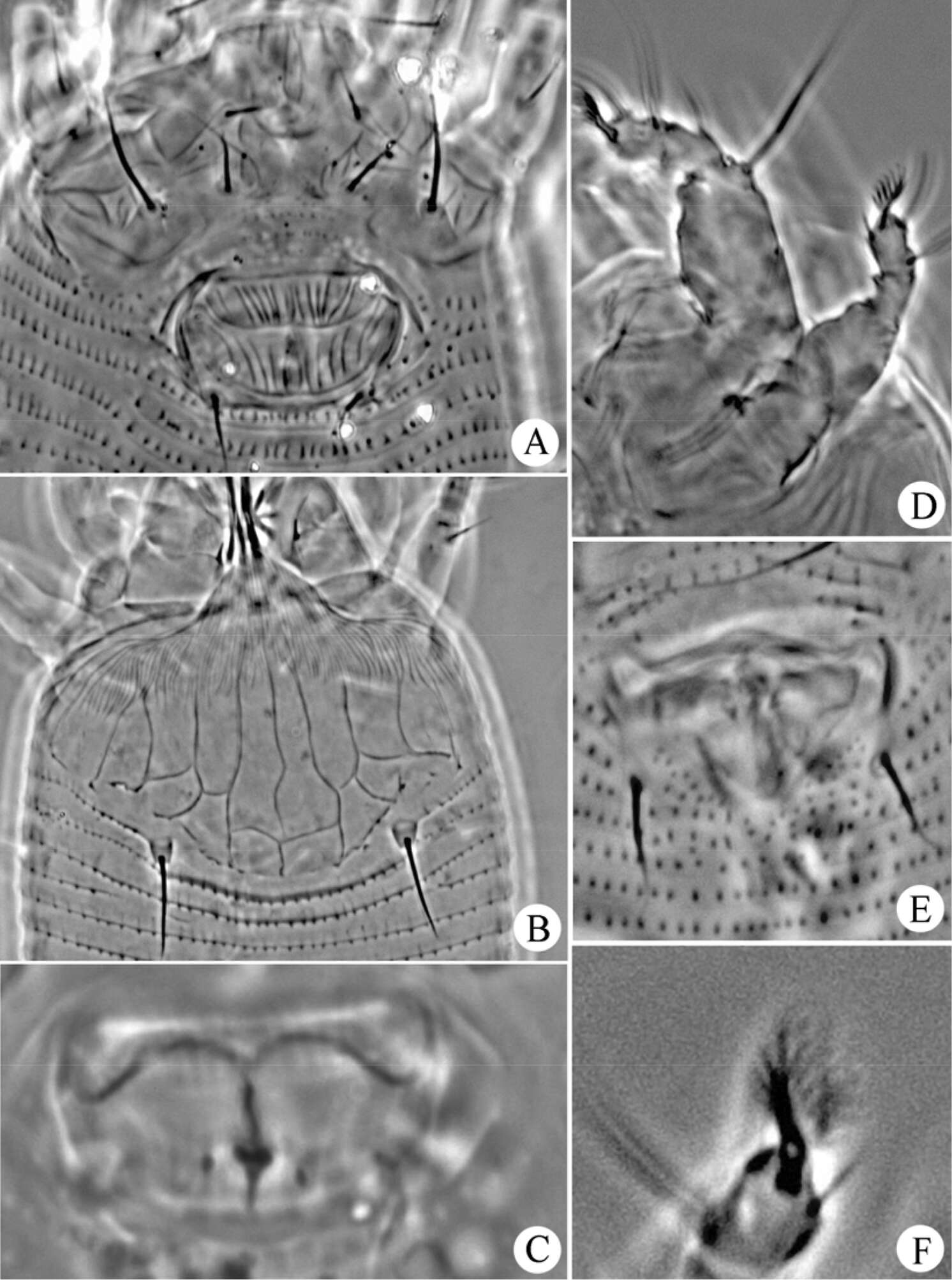
Figure 4.Gammaphytoptus striatilobus sp. n.: A coxae and female genitalia B prodorsal shield C female internal genitalia D leg I and leg II E male genitalia F empodium.
Parisa Lotfollahi, Enrico de Lillo, Karim Haddad Irani-Nejad
Zookeys
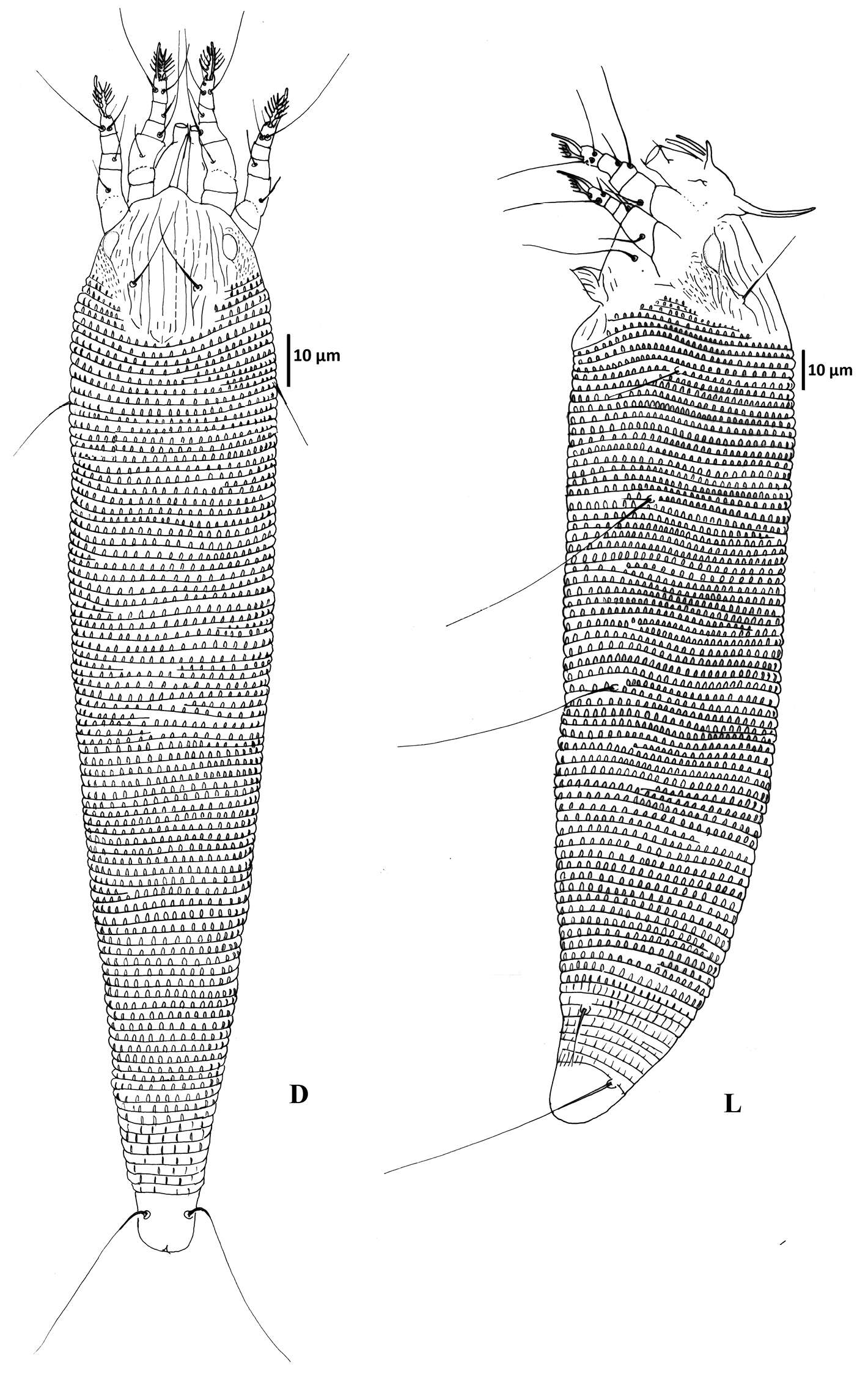
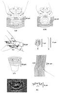
Figure 3.Schematic drawings of Notallus pesthae sp. n.: AD Dorsal view of anterior body region CG Female coxigenital region em Empodium GM Male genital region IG Internal female genitalia L Lateral view L1 Leg I. Scale bar: 10 μm for AD, CG, IG, GM, L, PM; 5 μm for L1; 2.5 μm for em.
Angsumarn Chandrapatya, Ploychompoo Konvipasruang, Carlos H. W. Flechtmann, Gilberto J. de Moraes
Zookeys
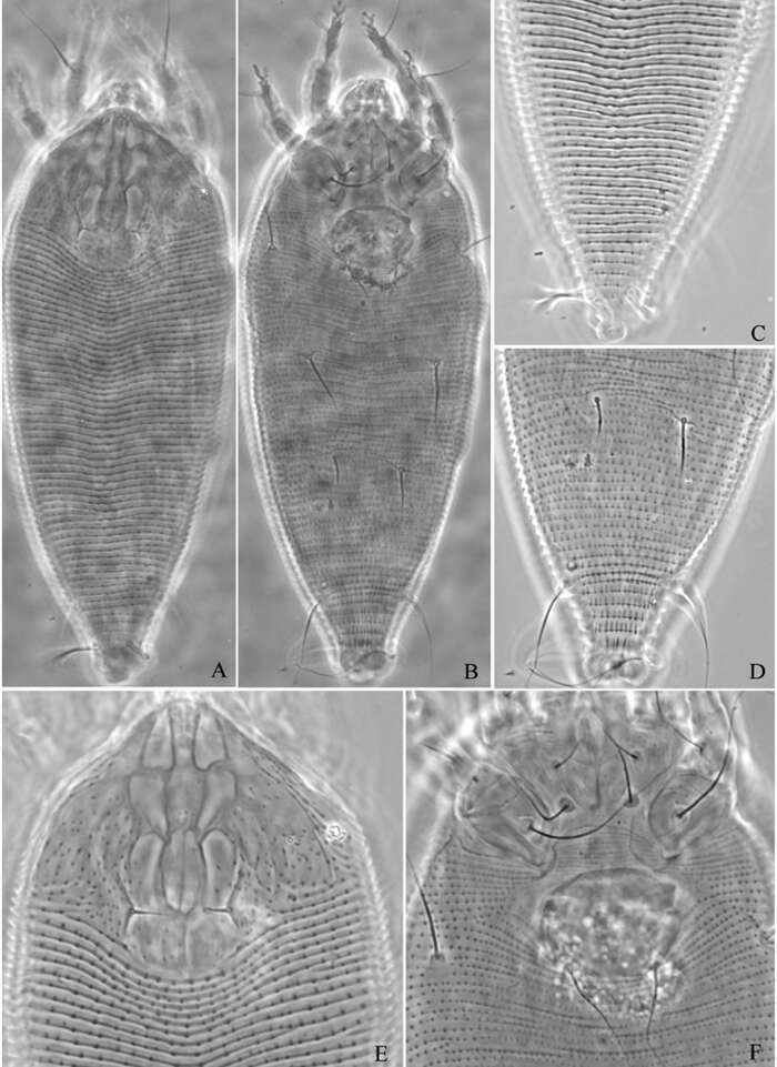
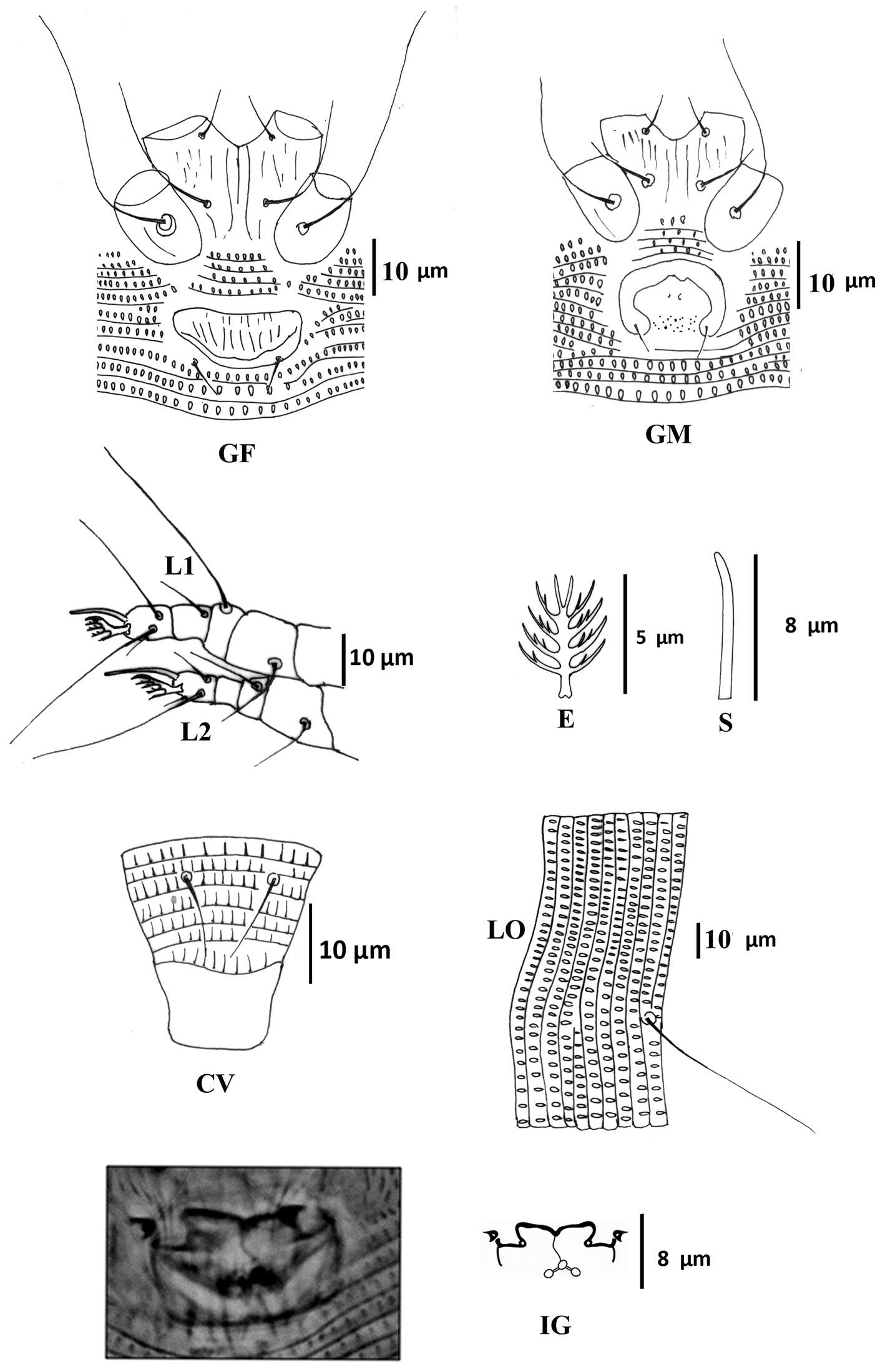
Figure 2.Colomerus novahebridensis Keifer. Female: CV ventral view of caudal region E empodium GF external female genitalia IG internal genitalia L1 leg I L2 leg II LO lateral opisthosoma S solenidion. Male: GM external male genitalia. Specimens collected in Thailand.