-
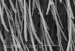
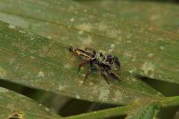
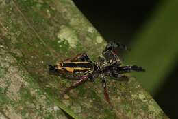
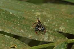
Under a magnification of 186x, 7X greater than PHIL 10075, this scanning electron micrograph (SEM) depicted the hairy dorsal surface of the anterior cephalothorax of a venomous brown recluse spider, Loxosceles reclusa, found inhabiting a Kentucky farm. These are not really hairs at all in the mammalian sense, which are composed of keratin, but are composed of chitin, and are known as setae, which are sensorial in nature, supplying the spider with information about changes in its environment such as changes in temperature, wind direction, and chemical queues such as pheromones and poisons. L. reclusa is sometimes referred to as the violin or fiddle spider, for on its cephalothorax one will see what appears to be coloration in the shape of these stringed instruments, which is quite evident in the color photograph PHIL 1125, depicting a live specimen. Also see PHIL 2224, and 6268 for additional brown recluse images.Created: 2007
-
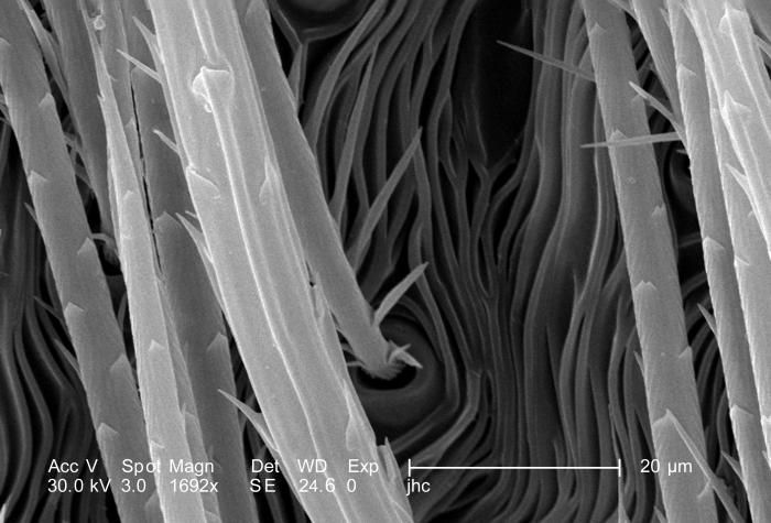
Under a high magnification of 1692x, 4X greater than PHIL 10080, this scanning electron micrograph (SEM) depicted the hairy dorsal surface of the abdominal exoskeleton of a venomous brown recluse spider, Loxosceles reclusa, found inhabiting a Kentucky farm. These are not really hairs at all, in the mammalian sense, which are composed of keratin, but are composed of chitin, and are known as setae, which are sensorial in nature, supplying the spider with information about changes in its environment such as changes in temperature, wind direction, and chemical queues such as pheromones and poisons. L. reclusa is sometimes referred to as the violin or fiddle spider, for on its cephalothorax one will see what appears to be coloration in the shape of these stringed instruments, which is quite evident in the color photograph PHIL 1125, depicting a live specimen. Also see PHIL 2224, and 6268 for additional brown recluse images.Created: 2007
-
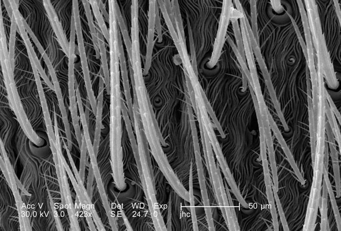
Under a magnification of 423x, 8X greater than PHIL 10079, this scanning electron micrograph (SEM) depicted the hairy dorsal surface of the abdominal exoskeleton of a venomous brown recluse spider, Loxosceles reclusa, found inhabiting a Kentucky farm. These are not really hairs at all, in the mammalian sense, which are composed of keratin, but are composed of chitin, and are known as setae, which are sensorial in nature, supplying the spider with information about changes in its environment such as changes in temperature, wind direction, and chemical queues such as pheromones and poisons. L. reclusa is sometimes referred to as the violin or fiddle spider, for on its cephalothorax one will see what appears to be coloration in the shape of these stringed instruments, which is quite evident in the color photograph PHIL 1125, depicting a live specimen. Also see PHIL 2224, and 6268 for additional brown recluse images.Created: 2007
-
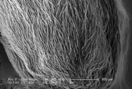
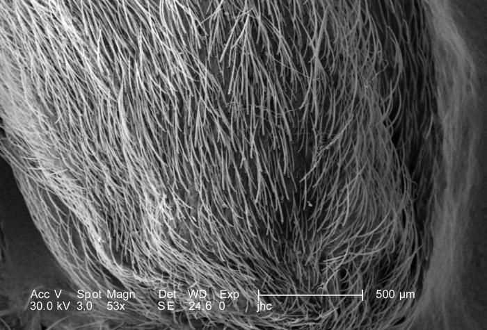
Under a very low magnification of 53x, this scanning electron micrograph (SEM) depicted the hairy dorsal surface of the abdominal exoskeleton of a venomous brown recluse spider, Loxosceles reclusa, found inhabiting a Kentucky farm. These are not really hairs at all, in the mammalian sense, which are composed of keratin, but are composed of chitin, and are known as setae. Chitin is a molecule made up of bound units of acetylglucosamine, joined in such a way as to allow for increased points at which hydrogen bonding can occur. In this way chitin provides increased strength, and durability as an exoskeletal foundation. L. reclusa is sometimes referred to as the violin or fiddle spider, for on its cephalothorax one will see what appears to be coloration in the shape of these stringed instruments, which is quite evident in the color photograph PHIL 1125, depicting a live specimen. Also see PHIL 2224, and 6268 for additional brown recluse images.Created: 2007
-
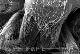
Under a high magnification of 6922x this scanning electron micrograph (SEM) depicted the striated texture found on the exoskeletal surface of a venomous brown recluse spider, Loxosceles reclusa, found inhabiting a Kentucky farm. As arthropods, spiders possess an exoskeleton composed of chitin, which is a molecule made up of bound units of acetylglucosamine, joined in such a way as to allow for increased points at which hydrogen bonding can occur. In this way chitin provides increased strength, and durability as an exoskeletal foundation. L. reclusa is sometimes referred to as the violin or fiddle spider, for on its cephalothorax one will see what appears to be coloration in the shape of these stringed instruments, which is quite evident in the color photograph PHIL 1125, depicting a live specimen. Also see PHIL 2224, and 6268 for additional brown recluse images.Created: 2007
-
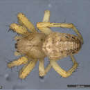
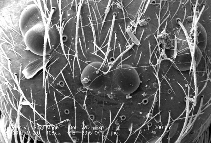
Under a magnification of 108x, approximately twice that of PHIL 10076, this scanning electron micrograph (SEM) depicted the dorsal cephalothorax, i.e., a combination of its head and thoracic regions, of a venomous brown recluse spider, Loxosceles reclusa, found inhabiting a Kentucky farm. Most spiders possess eight eyes (4 pairs), however, as is evidenced in this image, recluse spiders only possess 3 pairs. L. reclusa is sometimes referred to as the violin or fiddle spider, for on its cephalothorax one will see what appears to be coloration in the shape of these stringed instruments, which is quite evident in the color photograph PHIL 1125, depicting a live specimen. Youll also note this spiders four pairs of jointed legs, which places it in the Phylum, Arthropoda, and the Class, Arachnida. Also see PHIL 2224, and 6268 for additional brown recluse images.Created: 2007
-
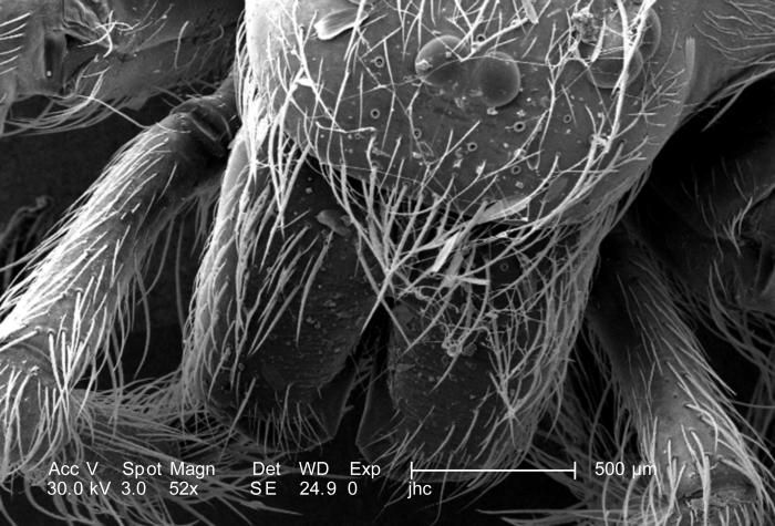
Under a low magnification of only 52x, approximately twice that of PHIL 10075, this scanning electron micrograph (SEM) depicted the dorsal cephalothorax, i.e., a combination of its head and thoracic regions, of a venomous brown recluse spider, Loxosceles reclusa, found inhabiting a Kentucky farm. Most spiders possess eight eyes (4 pairs), however, recluse spiders only possess six. L. reclusa is sometimes referred to as the violin or fiddle spider, for on its cephalothorax one will see what appears to be coloration in the shape of these stringed instruments, which is quite evident in the color photograph PHIL 1125, depicting a live specimen. Youll also note this spiders four pairs of jointed legs, which places it in the Phylum, Arthropoda, and the Class, Arachnida. Also see PHIL 2224, and 6268 for additional brown recluse images.Created: 2007
-
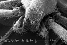
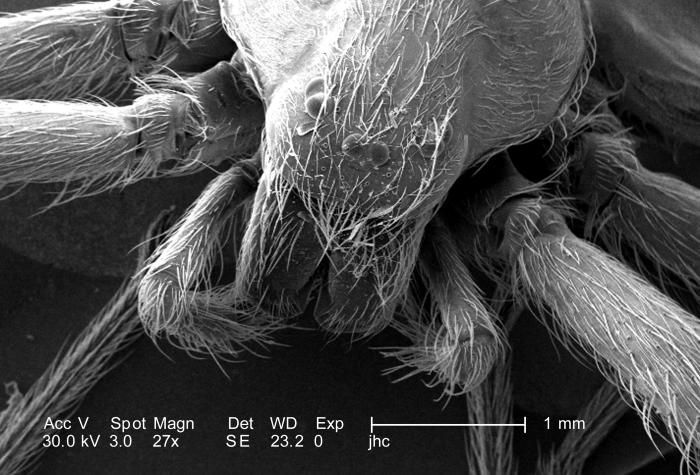
Under a low magnification of only 27x, twice that of PHIL 10074, this scanning electron micrograph (SEM) depicted the dorsal cephalothorax, i.e., a combination of its head and thoracic regions, of a venomous brown recluse spider, Loxosceles reclusa found inhabiting a Kentucky farm. Most spiders possess eight eyes (4 pairs), however, as is evidenced in this image, recluse spiders only possess six. L. reclusa is sometimes referred to as the violin or fiddle spider, for on its cephalothorax one will see what appears to be coloration in the shape of these stringed instruments, which is quite evident in the color photograph PHIL 1125, depicting a live specimen. Youll also note this spiders four pairs of jointed legs, which places it in the Phylum, Arthropoda, and the Class, Arachnida. Also see PHIL 2224, and 6268 for additional brown recluse images.Created: 2007
-
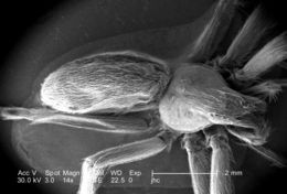
Under a very low magnification of only 14x, this scanning electron micrograph (SEM) depicted the dorsal surface of a venomous brown recluse spider, Loxosceles reclusa, found inhabiting a Kentucky farm. Most spiders possess eight eyes (4 pairs), however, recluse spiders only possess 3 pairs. L. reclusa is sometimes referred to as the violin or fiddle spider, for on its cephalothorax, i.e., a combination of its head and thoracic regions, one will see what appears to be coloration in the shape of these stringed instruments, which is quite evident in the color photograph PHIL 1125, depicting a live specimen. Youll also note this spiders four pairs of jointed legs, which places it in the Phylum, Arthropoda, and the Class, Arachnida. Also see PHIL 2224, and 6268 for additional brown recluse images.Created: 2007
-
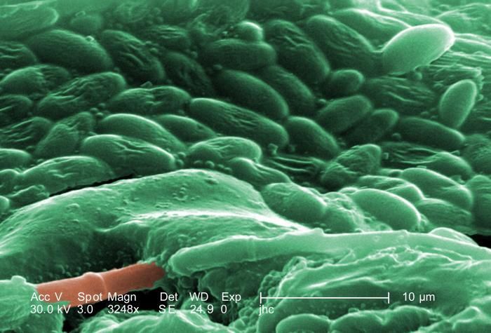
Under three increasingly greater magnifications, this being the highest at 3248X (see PHIL 10089, 10090), what is depicted here is an unidentified pore located on the dorsal abdomen of a venomous brown recluse spider, Loxosceles reclusa, found inhabiting a Kentucky farm. Note the material surrounding the pores orifice, and as the magnification increases, it becomes evident that the material is composed of an unidentified bacterial biofilm. It is not known if these were existing symbiotically upon the spiders exoskeleton, or if they were pathologic in nature, signifying manifestations of a progressive disease process? See PHIL 10088 for a black and white version of this image.L. reclusa is sometimes referred to as the violin or fiddle spider, for on its cephalothorax one will see what appears to be coloration in the shape of these stringed instruments, which is quite evident in the color photograph PHIL 1125, depicting a live specimen.Created: 2007
-
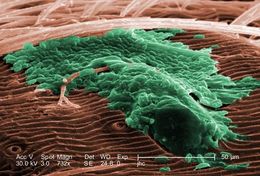
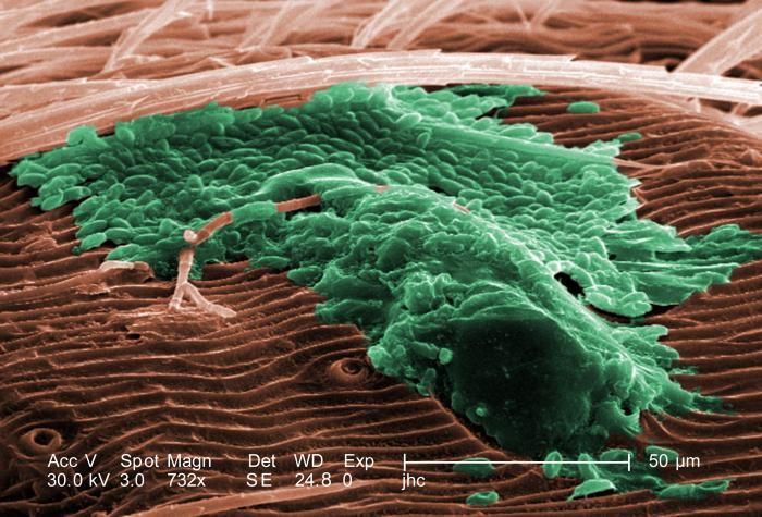
Under three increasingly greater magnifications, this being midway at 732X (see PHIL 10089, 10091), what is depicted here is an unidentified pore located on the dorsal abdomen of a venomous brown recluse spider, Loxosceles reclusa, found inhabiting a Kentucky farm. Note the material surrounding the pores orifice, and as the magnification increases, it becomes evident that the material is composed of an unidentified bacterial biofilm. It is not known if these were existing symbiotically upon the spiders exoskeleton, or if they were pathologic in nature, signifying manifestations of a progressive disease process? See PHIL 10087 for a black and white version of this image.L. reclusa is sometimes referred to as the violin or fiddle spider, for on its cephalothorax one will see what appears to be coloration in the shape of these stringed instruments, which is quite evident in the color photograph PHIL 1125, depicting a live specimen.Created: 2007
-
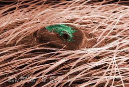
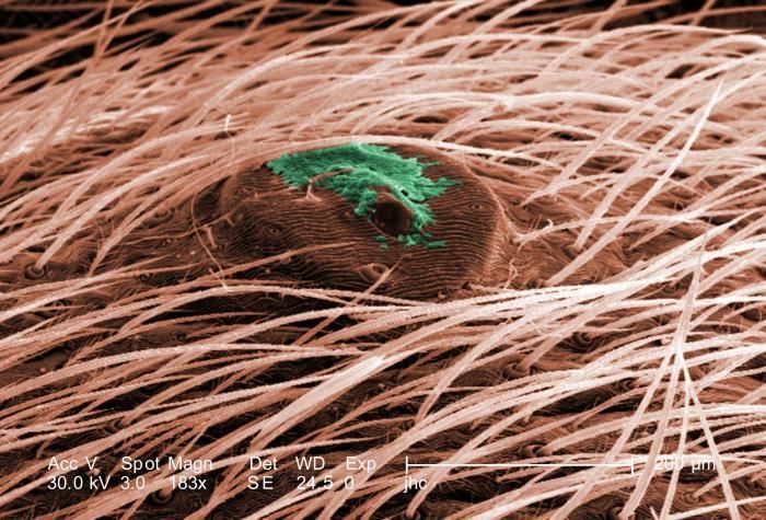
Under three increasingly greater magnifications, this being the lowest at 183X (see PHIL 10089, 10090), what is depicted here is an unidentified pore located on the dorsal abdomen of a venomous brown recluse spider, Loxosceles reclusa, found inhabiting a Kentucky farm. Note the material surrounding the pores orifice, and as the magnification increases, it becomes evident that the material is composed of an unidentified bacterial biofilm. It is not known if these were existing symbiotically upon the spiders exoskeleton, or if they were pathologic in nature, signifying manifestations of a progressive disease process? See PHIL 10086 for a black and white version of this image.L. reclusa is sometimes referred to as the violin or fiddle spider, for on its cephalothorax one will see what appears to be coloration in the shape of these stringed instruments, which is quite evident in the color photograph PHIL 1125, depicting a live specimen.Created: 2007
-
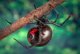
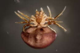
This 2007 photograph, captured by CDC biomedical photographer, James Gathany, depicted a female black widow spider, Latrodectus mactans, as she was in the process of spinning her web upon a tree branch. Youll note the characteristic red hourglass located on her inferior abdominal surface, which can vary in coloration from yellowish, to shades of orange and red, and at times, can even be white. The females body is an overall shiny jet-black in color. This spider was found on a farm, here in the state of Georgia.Created: 2007
-
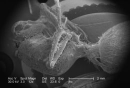
Under a very low magnification of only 12X, this scanning electron micrograph (SEM) depicted a rather ominous scene, for entangled in this brown recluse spider web was the exoskeletal remains of an unidentified insect, which was believed to be an ant. Known as spider silk, the strands of silk are produced by the spiders spinnerets, which are glands located in the distal tip of its abdomen. Once the prey has become entangled in the web, the spider will cautiously, though aggressively, approach the prey, subduing it with a neurotoxic bite, which also contains proteolytic, or protein-destroying enzymes, and further enwraps the prey in a web cocoon like the one seen here.Created: 2007
-
-
-
-
-
-
-
-
-
-