-
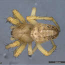
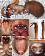
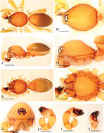
Figures 1–6.Copulatory organs of Selenops arikok sp. n. female holotype from Arikok National Park, Aruba (EME sel_068) 1–2 Selenops curazao Alayón-García male holotype from CarMaBI, Curaçao, Netherlands Antilles (MCZ) 3–4 female paratype from Piscadera Baai building, Curaçao, Netherlands Antilles (MCZ) 5–6, 1, 5 epigyne, ventral view 2, 6 spermathecae, dorsal view 3 male pedipalp, ventral view 4 male pedipalp, retrolateral view. Scale bar = 0.40 mm (1–2), 0.30 mm (3–6). Abbreviations: S = septum, MF = median field, EP = epigynal pockets, FD = fertilization duct, SP = spermathecae, PF = posterodorsal fold, C = conductor, CY = cymbium, MA = median apophysis, E = embolus, RTA = retrolateral tibial apophysis, VRTA = ventral retrolateral tibial apophysis, DRTA = dorsal retrolateral tibial apophysis.
-
Jeremy Miller, Cahyo Rahmadi
Zookeys
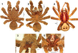
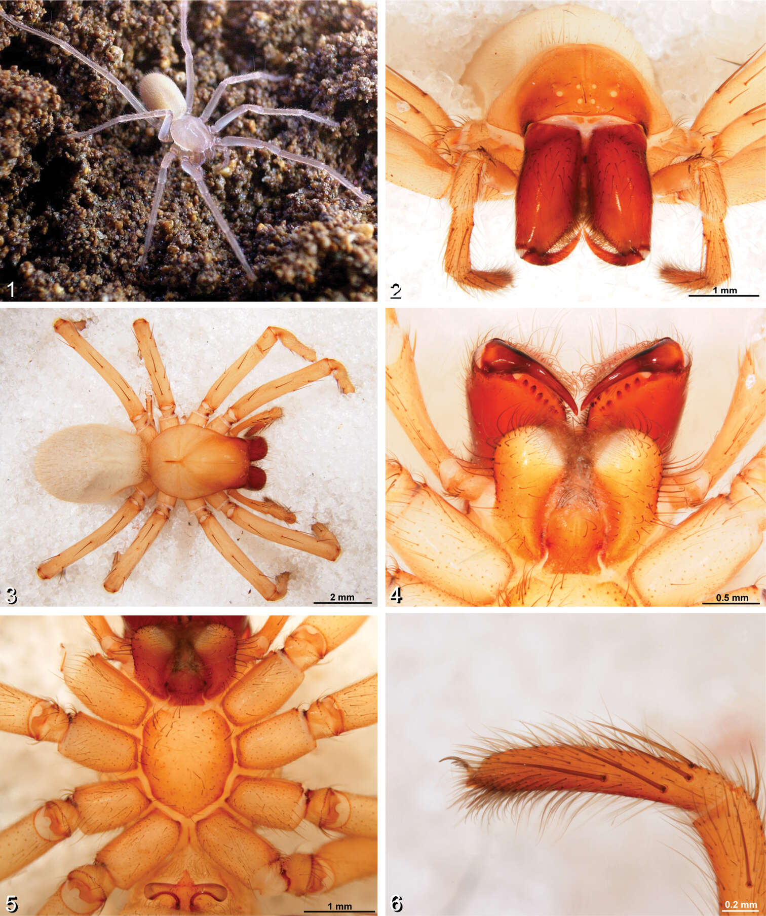
Figures 1–6.Amauropelma matakecil sp. n. 1 female habitus 2–6 habitus of female holotype (MZB.Aran.500) 1 Portrait of live specimen in natural habitat from Gua Nguwik, Central Java (Photo S. Harjanto) 2 Anterior view 3 Dorsal view 4 Ventral view showing labium, endites, and chelicerae 5 Ventral view showing sternum, coxae, and trochanters 6 Left pedipalpal, retrolateral view.
-
Jeremy A. Miller, Charles E. Griswold, Nikolaj Scharff, Milan Řezáč, Tamás Szűts, Mohammad Marhabaie
Zookeys
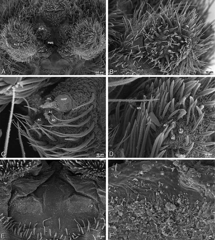
Figure 78.A–F Seothyra henscheli from Gobabeb Station, Namibia (SMN 40828, NMN), scanning electron micrographs of male spinnerets. A overview B right ALS C left PMS D left PLS E vestigial cribellum F detail of vestigial cribellum. AC aciniform gland spigot ALS anterior lateral spinneret mAP minor ampullate gland spigot MS modified spigot PI piriform gland spigot PLS posterior lateral spinneret PMS posterior median spinneret.
-
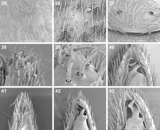
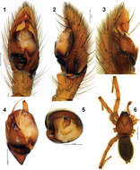
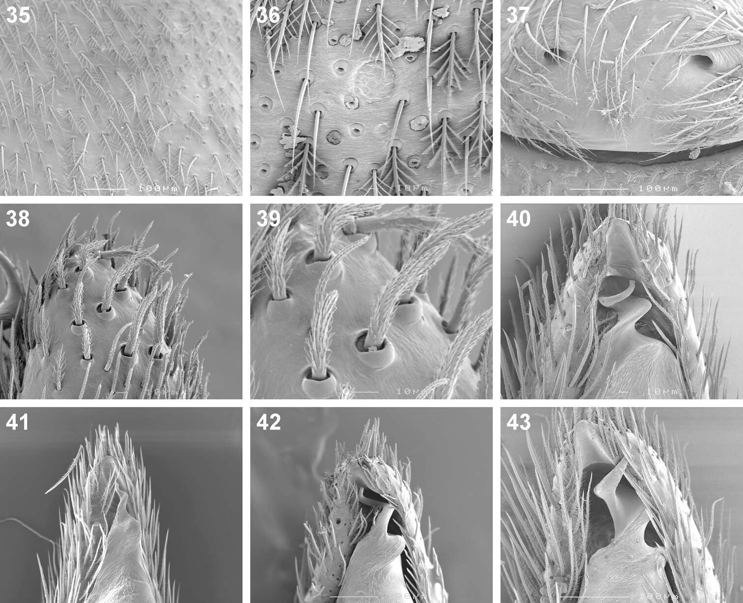
Figures 35–43.Scanning electron microscope photographs of Cambalida dippenaarae sp. n. (35–39, 42), Cambalida compressa sp. n. (40), Cambalida deminuta (Simon, 1909) (41) and Cambalida loricifera (Simon, 1885) (43): 35 female, dorsal abdominal surface 36 dorsal abdominal sigillum and detail of plumose setae 37 female epigyne 38 thickened setae at dorsal distal end of male palpal cymbium 39 detail of modified setae 40–43 male emboli.
-
Figures 1–6.General habitus photographs of Copa flavoplumosa Simon, 1885 (1–4) and Copa kei sp. n. (5, 6): 1 female from Lesideng Research Camp, Botswana 2 female from Livingtone, Zambia 3 male and 4 female from Wildlives Game Farm, Zambia 5 female from Hogsback, South Africa 6 male from Cwebe Nature Reserve, South Africa.
-
Figures 1–6.Mallinella sphaerica sp. n., 1 male habitus, dorsal view 2 female habitus, dorsal view 3 male ocular area, frontal view 4 female, ocular area, frontal view 5 male, posterior ventral spines, ventral view 6 female, posterior ventral spines, ventral view. Scale bars: 2 mm (1–2); 0.5 mm (3–4); 0.2 mm (5–6).
-
Dan Quan, Jian Chen, Jie Liu
Zookeys
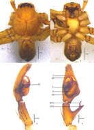
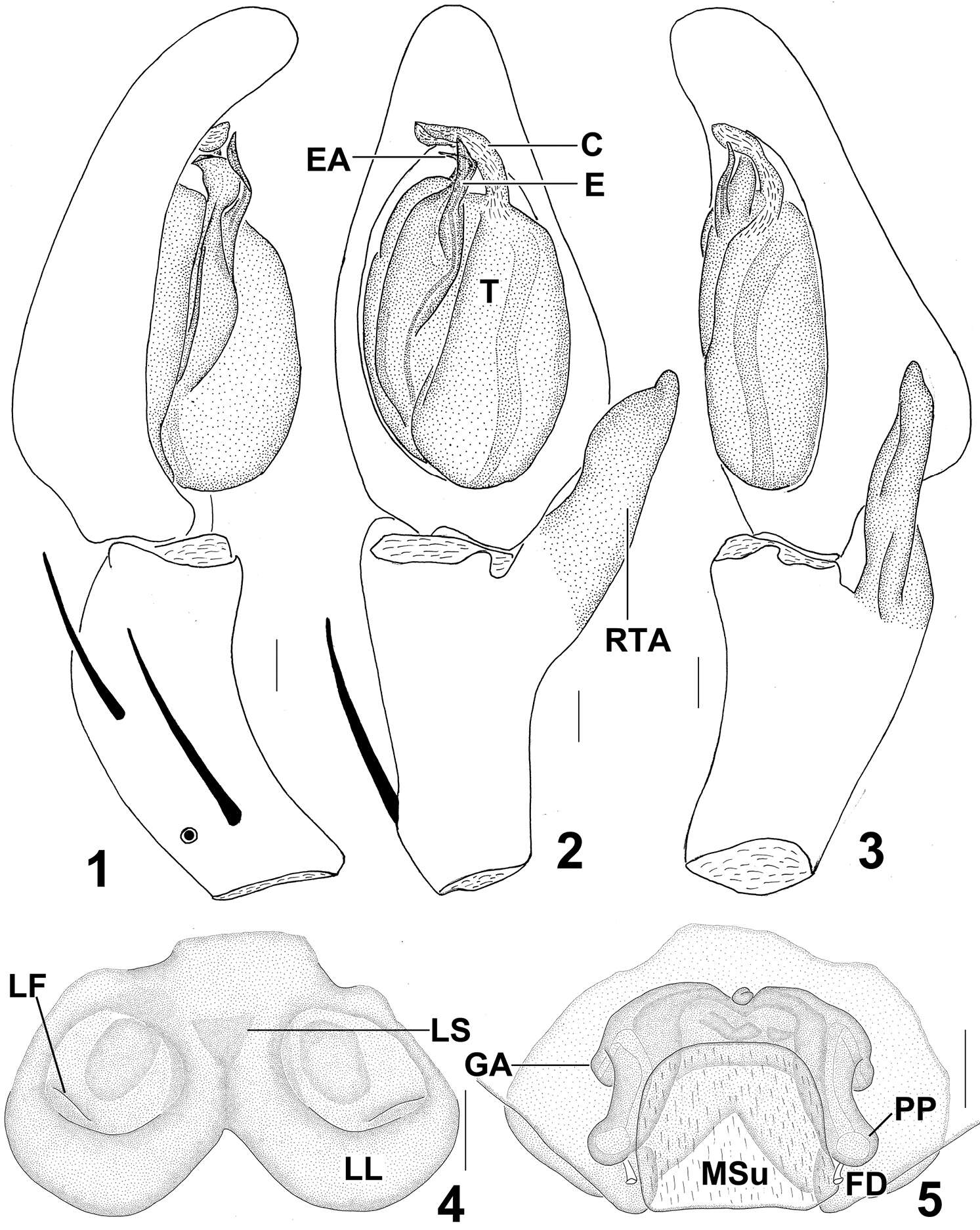
Figures 1–5.Sinopoda serrata (Wang, 1990), from Tiantangzhai National Forest Park (Hubei Province, China). 1 Left male palp, prolateral view 2 Left male palp, ventral view 3 Left male palp, retrolateral view 4 Epigyne, ventral view 5 Vulva, dorsal view. Scales = 0.2 mm. C conductor, E embolus, EA embolic apophysis, FD fertilization duct, GA glandular appendage, LF lateral furrow, LL lateral lobes, LS lobal septum, MSu membranous sac unexpanded, RTA retrolateral tibial apophysis, PP posterior part of spermathecae, T tegulum.
-
Peter Michalik, Luis Piacentini, Elisabeth Lipke, Martin J. Ramírez
Zookeys
Figure 3.Somatic characters of the female of Progradungula otwayensis. A Lateral view of prosoma and opisthosoma (ZIMG II/28128) B Dorsal view of prosoma (MV) C Ventral view of Prosoma (MV) D Frontal view of ocular area (ZIMG II/28128) E Ventral view of opisthosoma F Tarsus of leg I G Tarsus of leg IV H Calamistrum. I Ventral view of spinnerets. Scale bar in F–H is 500 µm.
-
Yuri M. Marusik, Mikhail M. Omelko
Zookeys
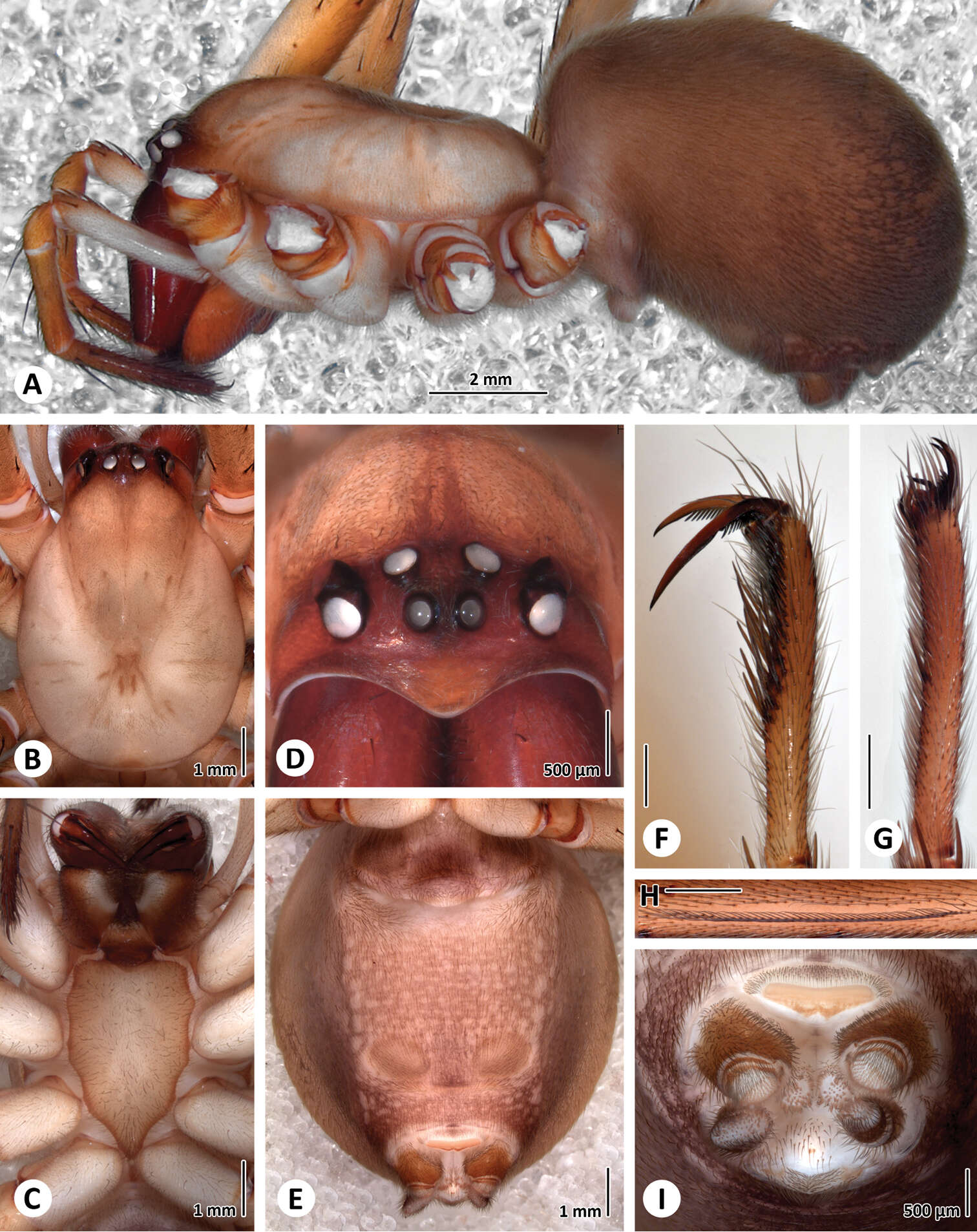
Figures 1–5.General appearance of males of Cryptothele verrucosa (1–3) and Cryptothele alluaudi (4–5). 1, 4 dorsal 2 ventral 3, 5 frontal 4–5 after Marusik and Omelko (2012). Abbreviations: AL anterior lateral eye; AM anterior median eye; At anal tubercle, Sn spinneret.
-
Feng Zhang, Bao-Shi Zhang, Zhi-Sheng Zhang
Zookeys
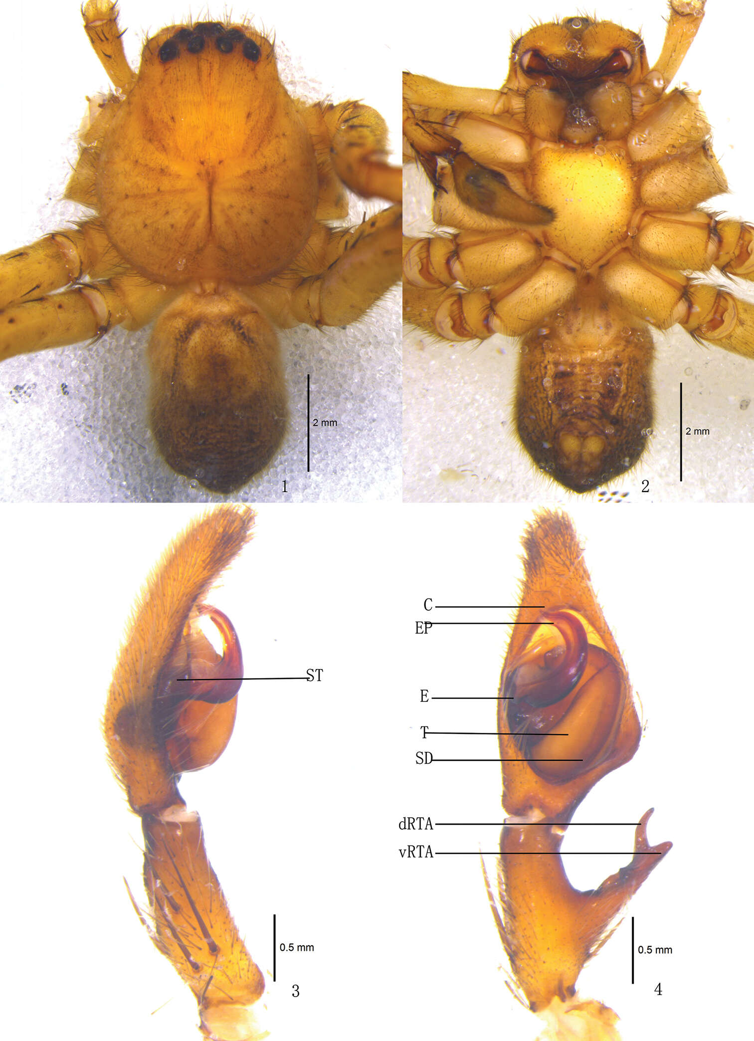
Figures 1–4.Pseudopoda acuminata sp. n., Male (SP–SC–03–0050): 1–2 Body (1 dorsal 2 ventral) 3–4 Left palp (3 prolateral 4 ventral). Abbreviations: C conductor; dRTA dorsal branch of retrolateral tibial apophysis; E embolus; EP embolic projection; SD sperm duct; ST subtegulum; T tegulum; vRTA ventral branch of retrolateral tibial apophysis. Scale bars: 2 mm (1–2); 0.5 mm (3–4).
-
Ning Sun, Yuri M. Marusik, Lihong Tu
Zookeys
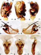
Figure 2.Acanoides beijingensis sp. n. A male palp, prolateral B male palp, prolateral, with embolic division removed C male palp, retrolateral D embolic division, ventral E embolic division, dorsal F epigynum, ventral G epigynum, dorsal H epigynum, lateral. CG copulatory groove; CO copulatory opening; DP dorsal plate; EA extensible area of epigynal basal part; EM embolic membrane; EP embolus proper; FG fertilization groove; FiG Fickert’s gland; LC lamella characteristica; MP median plate; P paracymbium; PCA proximal cymbial apophysis; R radix; S spermathecae; TA terminal apophysis; TH thumb of embolus; VP ventral plate. [Scale bars: mm].
-
Figure 1.Sinamma oxycera gen. n. & sp. n., male holotype (A–B, E, G) and female paratype (C–D, F, H). A–F Habitus G, H Prosoma. A, C dorsal view B, D ventral view E, F lateral view G, H anterior view.
-
Figure 4.Xyphinus hwangi sp. n., male. A, C, E habitus, dorsal, lateral and ventral views B, D, F, G prosoma, dorsal, lateral, ventral and anterior views H–J left palp, retrolateral, prolateral and dorsal views. Scale bars: A, C, E = 0.4 mm; B, D, F–J= 0.2 mm.
-
Dmitri V. Logunov, Yuri M. Marusik
Zookeys
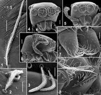
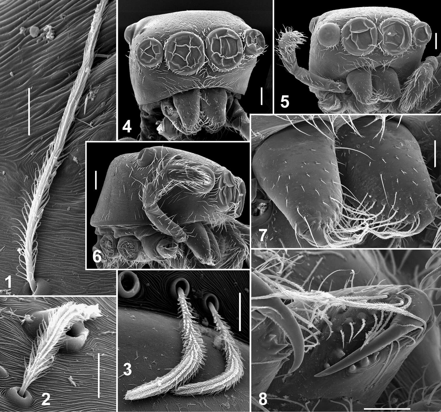
Figures 1–8.Somatic characters of Eupoa lehtineni sp. n. 1–3 plumose scales on female carapace. 4 male carapace, frontal view 5 female carapace, frontal view 6 ditto, lateral view 7 female chelicerae, frontal view 8 female fang and cheliceral teeth. Scale bars: 10 μm (1–3), 50 μm (7–8), 0.1 mm (4–6).
-
Carles Ribera, Mert Elverici, Kadir Boğaç Kunt, Recep Sulhi Özkütük
Zookeys
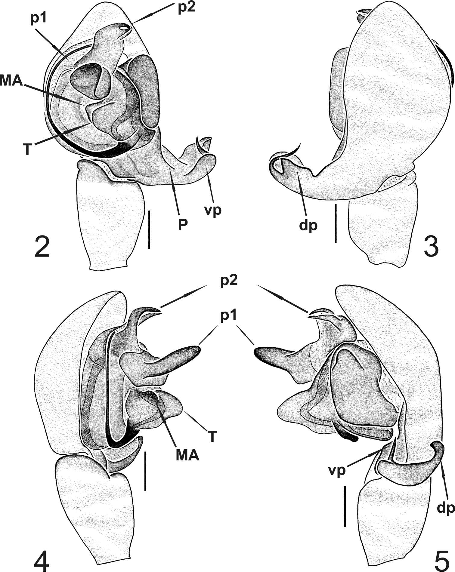
Figures 2–5.Typhlonesticus gocmeni sp. n. male palp. 2 ventral view 3 dorsal view 4 prolateral view 5 retrolateral view. Abbreviations: T = tegulum, MA = median apophysis, p1 = process 1 of TTA, p2 = process 2 of TTA, P = paracymbium, vp = ventral process of paracymbium, dp = dorsal process of paracymbium. Scale bars 0.1 mm.
-
Yuri M. Marusik, Alexander A. Fomichev, Mikhail M. Omelko
Zookeys
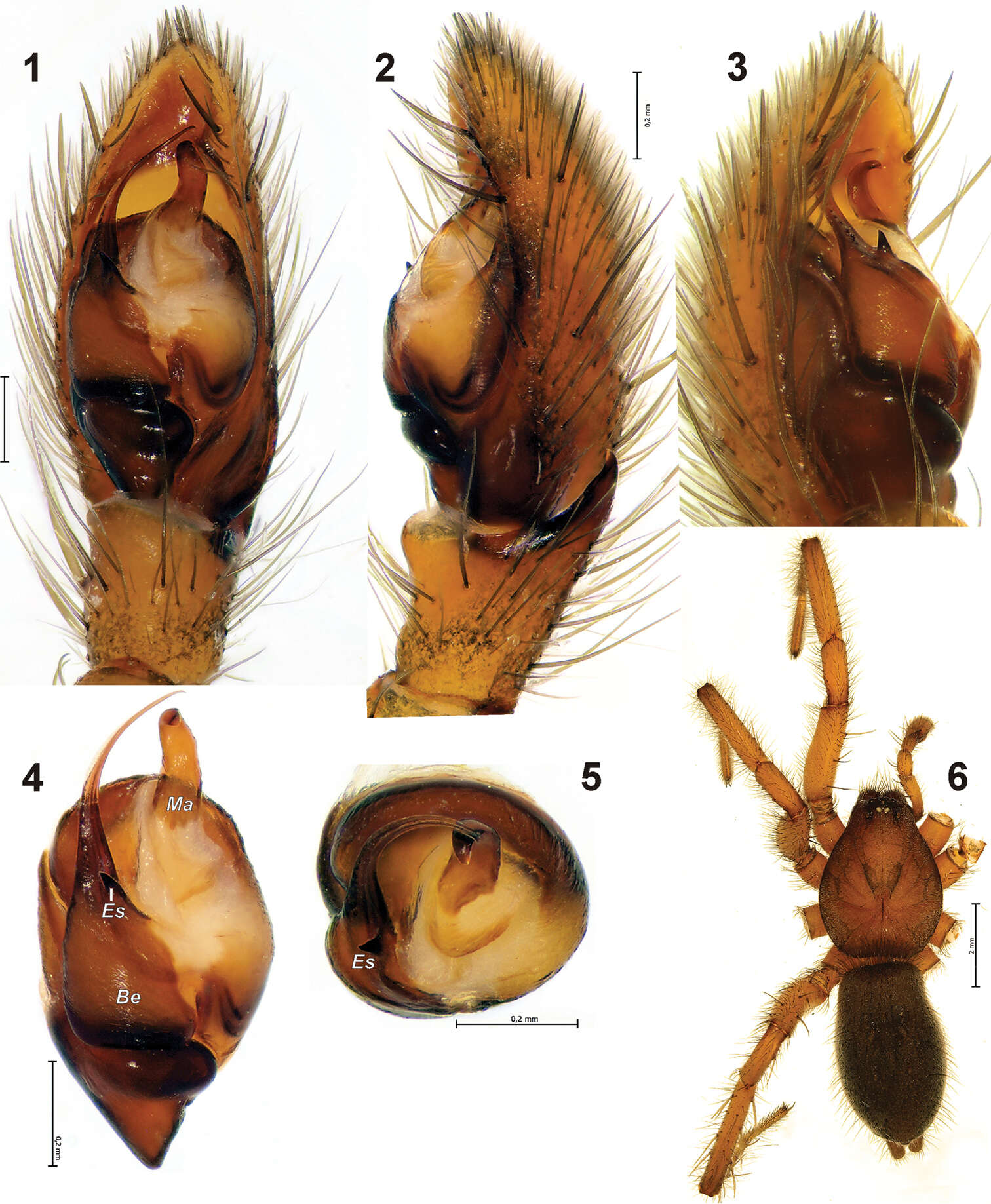
Figures 1–6.Holotype of Gnaphosa khovdensis sp. n. 1–3 male palp, ventral, retro and prolateral 4–5 bulbus, ventral and from above 6 habitus. Scale = 0.2 mm if not otherwise indicated. Be – base of embolus; Es – embolic spine; Ma – median apophysis.
-
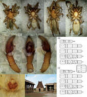
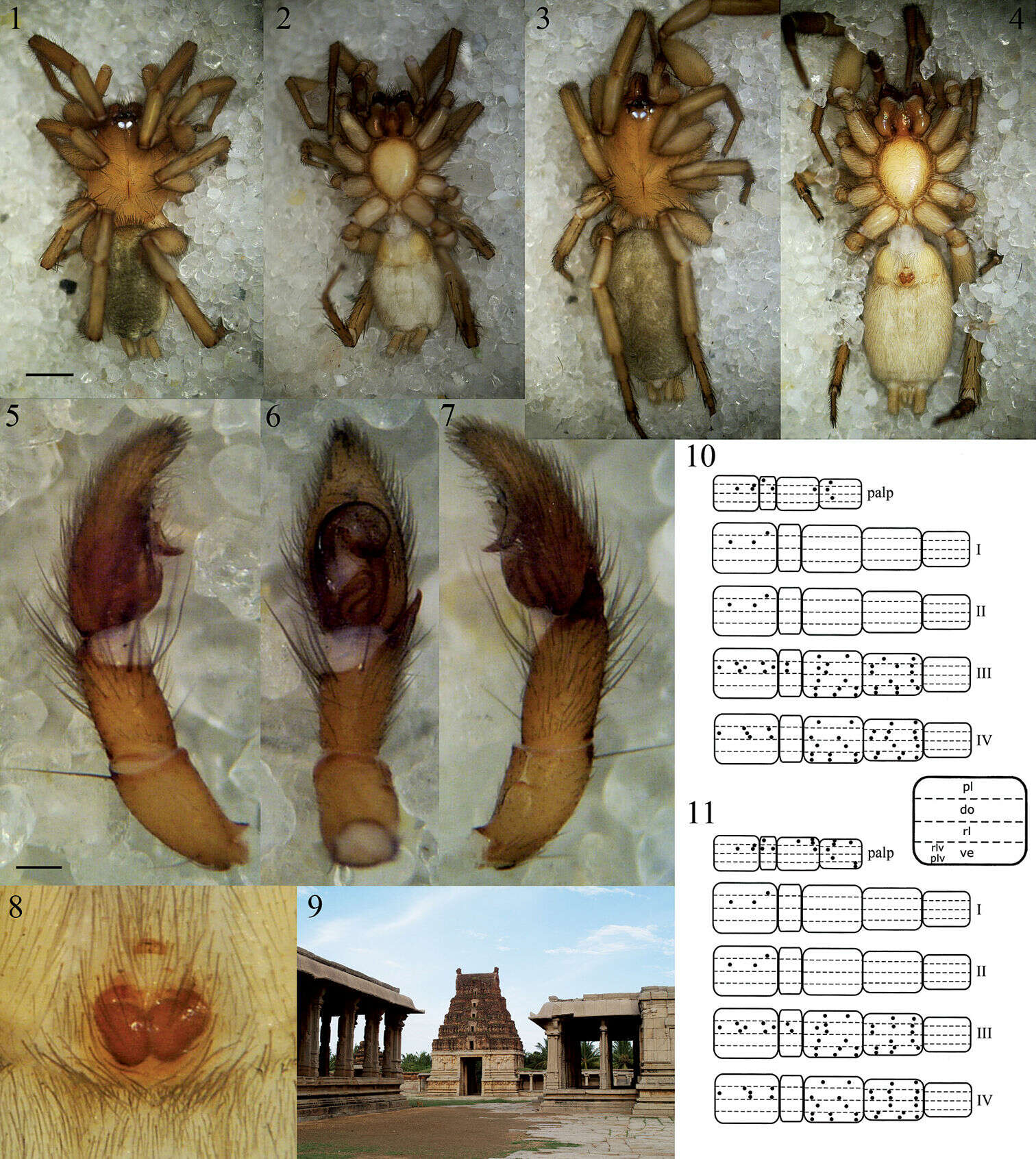
Figures 1–11.Heser vijayanagara sp. n. 1 Male holotype, dorsal 2 Male holotype, ventral 3 Female allotype, dorsal 4 Female allotype, ventral 5 Male palp, prolateral 6 Male palp, ventral 7 Male palp, retrolateral 8 Epigyne, ventral 9 Pattabhirama temple in close vicinity of the locus typicus, giving a good impression of the type of terrain where the type specimens were found 10 Male leg spination diagram, legend below right 11 Female leg spination diagram. Scale bars: 1–4: 1.0; 5–8: 0.25.
-
Sarah C. Crews, Mark S. Harvey
Zookeys
Figures 15–22.Copulatory organs of Karaops monteithi sp. n., female holotype from Lankelly Creek, Coen District, North Queensland, Australia (QM S61052) (15–16), Karaops alanlongbottomi sp. n., male holotype from northwest tip of Degerando Island, Champagny Islands, Western Australia, Australia (WAM T93/1330) (17–18), Karaops keithlongbottomi sp. n., male holotype from Drysdale River Station, Western Australia, Australia (WAM T55002) (19–20), and Karaops larryoo sp. n. male holotype from north of Larryoo, Drysdale River National Park, Western Australia, Australia (WAM T93/1333) (21–22). 15 epigyne, ventral view 16 spermathecae, dorsal view 17, 19, 21 male pedipalp, ventral view 18, 20, 22 male pedipalp, retrolateral view. Scale bar: (15–16) 0.25 mm, (17–22)0.50 mm. Abbreviation: C = conductor.
-
Jimena, Andalucia, Spain
-
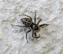
We saw this girl on a salt lake and she could run like the wind. Photo: JeaniD: Artoriopsis Expolita (Polished Wolf Spider) Ethan YeomanExplore 16 October 2019 #5
-
-
The Settlement, Christmas Island
-
Inhabitant no. 6 in our room at Playa Giron, Cuba
-
Ipswich, England, United Kingdom