-
-
-
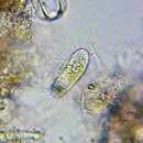
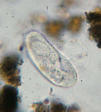
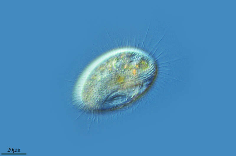
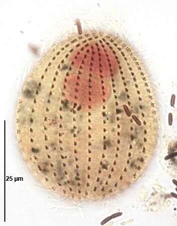
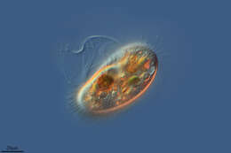
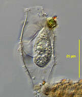
[taxonomy:genus=Cyclidium]
Date:
12 Aug 2011
Location:
Permanently wet longkang beside NUS Enterprise Incubator, under shade of large banyan tree. Drain full of leaf litter and shed banyan roots. Water clear, not green, grey-brown floc when bottom and litter stirred.
Microscope:
Bright-field with closed condenser aperture.
Camera:
Nikon D7000
Collector:
Brandon Seah
-
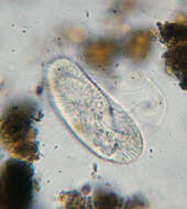
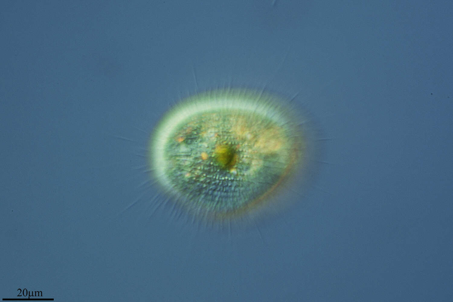
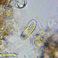
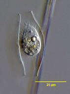
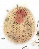
Longitude (deg): -0.7. Latitude (deg): 51.1. Longitude (deg/min): 0° 50' W. Latitude (deg/min): 51° 10' N. Vice county name: Surrey. Vice county no.: 17. Country: England. Associated species: Marchantia polymorpha. Identified by: Malcolm Storey. Comment: with Marchantia polymorpha. Category: standard photograph or close-up. Photographic equipment used: "35mm transparencies (on a variety of films, but Agfa CT18 in the 1960's to early 1980's followed by Fujichrome in the late 1980's.) Transparencies scanned with Minolta Dimage Scan Dual II AF-2820U transparency scanner.".
-
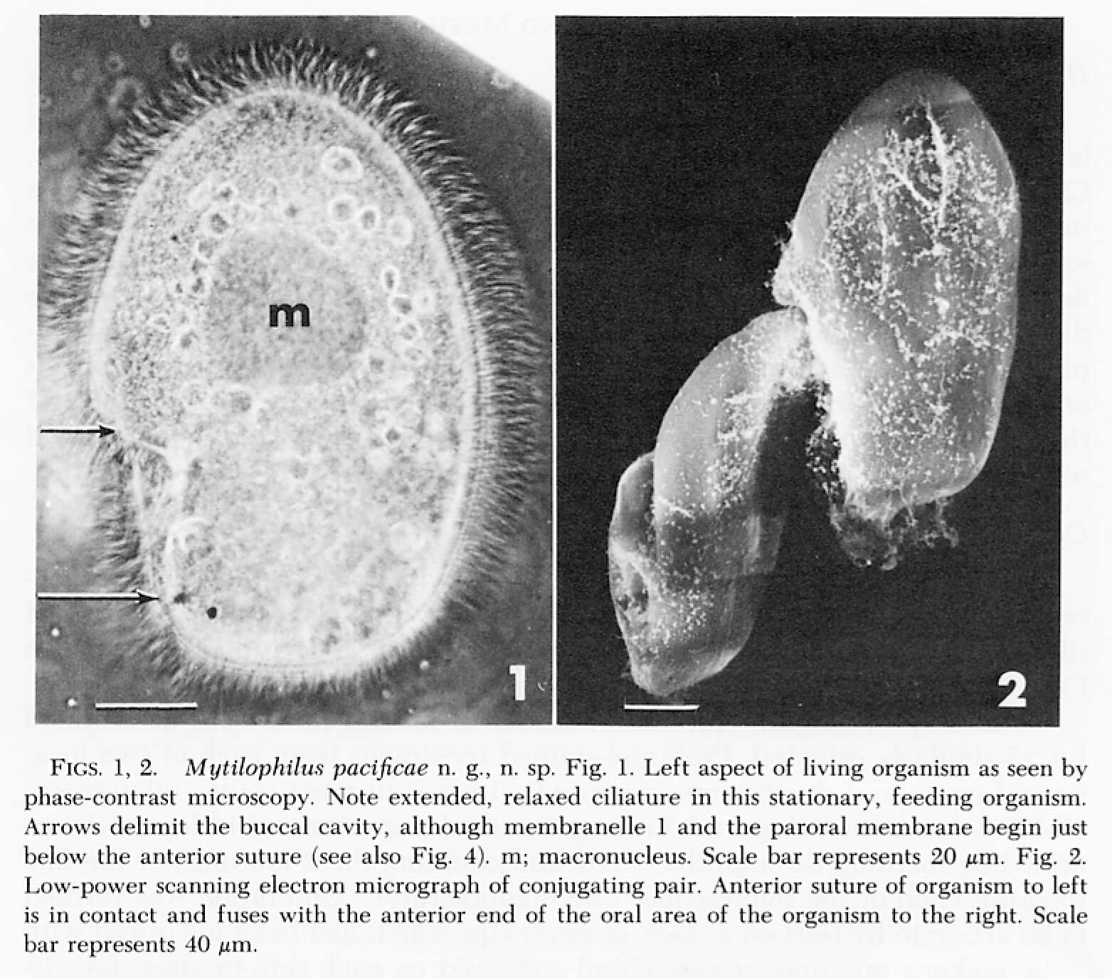
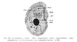
The species now known as Peniculistoma mytili was first described as Conchopthirus mytili by William De Morgan in 1925 in an article in the Journal of the Marine Biological Association (vol 13, 600-660).
-
Image of Peniculistoma mytili (endocommensal of the blue mussel Mytilis edulis) using DIC optics. Image by G. A. Antipa.
-
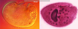
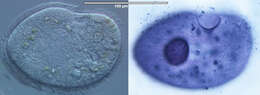
Left image: Live specimen imaged using DIC optics; Right image: protargol-stained specimen. Images by G. A. Antipa
-
Canencia, Madrid, Spain
-
Galende, Castille and Leon, Spain
-
Hoyo de Manzanares, Madrid, Spain
-
Ribadelago de Franco, Castille and Leon, Spain
-
Hoyo de Manzanares, Madrid, Spain
-
Canencia, Madrid, Spain
-
Hoyo de Manzanares, Madrid, Spain
-
Logroo, La Rioja, Espaa
-
Talveila, Castille and Leon, Spain
-
Images from the species description by Antipa & Dolan (1985)
-
Left panel: live specimen imaged with DIC optics (image by G.A. Antipa); right panel: protargol-stained specimen (image by D.H. Lynn).
-
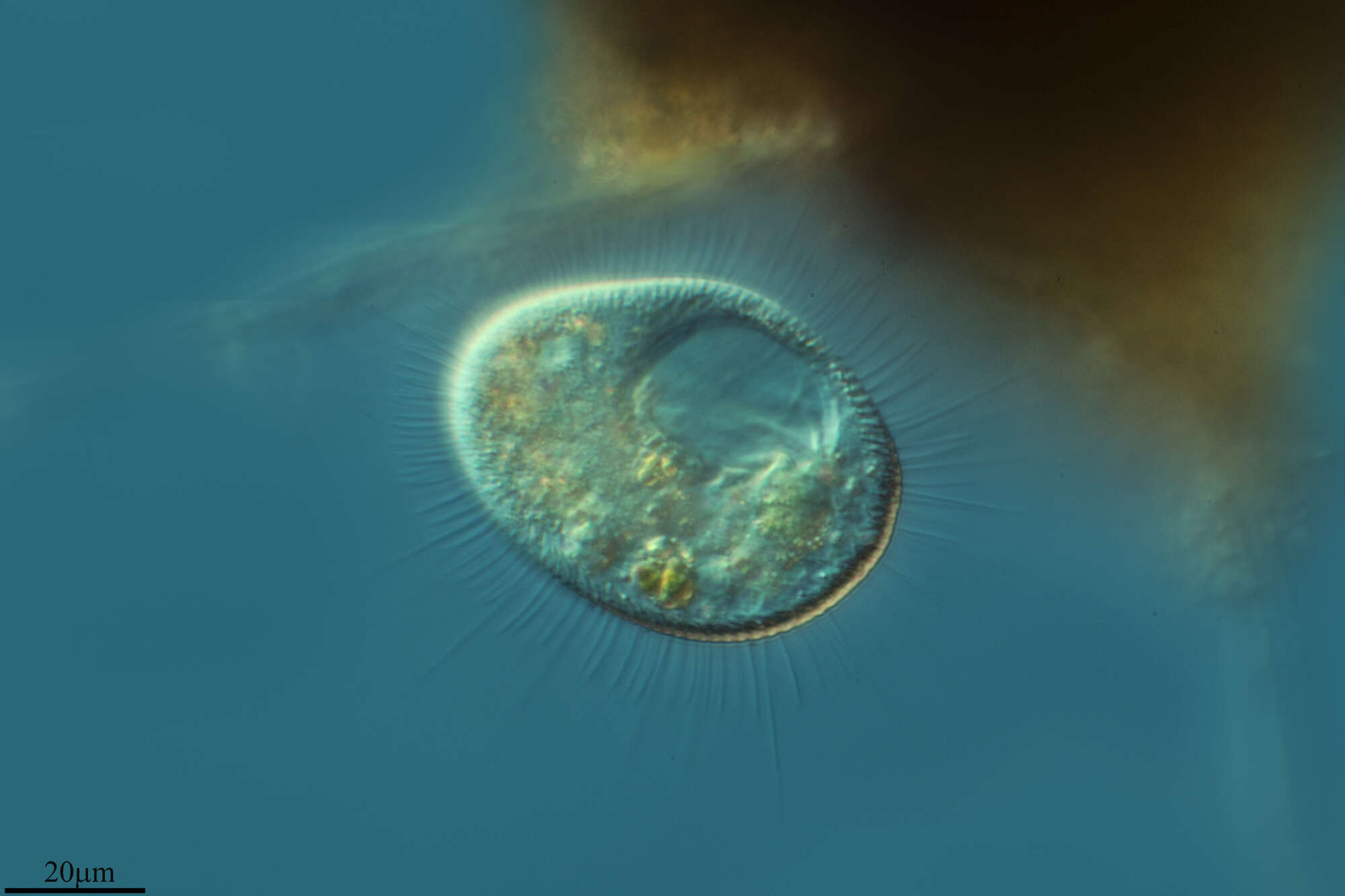
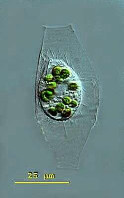
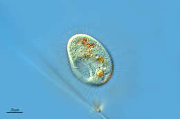
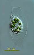
Calyptotricha (kah-lip-toe-trike-a) pleuronemoides is an ovoid to pyriform ciliate. The ciliate forms a transparent lorica. The lorica is tube-like and has apertures at both ends. The middle the tube can have parallel sides or a central bulbous region in which the ciliate is housed. The undulating membrane of the oral aperture stretches down the right side of the body to form a pouch in the posterior body half. Extrusomes are present. There is a conspicuous caudal cilium. Contractile vacuole in posterior body region. The macronucleus is spherical with attached micronuclei. Several endosymbiotic algae are visible and the conspicuous caudal cilium. Ciliate measuring 28 microns, lorica 64 microns. This specimen was collected in freshwater ponds near Konstanz, Germany. Differencial interference contrast.
-
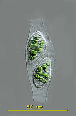
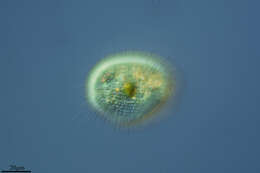
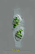
Calyptotricha (kah-lip-toe-trike-a) pleuronemoides is an ovoid to pyriform ciliate. The ciliate forms a transparent lorica. The lorica is tube-like and has apertures at both ends. The middle the tube can have parallel sides or a central bulbous region in which the ciliate is housed. The undulating membrane of the oral aperture stretches down the right side of the body to form a pouch in the posterior body half. Extrusomes are present. There is a conspicuous caudal cilium. Contractile vacuole in posterior body region. The macronucleus is spherical with attached micronuclei. This image taken shortly after cell division when there are two specimens in the lorica. Lorica measuring 68 microns. This specimen was collected in freshwater ponds near Konstanz, Germany. Differential interference contrast.
-
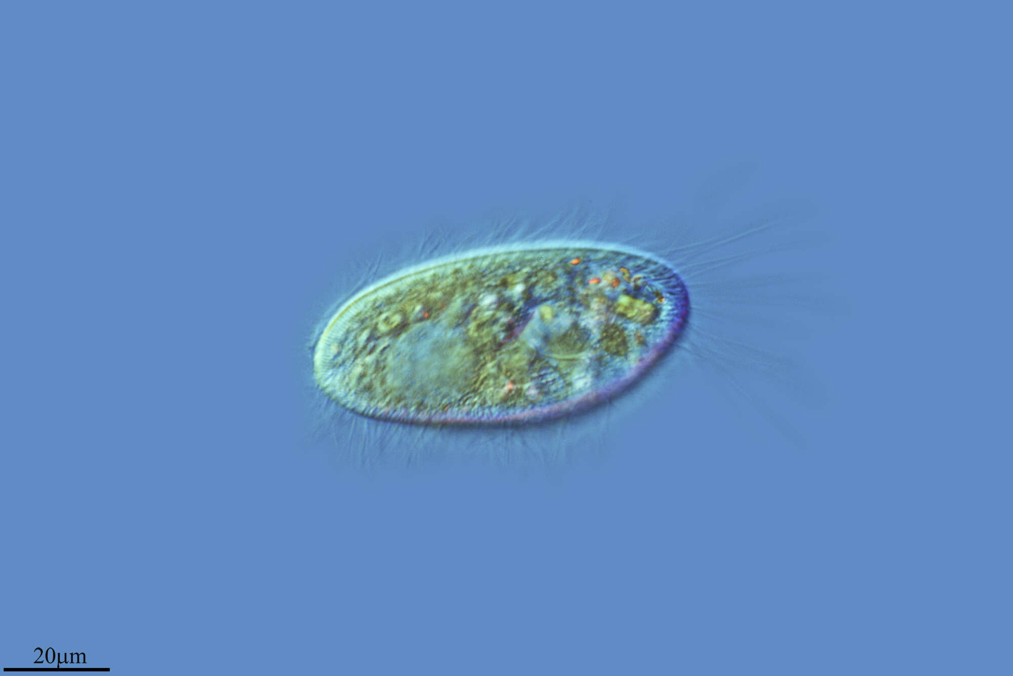
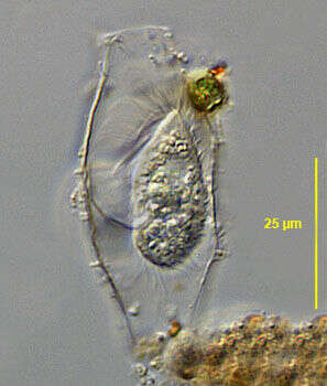
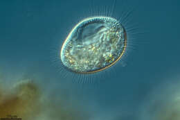
Portrait of the loricate pleuronematid ciliate, Calyptotricha pleuronemoides (Phillips, 1882). The transparent lorica of this species is open at both ends and dilated in the center where the cell resides. The cell is bluntly pointed anteriorly and broadly rounded posteriorly. The peristome is about 3/4 cell length. There is a prominent undulating membrane on the right margin of the peristome curving around its posterior end to form a shallow pouch (seen well here). There are three inconspicuous adoral membranelles. The longitudinal somatic kineties are uniformly distributed. There is a preoral and postoral suture. There is a single long caudal cilium. There is a single posterior contractile vacuole. The spherical macronucleus is centrally located. C. lanuginosa is similar in appearance of the cell except that it has two long anterior apical cilia and a cylindrical lorica with parallel sides. Collected from a freshwater dredge pond near Idaho City, Idaho June 2003. DIC.
-
Portrait of the loricate pleuronematid ciliate, Calyptotricha pleuronemoides (Phillips, 1882). The transparent lorica of this species is open at both ends and dilated in the center where the cell resides. The cell is bluntly pointed anteriorly and broadly rounded posteriorly. The peristome is about 3/4 cell length. There is a prominent undulating membrane on the right margin of the peristome curving around its posterior end to form a shallow pouch. There are three inconspicuous adoral membranelles. The longitudinal somatic kineties are uniformly distributed. There is a preoral and postoral suture. There is a single long caudal cilium. There is a single posterior contractile vacuole. The spherical macronucleus is centrally located. C. lanuginosa is similar in appearance of the cell except that it has two long anterior apical cilia and a cylindrical lorica with parallel sides. Collected from a freshwater dredge pond near Idaho City, Idaho June 2003. DIC.
-
Dorsal infraciliature of Calyptotricha pleuronemoides (PHILLIPS,1882). Collected from organically enriched stagnant water at the edge of a freshwater stream near Boise, Idaho.Stained by the silver carbonate technique (Foissner,W. Europ. J. Protistol.27:313-330;1991).Brightfield.