À propos
Éducation
Discuter
TraitBank
Se connecter
S’inscrire
Langue
Deutsch
English
Español
français
italiano
Nederlands
Piemontèis
Português do Brasil
suomi
Türkçe
Čeština
Ελληνικά
македонски
Українська
العربية
简体中文
繁體中文
noms dans le fil d’Ariane
vernaculaire
scientifique
À propos
Éducation
Discuter
TraitBank
Se connecter
S’inscrire
fr
Deutsch
English
Español
français
italiano
Nederlands
Piemontèis
Português do Brasil
suomi
Türkçe
Čeština
Ελληνικά
македонски
Українська
العربية
简体中文
繁體中文
noms dans le fil d’Ariane
vernaculaire
scientifique
Life
»
…
»
Alveolata
»
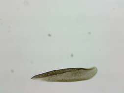
Ciliophora
Life
»
Organismes Cellulaires
»
Eucaryotes
»
SAR (Stramenopiles, Alveolates, Rhizaria)
»
Alveolata
»
Ciliophora
«
Postciliodesmatophora
recueillir
vue d’ensemble
données
média
articles
noms
licence
n’importe quelle licence
CC-BY
CC-BY-NC
CC-BY-NC-SA
CC-BY-SA
No copyright
type
n’importe quel type
image
vidéo
fournisseur
n’importe quel fournisseur
iNaturalist
Wikimedia Commons
Barcode of Life Data Systems
Flickr Group
BioImages, the virtual fieldguide, UK
Freshwater and Marine Image Bank U Washington
micro*scope
protisten.de
vimeo
1
2
3
4
5
…
Last »
cc-publicdomain
fiable
cc-publicdomain
fiable
cc-publicdomain
fiable
cc-by-nc-4.0
fiable
cc-by-nc-4.0
fiable
cc-by-nc-4.0
fiable
cc-by-nc-4.0
fiable
cc-by-nc-4.0
fiable
cc-by-nc-4.0
fiable
cc-by-nc-4.0
fiable
cc-by-nc-4.0
fiable
cc-by-nc-4.0
fiable
cc-by-nc-4.0
fiable
cc-by-nc-4.0
fiable
cc-by-nc-4.0
fiable
cc-by-nc-4.0
fiable
cc-by-nc-4.0
fiable
cc-by-nc-4.0
fiable
cc-by-nc-4.0
fiable
cc-by-nc-4.0
fiable
cc-by-4.0
fiable
cc-by-4.0
fiable
cc-by-nc-4.0
fiable
cc-by-nc-4.0
fiable
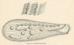
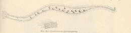
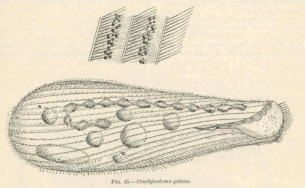
Condylostoma patens. 1902. Condylostoma.
cc-publicdomain
Freshwater and Marine Image Bank U Washington
Condylostoma patens.
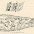
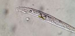
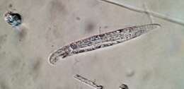
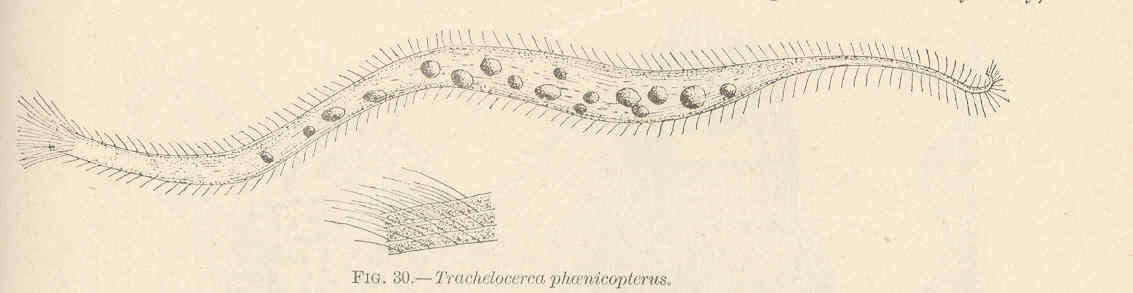
Trachelocerca phoenicopterus. 1902. Trachelocerca; Protozoa.
cc-publicdomain
Freshwater and Marine Image Bank U Washington
Trachelocerca phoenicopterus.
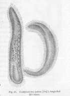
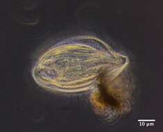
Condylostoma patens (Duj.), magnified. 1868. Condylostoma.
cc-publicdomain
Freshwater and Marine Image Bank U Washington
Condylostoma patens (Duj.), magnified....
"
cc-by-nc-4.0
peptolab
iNaturalist
"
cc-by-nc-4.0
peptolab
iNaturalist
"
cc-by-nc-4.0
peptolab
iNaturalist
"
cc-by-nc-4.0
peptolab
iNaturalist
"
cc-by-nc-4.0
peptolab
iNaturalist
"
cc-by-nc-4.0
peptolab
iNaturalist
"
cc-by-nc-4.0
peptolab
iNaturalist
"
cc-by-nc-4.0
peptolab
iNaturalist
"
cc-by-nc-4.0
peptolab
iNaturalist
"
cc-by-nc-4.0
peptolab
iNaturalist
"
cc-by-nc-4.0
peptolab
iNaturalist
"
cc-by-nc-4.0
peptolab
iNaturalist
"
cc-by-nc-4.0
David Flanagan
iNaturalist
"
cc-by-nc-4.0
David Flanagan
iNaturalist
"
cc-by-nc-4.0
David Flanagan
iNaturalist
"
cc-by-nc-4.0
David Flanagan
iNaturalist
"
cc-by-nc-4.0
David Flanagan
iNaturalist
"
cc-by-4.0
Don Loarie
iNaturalist
"
cc-by-4.0
Don Loarie
iNaturalist
"
cc-by-nc-4.0
peptolab
iNaturalist
"
cc-by-nc-4.0
peptolab
iNaturalist
1
2
3
4
5
…
Last »