-
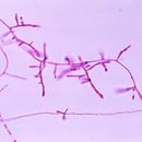
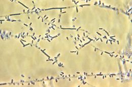
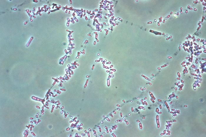
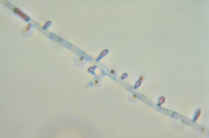
Magnified under a low magnification of 40X, this photomicrograph depicts the microconidia of the fungus Trichophyton mariatii.Created: 1979
-
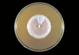
This photograph depicts the reverse view of a Petri dish within which a fungal colony of a Mexican isolate ofTrichophyton rubrum var. rodainii had been cultured atop a medium of Sabourauds agar.Created: 1977
-
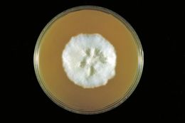
This photograph depicts the frontal view of a Petri dish within which a fungal colony of a Mexican isolate of Trichophyton rubrum var. rodainii had been cultured atop a medium of Sabourauds agar.Created: 1974
-
This photomicrograph revealed some of the ultrastructural morphology exhibited by Trichophyton rubrum fungal organisms.T. rubrum and T. tonsurans are two common dermatophytes. These two species are usually transmitted from person to person. Another common dermatophyte is Microsporum canis, which is transmitted from animals such as cats and dogs to people. Dermatophytes like to live on moist areas of the skin, such as places where there are skin folds. They can also contaminate items in the environment, such as clothing, towels and bedding.Created: 1974
-
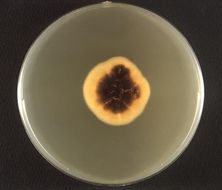
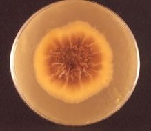
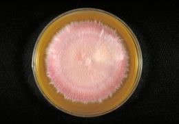
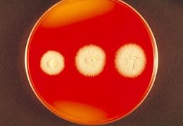
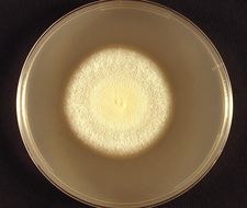
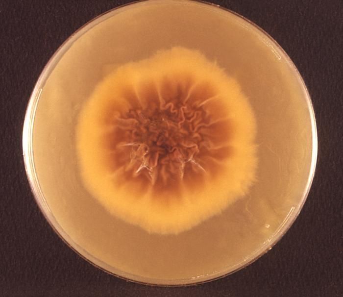
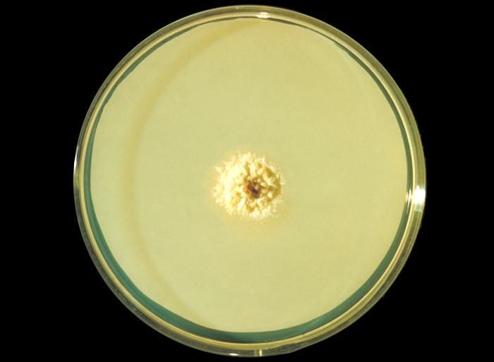
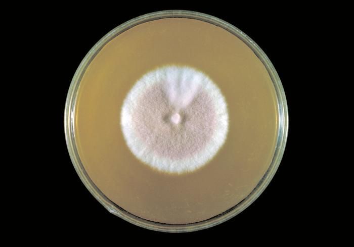
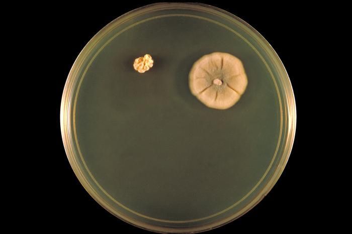
This photograph depicts the frontal view of a Petri dish within which a colony of the African form of the fungus, Trichophyton rubrum, had been cultured. Revealed is the colonial morphology, which in this case is both glaborous, i.e., flat to cottony, and raised and ruffled at its center. Its frontal coloration can range from a white to bright yellowish-beige, as it was here, and even to a red-violet coloration. From the reverse, or from the back, the colonies display a coloration that is a light yellowish to brown, or a reddish brown.Created: 1974
-
This photograph depicts the reverse view of a Petri dish within which fungal colonies of Trichophyton rubrum var. granulare had been cultured. From the front, (see PHIL 10540) the colonial morphology, which in the case of T. rubrum is said to be waxy, glaborous, i.e., flat to cottony, and display from a frontal perspective, a white to bright yellowish-beige, and even a red-violet coloration. From this reverse view, the colonies display a coloration that is a light yellowish to brown, or a reddish brown.Created: 1972
-
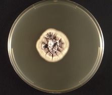
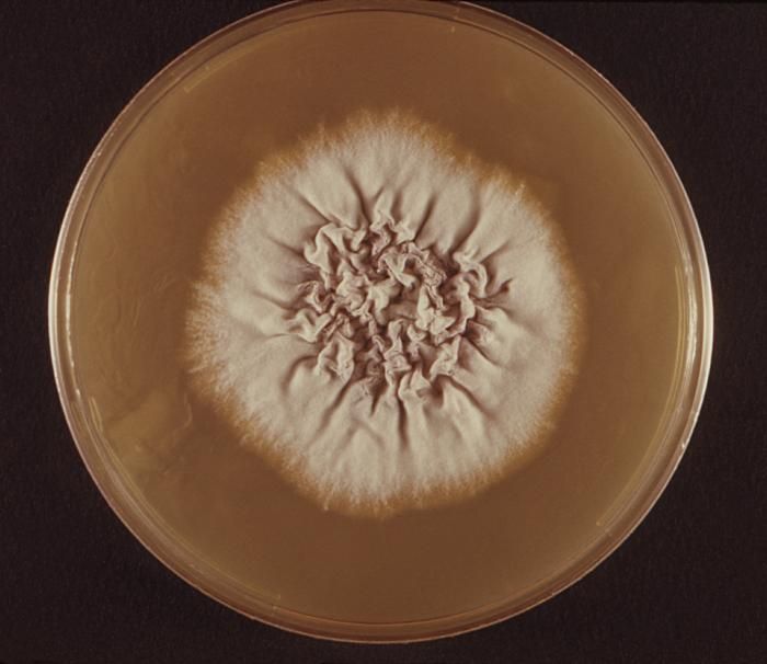
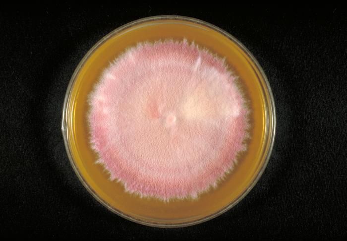
This photograph depicts the frontal view of a Petri dish within which fungal colonies of Trichophyton rubrum var. granulare had been cultured. Revealed is the colonial morphology, which in the case of T. rubrum is said to be waxy, glaborous, i.e., flat to cottony, and display from a frontal perspective, a white to bright yellowish-beige, and even a red-violet coloration. From the reverse (see PHIL 10541) or from the back, the colonies display a coloration that is a light yellowish to brown, or a reddish brown.Created: 1972
-
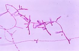
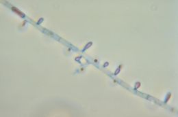
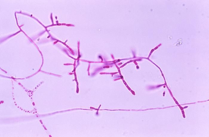
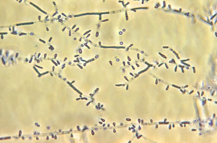
This photomicrograph reveals the microconidia of the fungus Trichophyton rubrum.Created: 1973
-
This photomicrograph reveals the microconidia of the fungus Trichophyton rubrum.Created: 1973
-
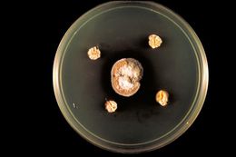
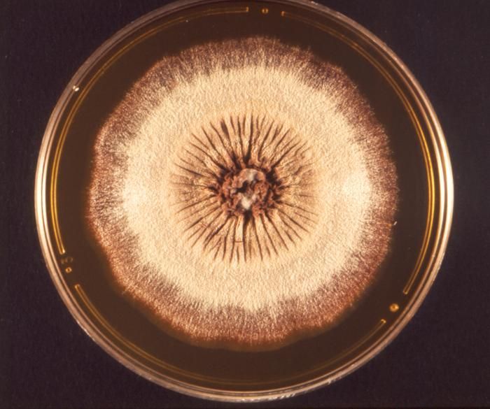
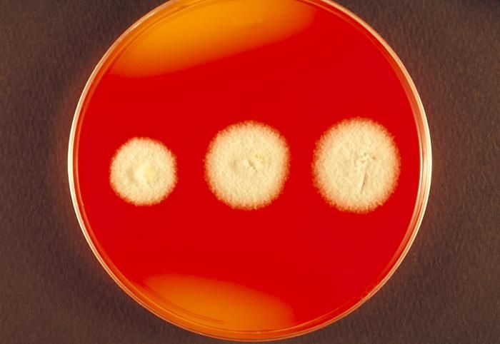
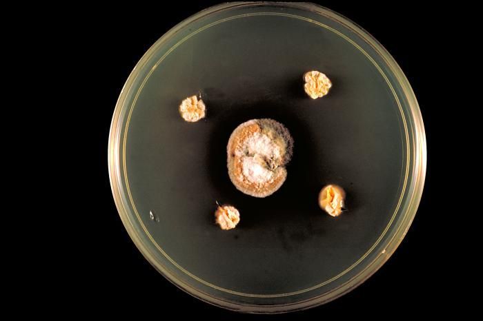
This was a Sabouraud's dextrose agar plate culture of the anthrophilic dermatophyteTrichophyton yaoundei at wk. 6.Created: 1962
-
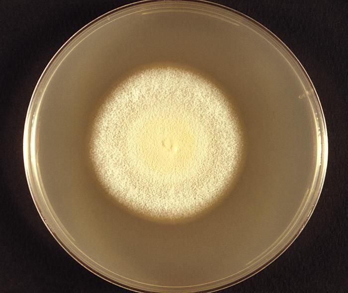
His hair-plate culture is growing the fungus Trichophyton terrestre.Created: 1963
-
This Sabourauds dextrose agar plate culture is growing T. terrestre fungus, rose-pigmented strain x231.Created: 1963
-
This Sabourauds dextrose agar plate culture is growing T. terrestre fungus, white strain x231, day 12.Created: 1963
-
This Sabourauds dextrose agar plate culture is growing T. terrestre fungus, strain x231 producing a red pigment.Created: 1963
-
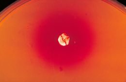
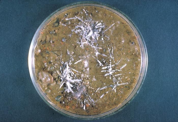
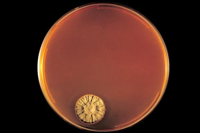
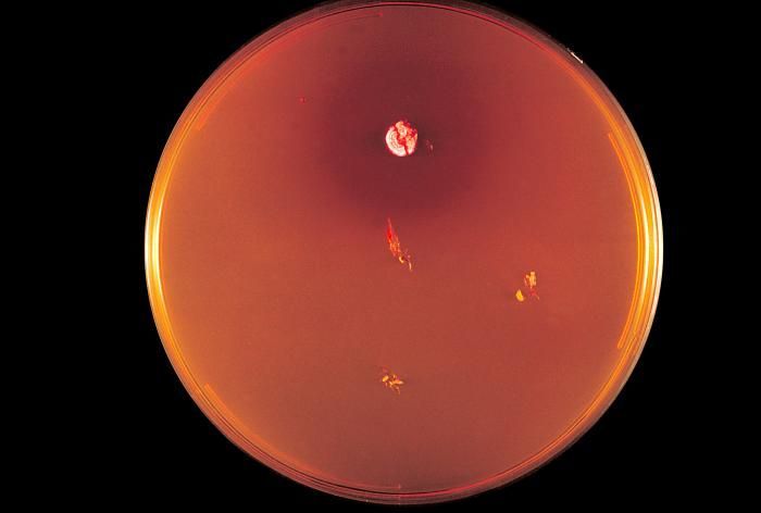
This image shows a DTM agar plate culture with colonies of a Trichophyton sp. fungal organism.Created: 1970
-
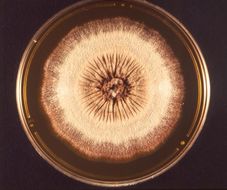
This was a DTM (Dermatophyte Test Medium) agar plate culture growing the fungus Trichophyton concentricum.Created: 1970
-
This is a DTM plate culture growing the dermatophytic fungus Trichophyton concentricum.Created: 1970
-
This was a DTM (Dermatophyte Test Medium) agar plate culture growing the fungus Trichophyton concentricum.Created: 1970
-
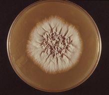
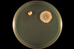
This image shows a C&C agar plate culture with colonies of the fungusTrichophyton concentricum.Created: 1970
-
This was a DTM (Dermatophyte Test Medium) agar plate culture growing the fungus Trichophyton concentricum.Created: 1970
-
This image shows a C&C agar plate culture with colonies of the fungusTrichophyton concentricum.Created: 1970
-
This image shows a C&C agar plate culture with colonies of the fungusTrichophyton concentricum.Created: 1970
-
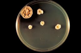
This image shows a DTM agar plate culture with colonies of the fungusTrichophyton concentricum.Created: 1970
-
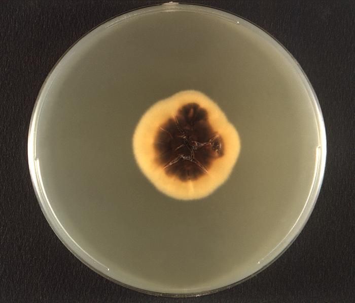
This photomicrograph revealed some of the morphology exhibited by a Trichophyton tonsurans fungal colony. Note the glaborous, or smooth velvety appearance of this colony, and early changes at its center, which in time will lead to its characteristically raised appearance, as well as its yellowish-beige colorationT. tonsurans and T. rubrum are two common dermatophytes. These two species are usually transmitted from person to person. Another common dermatophyte is Microsporum canis, which is transmitted from animals, including cats and dogs, to people. Dermatophytes like to live on moist areas of the skin, such as places where there are skin folds. They can also contaminate items in the environment, such as clothing, towels and bedding.Created: 1974