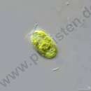
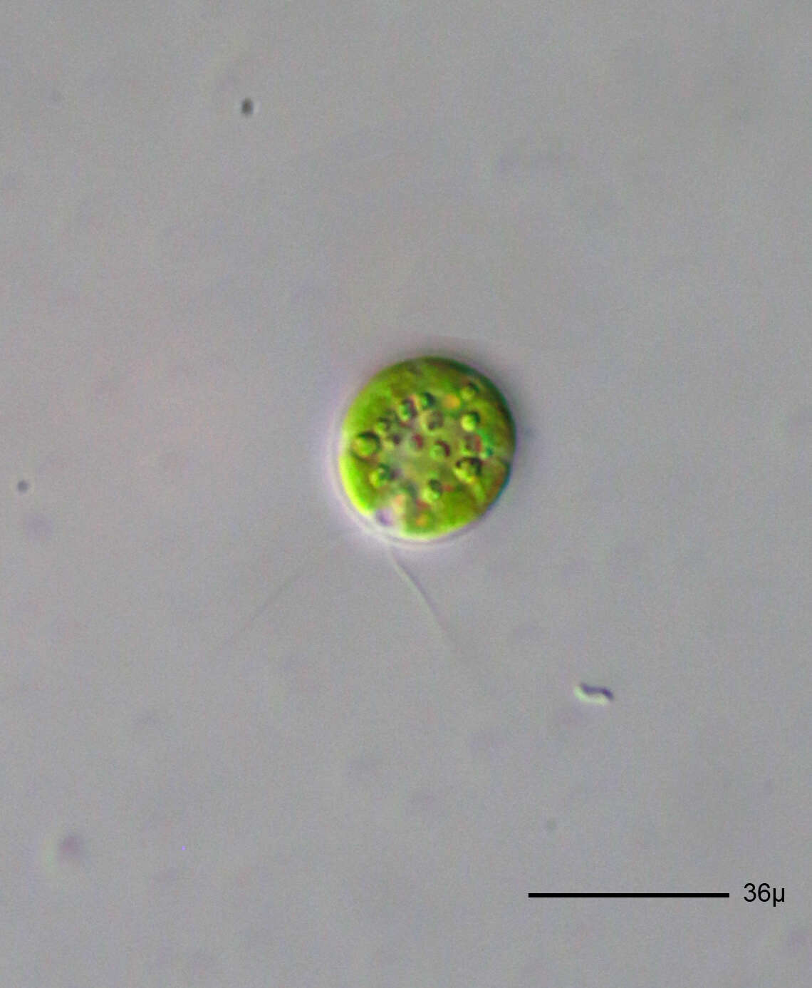
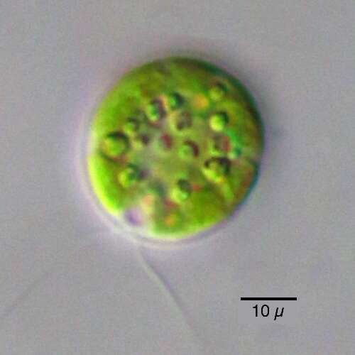
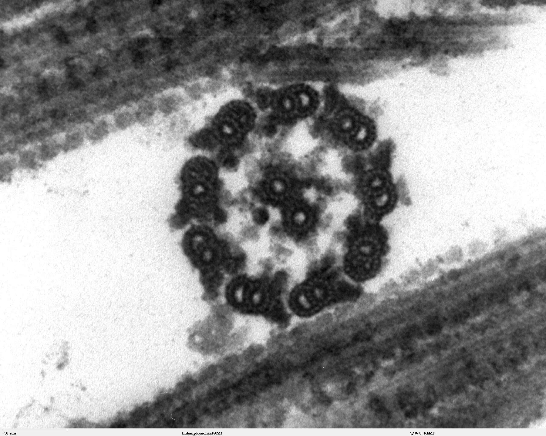
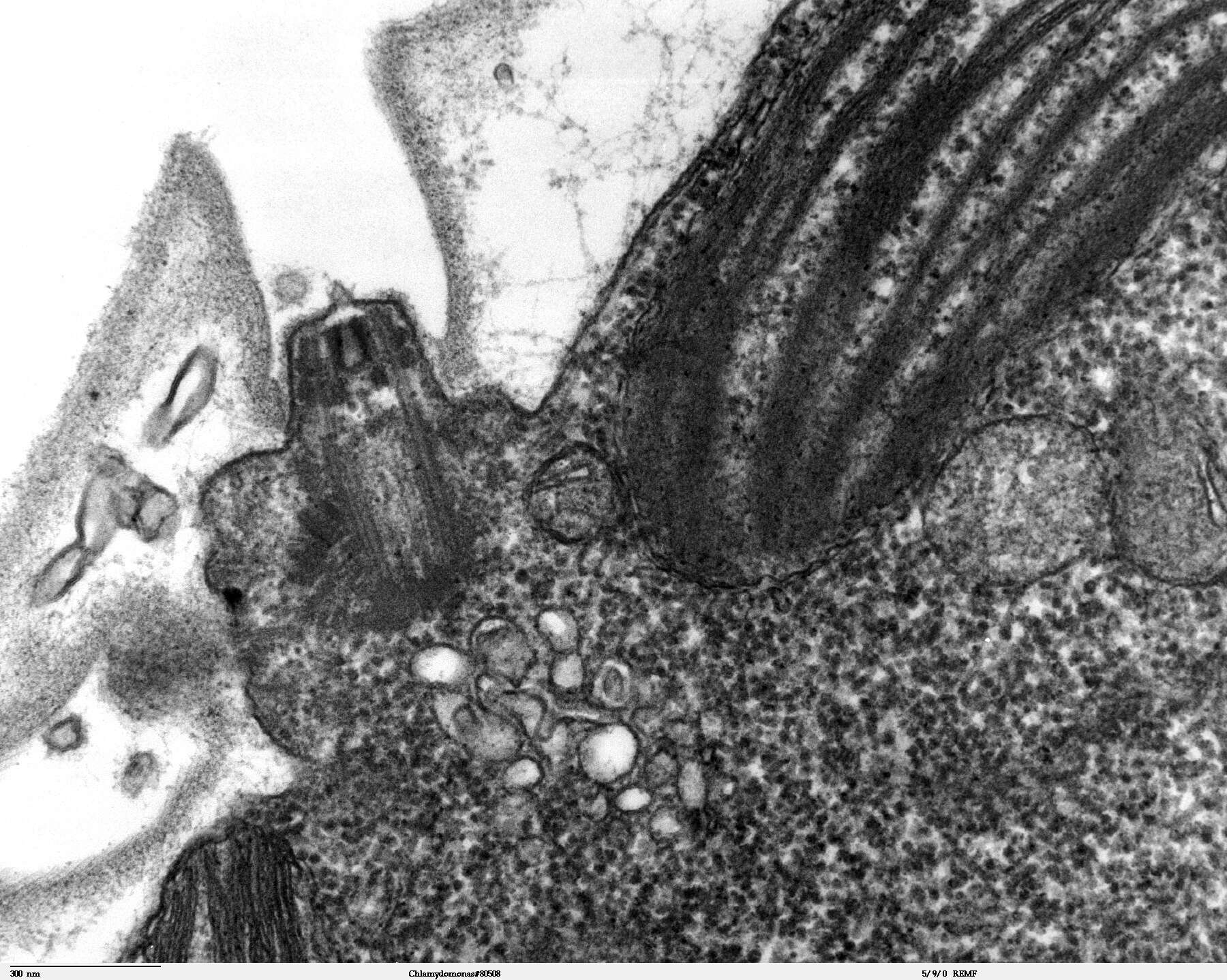
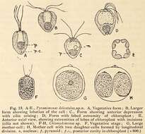
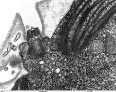
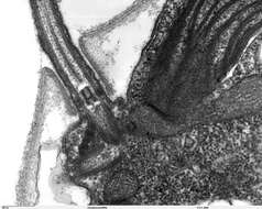
Description: Transmission electron microscope image, showing an example of green algae (Chlorophyta). Chlamydomanas reinhardtii is a unicellular flagellate used as a model system in molecular genetics work and flagellar motility studies. This image shows the flagellar apparatus, just after flagellar excision, which occurs at the transition zone(see area of flagella, with its fibers of the stellate structure). This image also shows components of the contractile vacuoles which are located just below the flagellar apparatus. JEOL 100CX TEM. Date: 7 October 2006. Source: Source and public domain notice at:
http://remf.dartmouth.edu/imagesindex.html http://remf.dartmouth.edu/images/algaeTEM/source/11.html. Author: Elizabeth Smith, Louisa Howard, Erin Dymek (Dartmouth Electron Microscope Facility, Dartmouth College). Permission(
Reusing this file): Released into the public domain.


