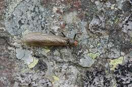
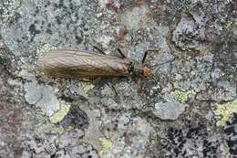
Hong–Liang Wang, Guo–Quan Wang, Wei–Hai Li
Zookeys
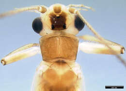
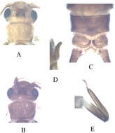
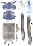
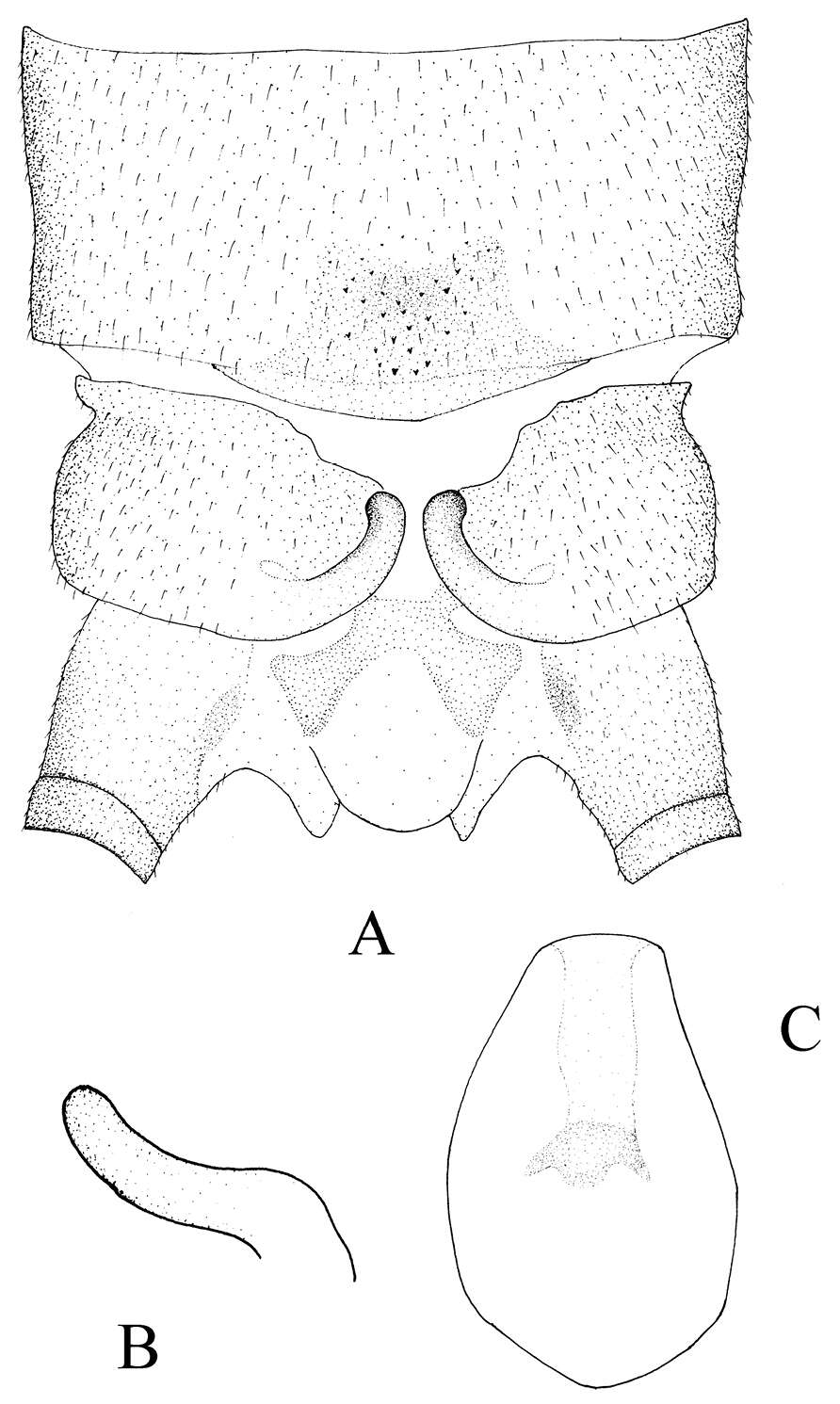
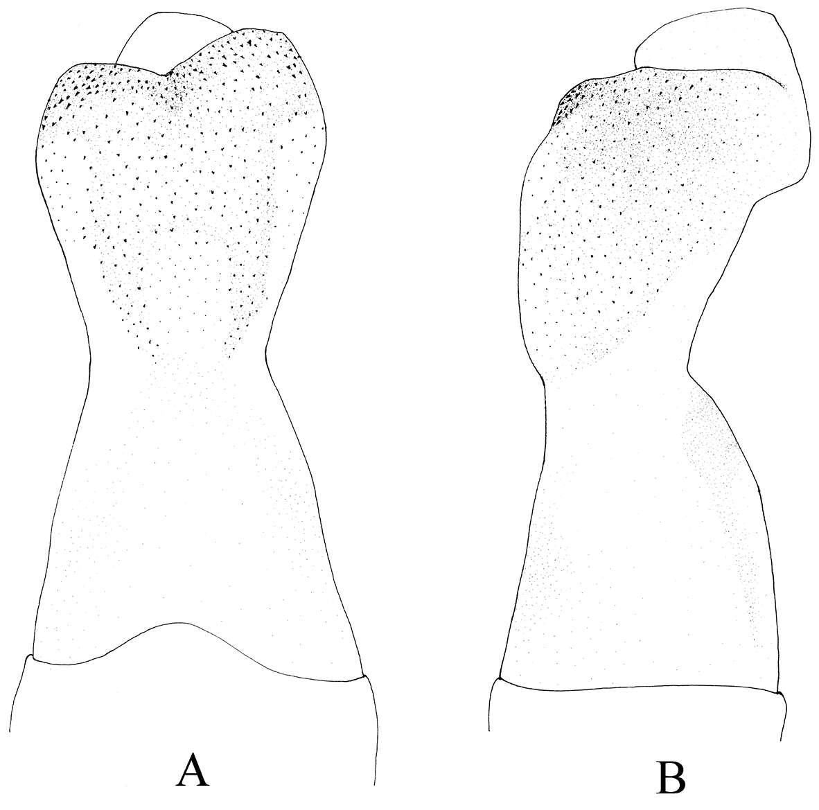
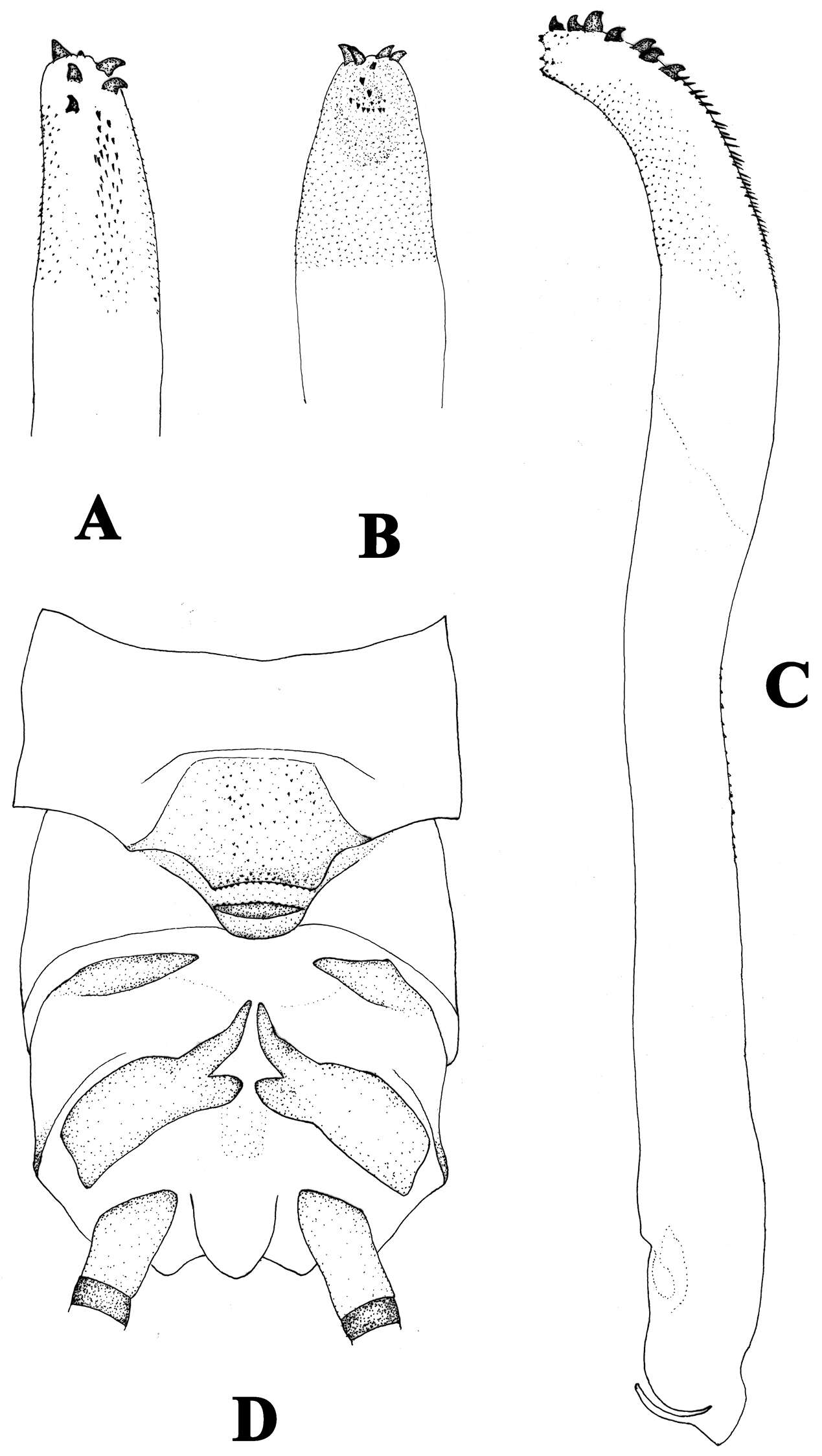
Figure 1.Kamimuria guangxia Li & Wang, sp. n. (male). A Head and pronotum, dorsal view (teneral specimen) B Head and pronotum, dorsal view (older specimen) C Terminalia, dorsal view D Hemitergal process, lateral view E Foreleg, lateral view.
Xue-Feng Qin, Dávid Murányi, Guo-Quan Wang, Wei-Hai Li
Zookeys
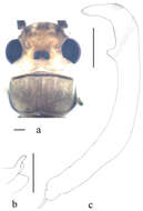
Figure 8.Neoperla xuansongae. Male a Head and pronotum, dorsal view b Aedeagus, lateral view with details in dorsal and ventral views. Scale bars: 0.5 mm.
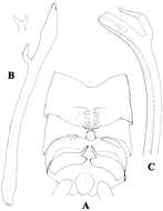
Figure 1.Neoperla latispina Wang & Li, sp. n. Male. a Head and pronotum, dorsal view b Terminalia, dorsal and lateral views c Aedeagus before eversion, lateral view d Aedeagus before eversion, dorsal view e foreleg femur.
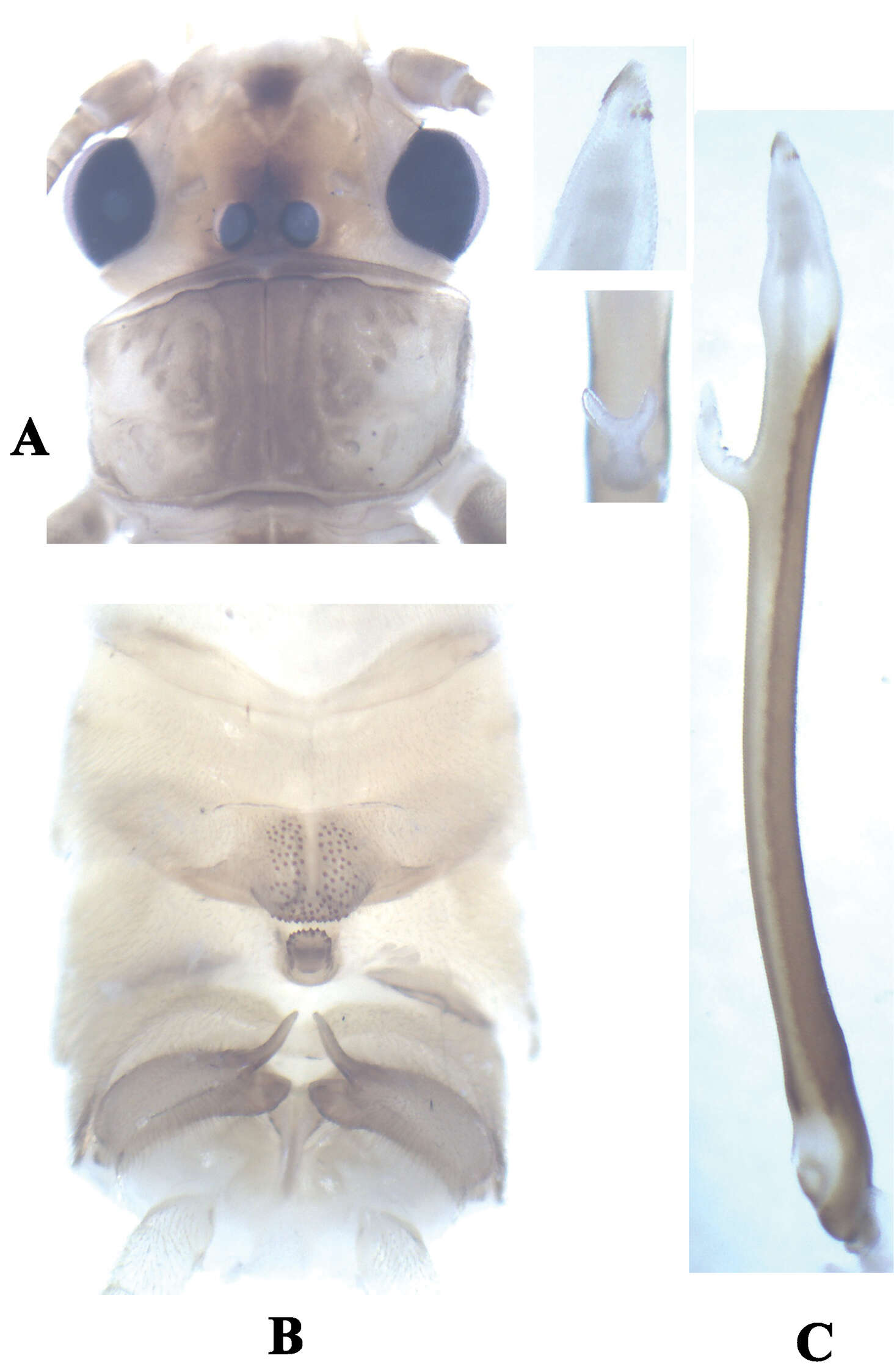
Figure 1.Neoperla nigromarginata Li & Zhang, sp. n. Male (a–e) a Head and pronotum, dorsal view b Terminalia, dorsal view c Terminalia, lateral view d Aedeagus before eversion, lateral view e Hindleg f Female subgenital fig, ventral view.
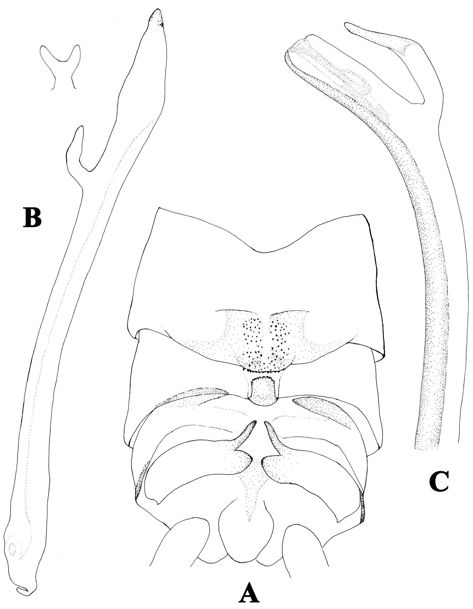
Figure 2.A–C Neoperla furcostyla Li and Qin,sp. n. (male). A Terminalia, dorsal view B Aedeagus, lateral view C Aedeagus of Neoperla forcipata Yang and Yang, lateral view.
Hong–Liang Wang, Guo–Quan Wang, Wei–Hai Li
Zookeys
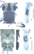
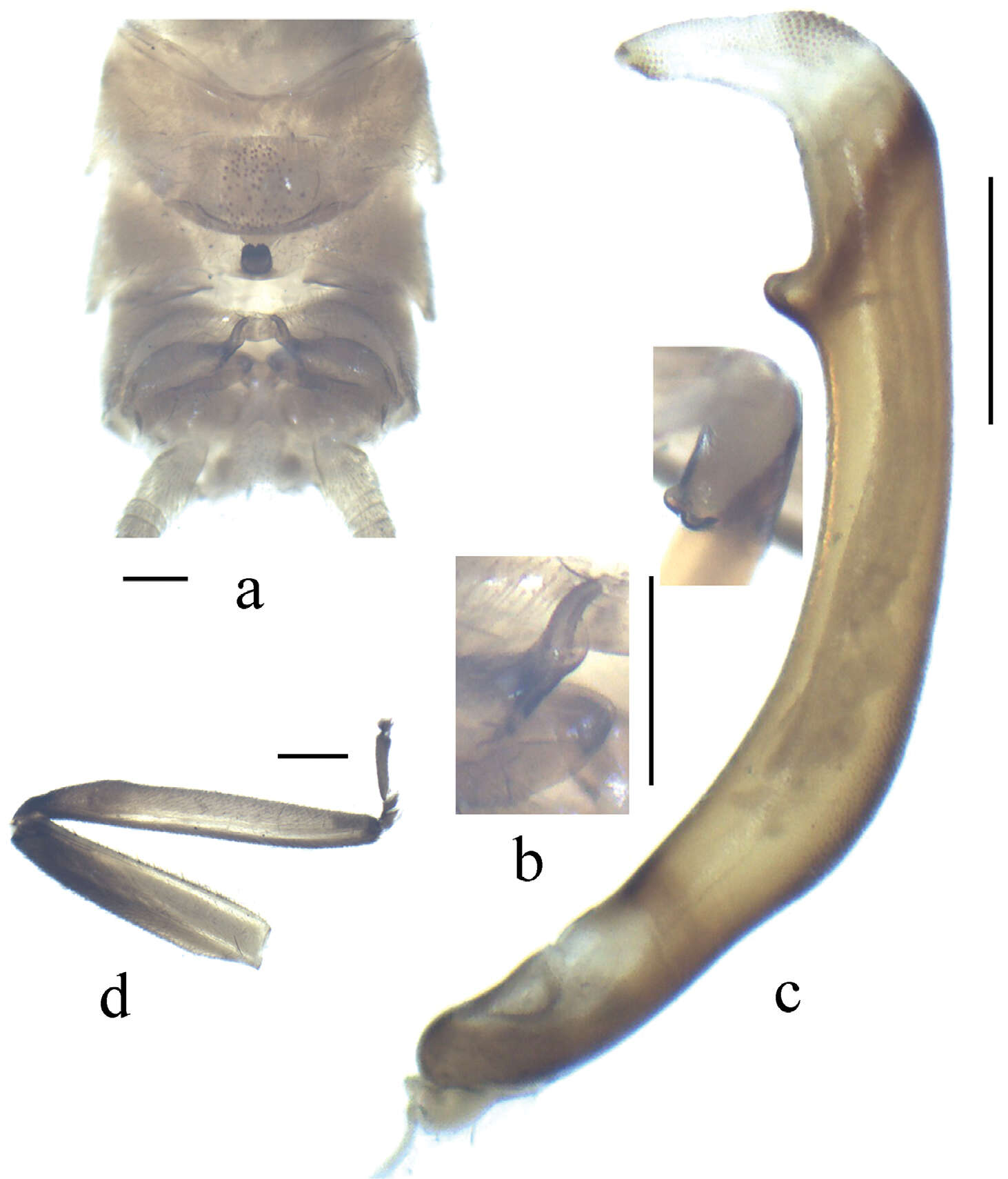
Figure 2.Kamimuria guangxia Li & Wang, sp. n. (male). A Terminalia, dorsal view B Hemitergal process, lateral view C Aedeagus before eversion, ventral view.
Xue-Feng Qin, Dávid Murányi, Guo-Quan Wang, Wei-Hai Li
Zookeys
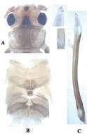
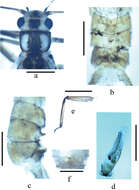
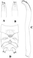
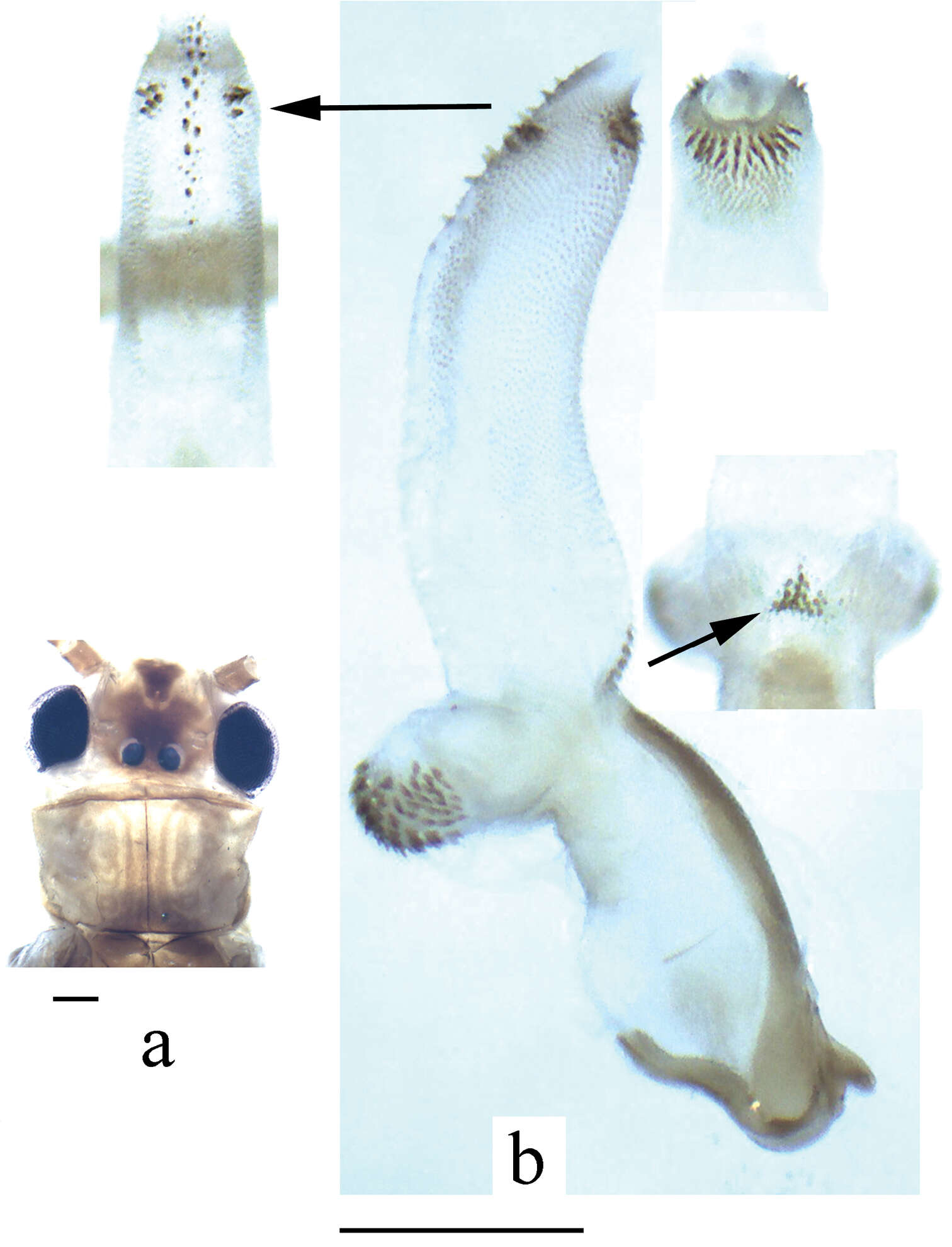
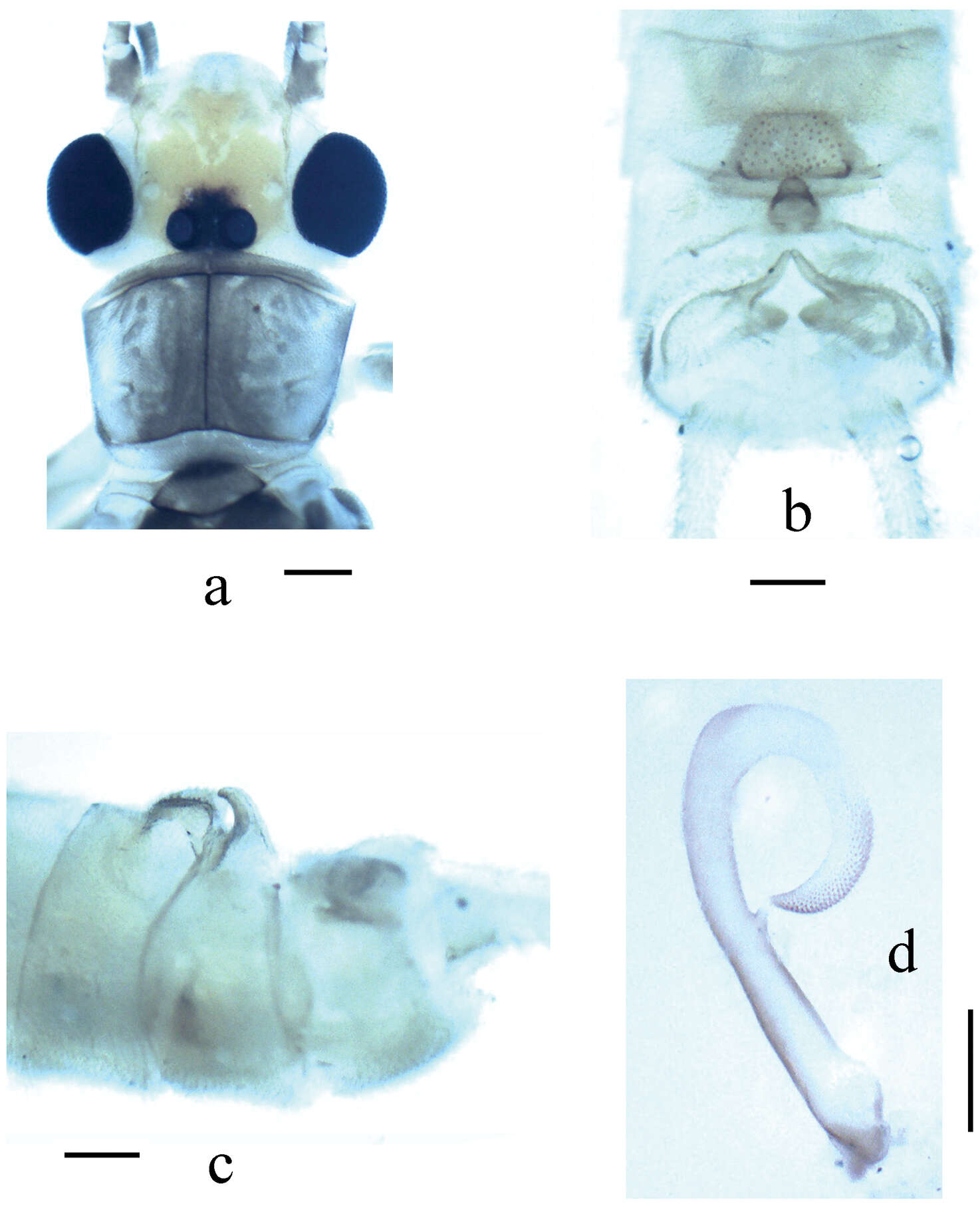
Figure 1.Neoperla brevistyla Li & Murányi, sp. n. Male a Head and pronotum, dorsal view b Hemitergal process, dorsal view c Aedeagus, lateral view. Scale bars: 0.5 mm.
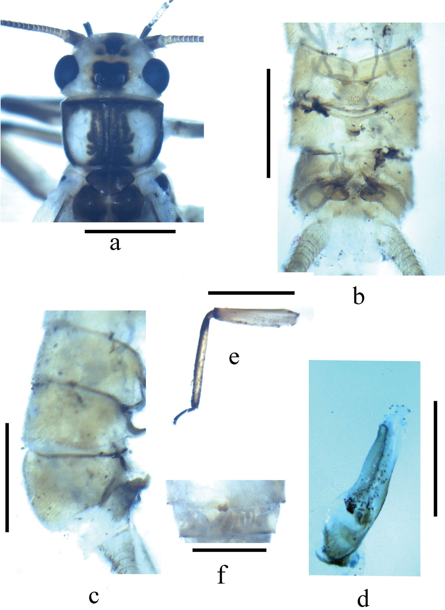
Figure 2.Neoperla nigromarginata Li & Zhang, sp. n. Male. a Dorsal aspect of aedeagal sac, top view b Aedeagus, lateral view. Note that the spines in b appear lightly pigmented and unclear, actually they are located on the lower surface of the sac, and are seen from beneath through the cuticle.
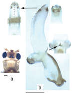
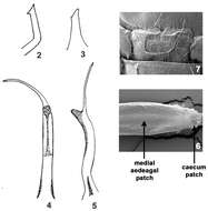
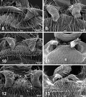
Figures 8–13.Male terminalia, posterodorsal view, SEM micrographs (8 Perlesta shubuta, USA, Mississippi, Clarke Co., Rolling Creek, 16 May 2011, 500X 9–13 Perlesta ephelida 9 USA, Alabama, Clay Co., Enitachopco Creek, 18 May 2008, 350X 10 USA, Indiana, Bartholomew Co., East Fork White River, 11 June 2000, 350X 11 USA, Maryland, Washington Co., Licking Creek, 12 July 1997, 350X 12 USA, Michigan, Calhoun Co., South Branch Kalamazoo River, 25 July 2006, 350X 13 USA, Missouri, Oregon Co., Eleven Point River, 27 June 2011, 200X.
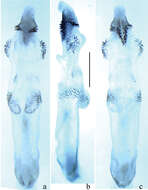
Figure 3.Neoperla similidella Li and Wang, sp. n. (male). A Head and pronotum, dorsal view B Terminalia, dorsal view C Aedeagus before eversion, lateral view D Aedeagus, lateral view E Aedeagal sac, dorsal view.
Xue-Feng Qin, Dávid Murányi, Guo-Quan Wang, Wei-Hai Li
Zookeys
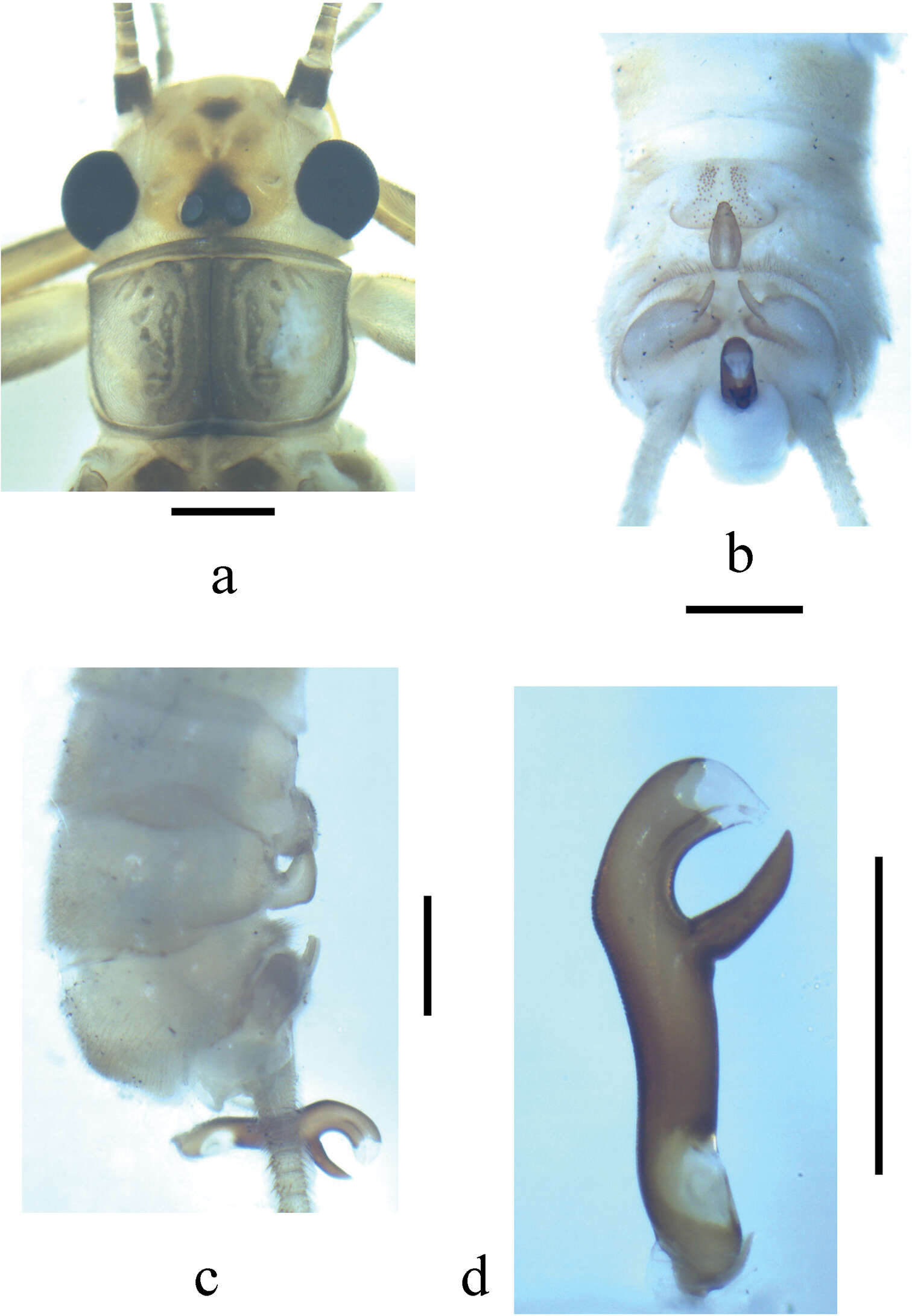
Figure 2.Neoperla brevistyla Li & Murányi,sp. n. Male a Terminalia, dorsal view b Hemitergal process, dorsal view c Aedeagus, lateral view d Foreleg, lateral view. Scale bars: 0.5 mm.
Figure 3.Neoperla mesospina Li and Wang, sp. n. Male. a Head and pronotum, dorsal view b Terminalia, dorsal view c Terminalia, lateral view d Aedeagus, lateral view.
Figure 3.Neoperla similiflavescens Li & Zhang, sp. n. Male. a Head and pronotum, dorsal view b Terminalia, dorsal view c Terminalia, lateral view d Aedeagus, lateral view.
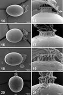
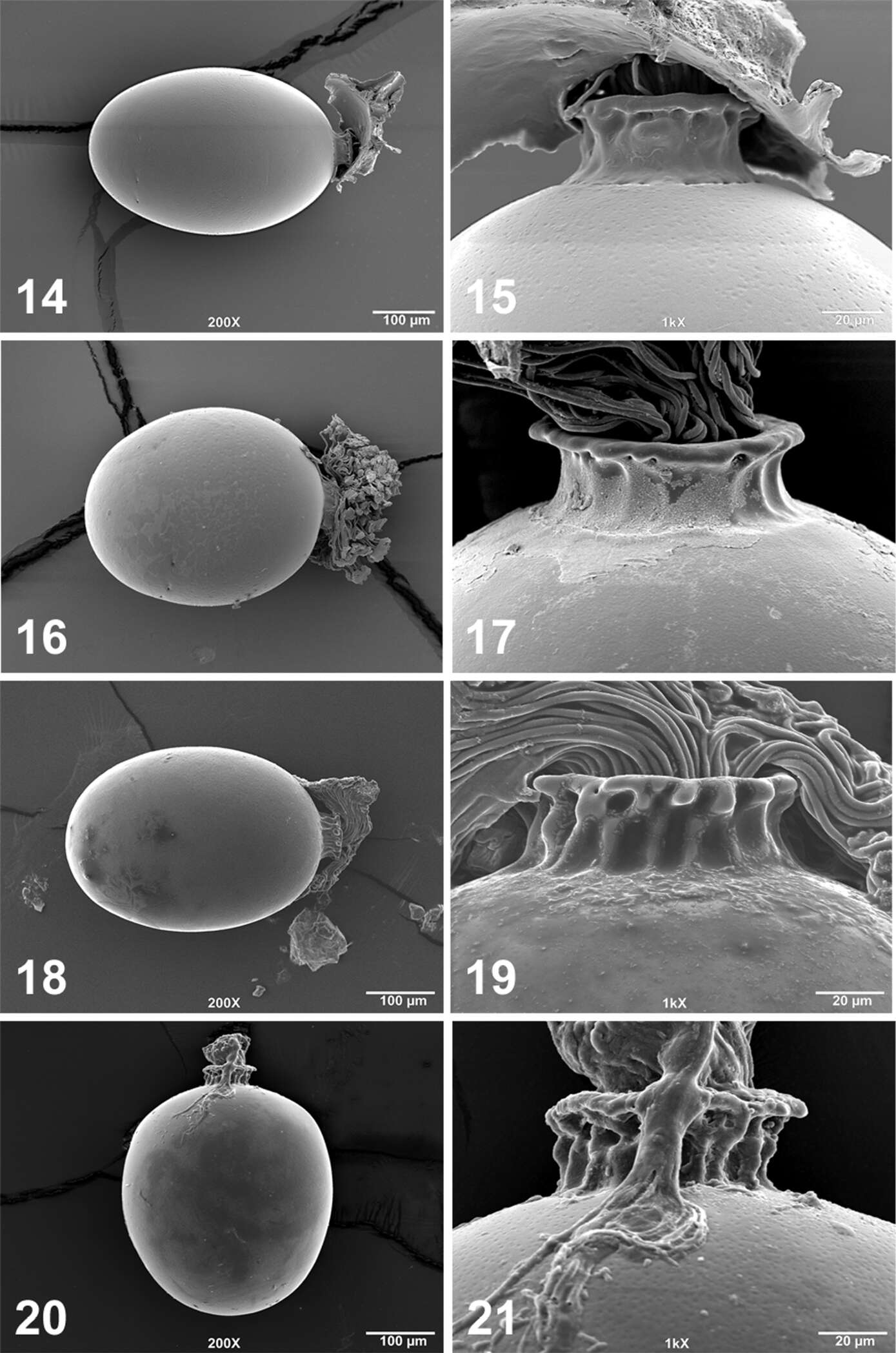
Figures 14–21.Perlesta ephelida egg SEM micrographs (14–15 USA, Kentucky, Warren Co., Trammel Fork, Drakes Creek, 24 May 1999 16–17 USA, Maryland, Washington Co., Licking Creek, 12 July 1997 18–19 USA, Michigan, Calhoun Co., South Branch Kalamazoo River, 25 July 2006 20–21 USA, Arkansas, Lawrence Co., Spring River, 27 June 2011) 14, 16, 18, 20 Entire egg, 200X; 15, 17, 19, 21. Anterior pole and collar, 1000X.
Figure 4.Neoperla similidella Li and Wang, sp. n. (male). A Aedeagal sac, dorsal view B Aedeagal sac, ventral view C Aedeagus, lateral view D Terminalia, dorsal view.