-
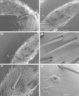
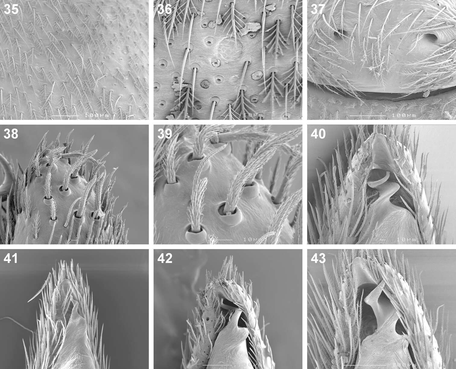
Figures 35–43.Scanning electron microscope photographs of Cambalida dippenaarae sp. n. (35–39, 42), Cambalida compressa sp. n. (40), Cambalida deminuta (Simon, 1909) (41) and Cambalida loricifera (Simon, 1885) (43): 35 female, dorsal abdominal surface 36 dorsal abdominal sigillum and detail of plumose setae 37 female epigyne 38 thickened setae at dorsal distal end of male palpal cymbium 39 detail of modified setae 40–43 male emboli.
-
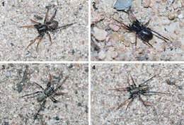
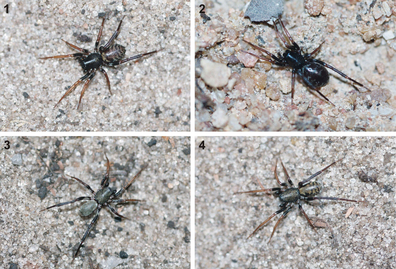
Figures 1–6.General habitus photographs of Copa flavoplumosa Simon, 1885 (1–4) and Copa kei sp. n. (5, 6): 1 female from Lesideng Research Camp, Botswana 2 female from Livingtone, Zambia 3 male and 4 female from Wildlives Game Farm, Zambia 5 female from Hogsback, South Africa 6 male from Cwebe Nature Reserve, South Africa.
-
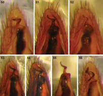
Figures 50–56.Digital microscope photographs of emboli of Afrotropical Cambalida species in ventral view: 50 Cambalida compressa sp. n. 51 Cambalida coriacea Simon, 1909 52 Cambalida deminuta (Simon, 1909) 53 Cambalida dippenaarae sp. n. 54 Cambalida fulvipes (Simon, 1896) 55 Cambalida griswoldi sp. n. 56 Cambalida loricifera (Simon, 1885). Scale bars = 0.1mm.
-
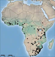
Figure 71.Distribution of Copa flavoplumosa Simon, 1885 in the Afrotropical Region.
-
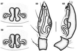
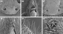
Figures 57–60.Genitalic morphology of Cambalida compressa sp. n.: 57 female epigyne, ventral view 58 same, dorsal view 59 male palp, ventral view 60 same, retrolateral view. Scale bars = 0.25mm.
-
Figures 7–12.Digital microscope photographs of Copa flavoplumosa Simon, 1885 from D.R. Congo (7–9) and Copa kei sp. n.from South Africa (10–12): 7, 10 female, dorsal habitus 8, 11 male, dorsal habitus 9, 12 sternum of female in ventral view. Scale bars = 1.0 mm.
-
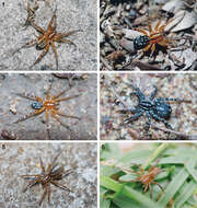
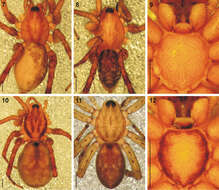
Figures 1–4.General habitus of Cambalida dippenaarae sp. n., indicating colour variations: 1 and 2 females and 3 male from Wildlives Game Farm, Zambia 4 male from Lesideng Research Camp, Botswana.
-
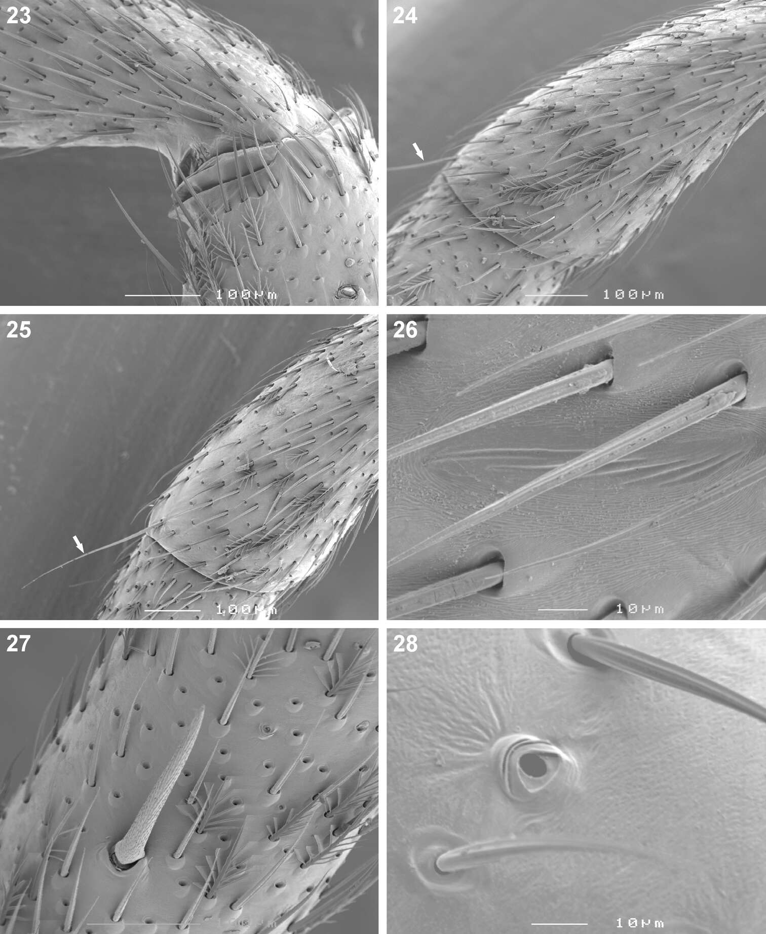
Figures 13–24.Scanning electron microscope photographs of Copa flavoplumosa Simon, 1885 female (13, 14, 16) and male (15, 17, 18) and Copa kei sp. n. female (19–24): 13 dorsal carapace setae 14, 15, 19 eye region and clypeus, anterolateral (14, 15) and anterior (19) views 16, 17, 20 cheliceral promarginal bent setae, anterior view 18, 22 mouthparts, ventral view 21 chelicerae, ventral view 23 serrula 24 femur, patella and tibia of leg II, indicating erect ventral setae on femora (EVS) and proximal and distal dorsal patellar setae (PS).
-
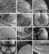
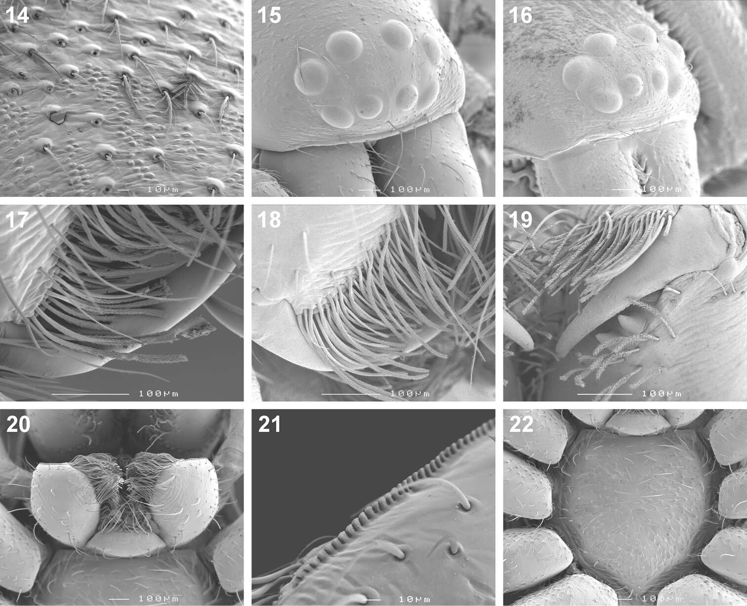
Figures 14–22.Scanning electron microscope photographs of Cambalida dippenaarae sp. n.female (14, 15, 17, 20–22) and male (16, 18, 19): 14 dorsal carapace setae 15, 16 eye region and clypeus, anterolateral view 17, 18 cheliceral promarginal bent setae, anterior view 19 chelicera, ventral view 20 mouthparts, ventral view 21 serrula 22 sternum.
-
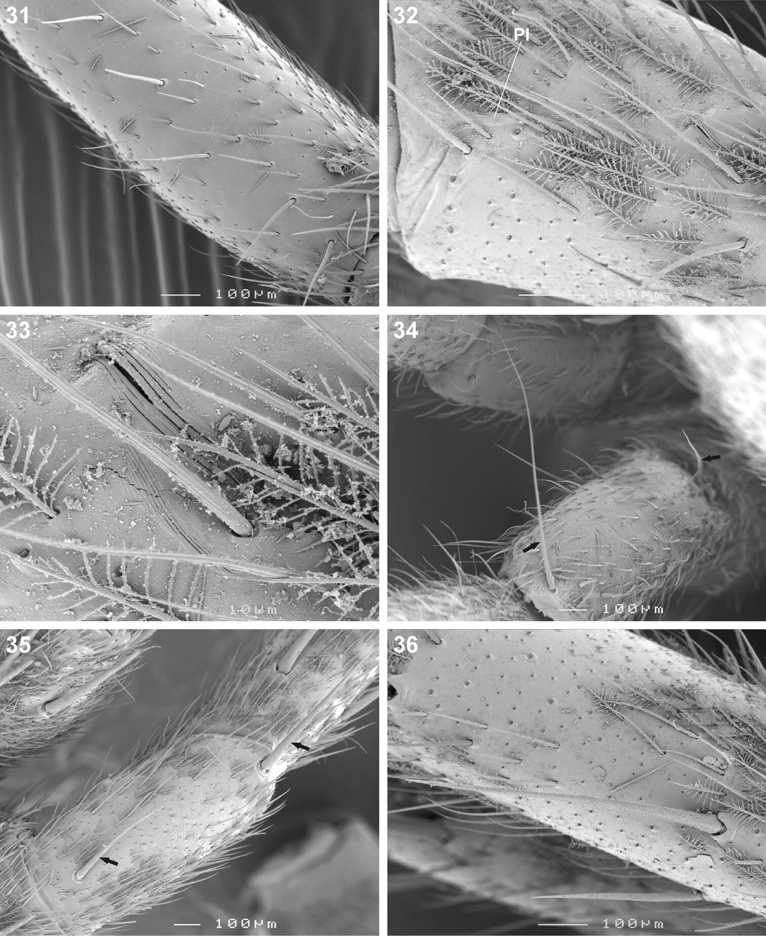
Figures 31–36.Scanning electron microscope photographs of Copa flavoplumosa Simon, 1885 male (31) and female (32–36): 31 femur I, erect ventral setae 32 patella II, indicating patellar indentation (PI) 33 same, detail of lyriform organ at proximal end of PI 34 patella II, arrows indicating proximal and distal dorsal patellar setae 35 patella III, arrows indicating proximal and distal dorsal patellar spines 36 tibia II, spines and feathery setae.
-
Figures 23–28.Scanning electron microscope photographs of Cambalida dippenaarae sp. n. female: 23 distal end of femur IV, plumose and short straight setae 24 patella III and 25 patella IV, arrows indicating long distal setae 26 leg II, detail of lyriform organ at proximal end of patellar indentation 27 tibia IV, spine and plumose setae 28 tibia II, trichobothrium base.
-
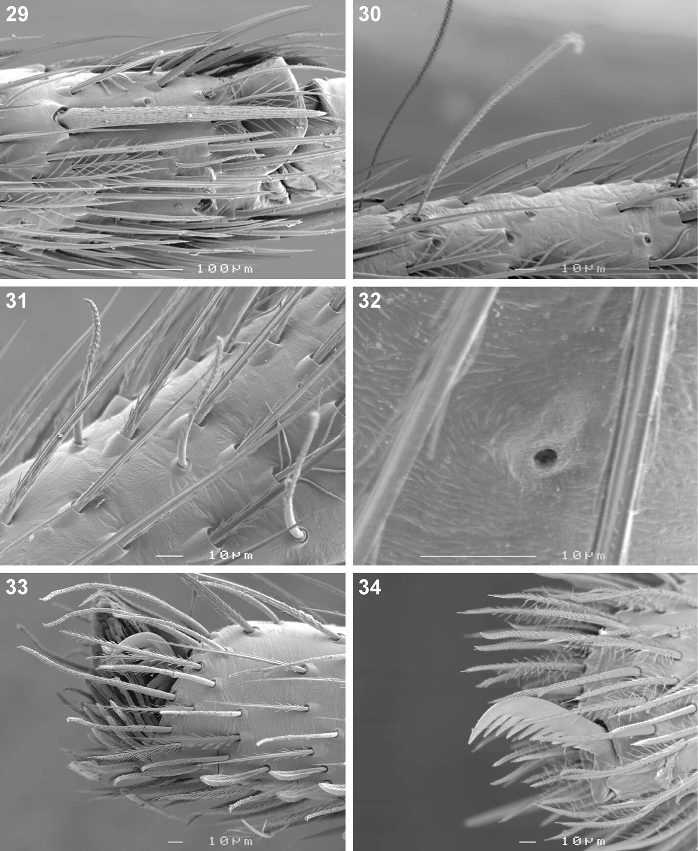
Figures 37–42.Scanning electron microscope photographs of Copa flavoplumosa Simon, 1885 female (37, 39–42) and male (38): 37 tibia I, long dorsal seta 38 tibia I, arrows indicating short erect setae 39 metatarsus II, spines and scopula 40 tarsus III 41 same, claw tuft and tarsal organ (arrow) 42 same, tarsal organ.
-
Figures 29–34.Scanning electron microscope photographs of Cambalida dippenaarae sp. n. female: 29 metatarsus IV, distal prolateral spine 30 tarsus IV, trichobothria 31 tarsus II, short erect setae 32 tarsus I, tarsal organ 33 same, claw tuft 34 palpal claw.
-
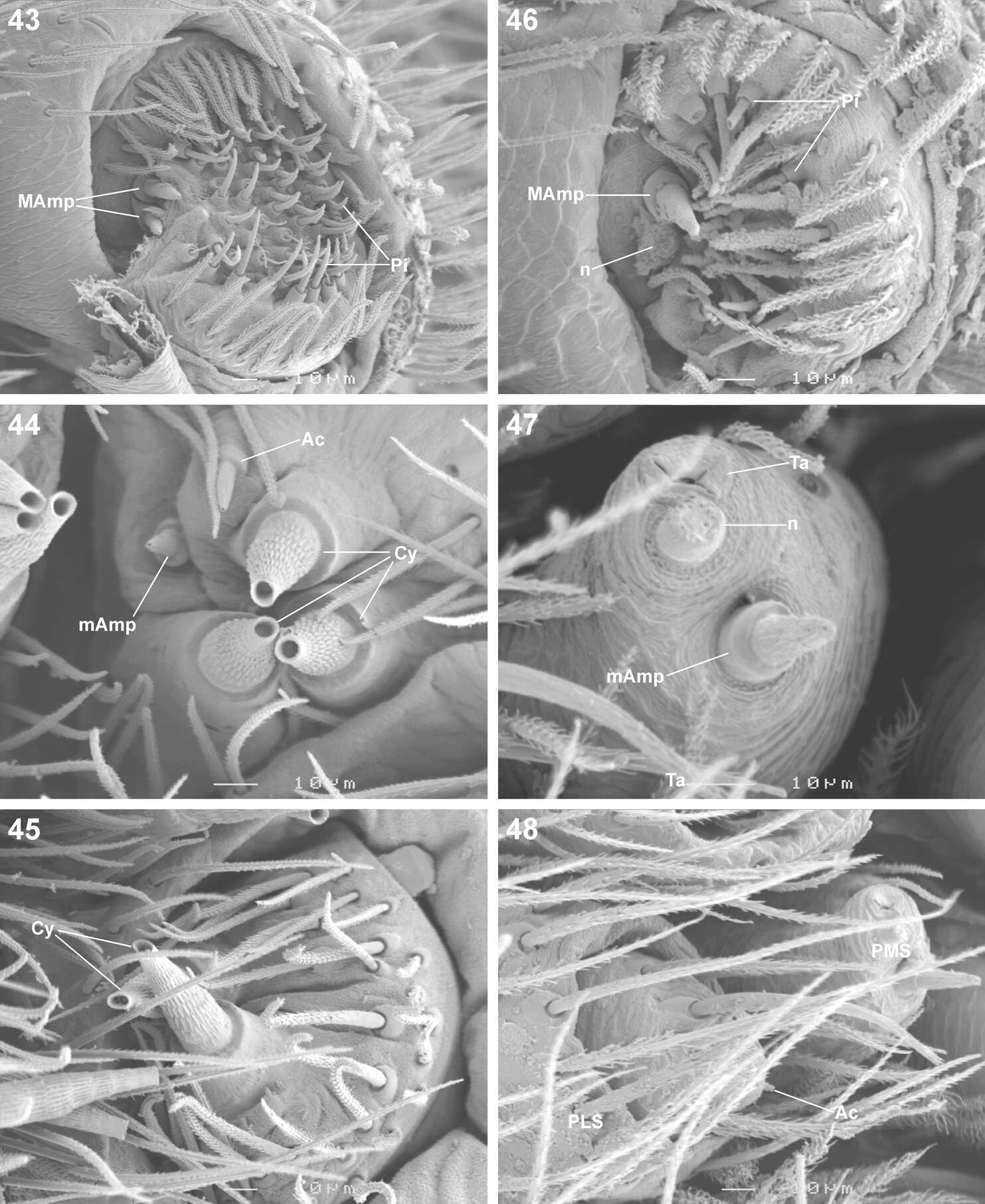
Figures 43–48.Scanning electron microscope photographs of Copa flavoplumosa Simon, 1885 female (43–45) and male (46–40) spinneret morphology: 43, 46 anterior lateral spinneret 44, 47 posterior median spinneret 45, 48 posterior lateral spinneret. Abbreviations: Ac aciniform gland spigot(s) Cy cylindrical gland spigot(s) MAmp major ampullate gland spigot(s) mAmp minor ampullate gland spigot(s) n nubbin Pi piriform gland spigot(s) PLS posterior lateral spinneret PMS posterior median spinneret ta tartipore.
-
Figures 35–43.Scanning electron microscope photographs of Cambalida dippenaarae sp. n. (35–39, 42), Cambalida compressa sp. n. (40), Cambalida deminuta (Simon, 1909) (41) and Cambalida loricifera (Simon, 1885) (43): 35 female, dorsal abdominal surface 36 dorsal abdominal sigillum and detail of plumose setae 37 female epigyne 38 thickened setae at dorsal distal end of male palpal cymbium 39 detail of modified setae 40–43 male emboli.
-
Figures 55–60.Scanning electron microscope photographs of Copa flavoplumosa Simon, 1885 (55, 56) and Copa kei sp. n. (57–60): 55, 57 female epigyne, ventral view 56, 58 male embolus, ventral view 59 male palpal cymbial setae 60 distal end of cymbium, retrolateral distal view.
-
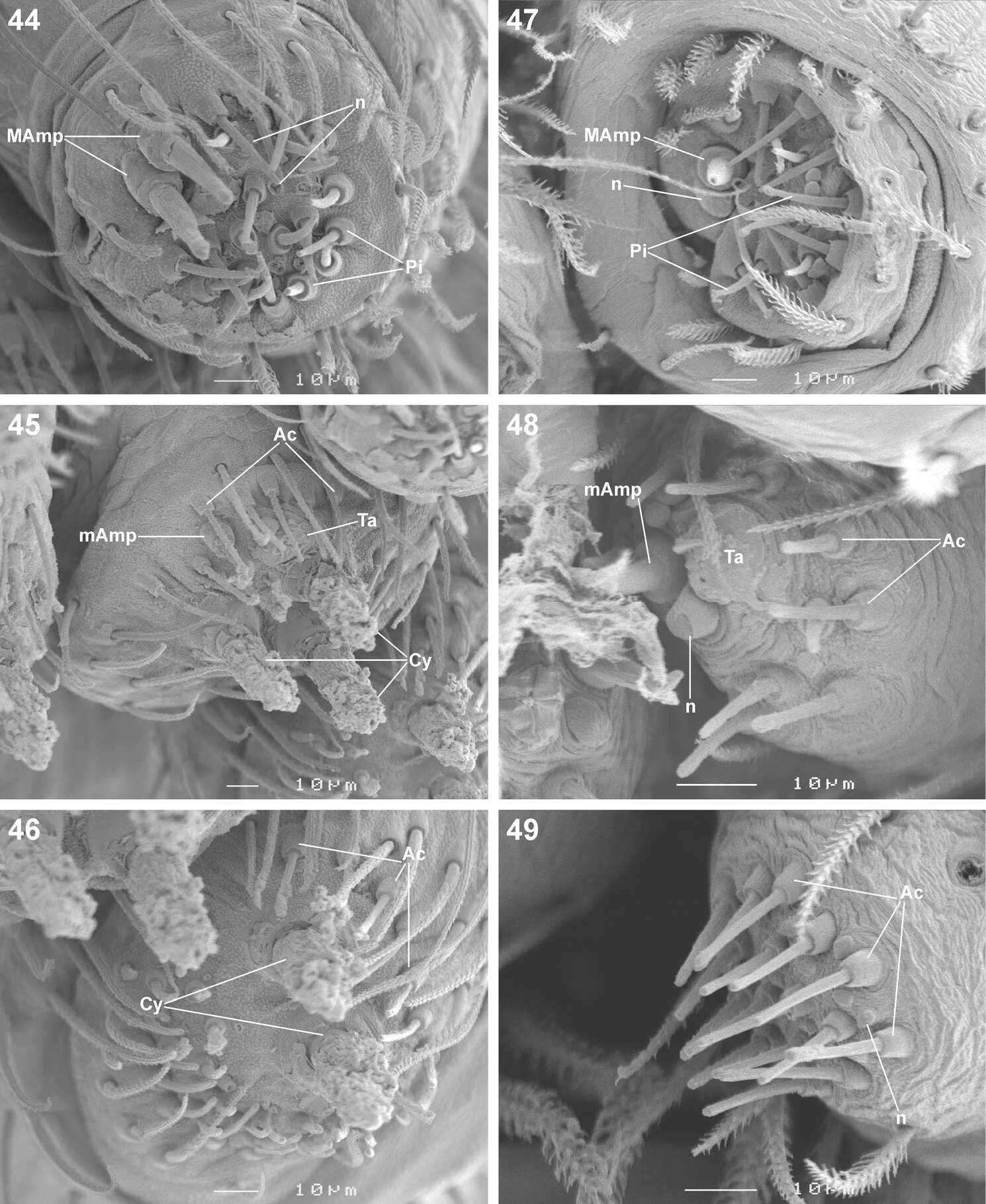
Figures 44–49.Scanning electron microscope photographs of Cambalida dippenaarae sp. n.female (44–46) and male (47–49) spinneret morphology: 44, 47 anterior lateral spinneret 45, 48 posterior median spinneret 46, 49 posterior lateral spinneret. Abbreviations: Ac aciniform gland spigot(s) Cy cylindrical gland spigot(s) MAmp major ampullate gland spigot(s) mAmp minor ampullate gland spigot(s) n nubbin(s) Pi piriform gland spigot(s) ta tartipore.
-
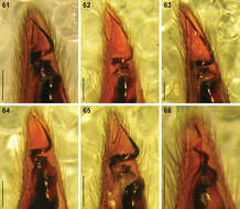
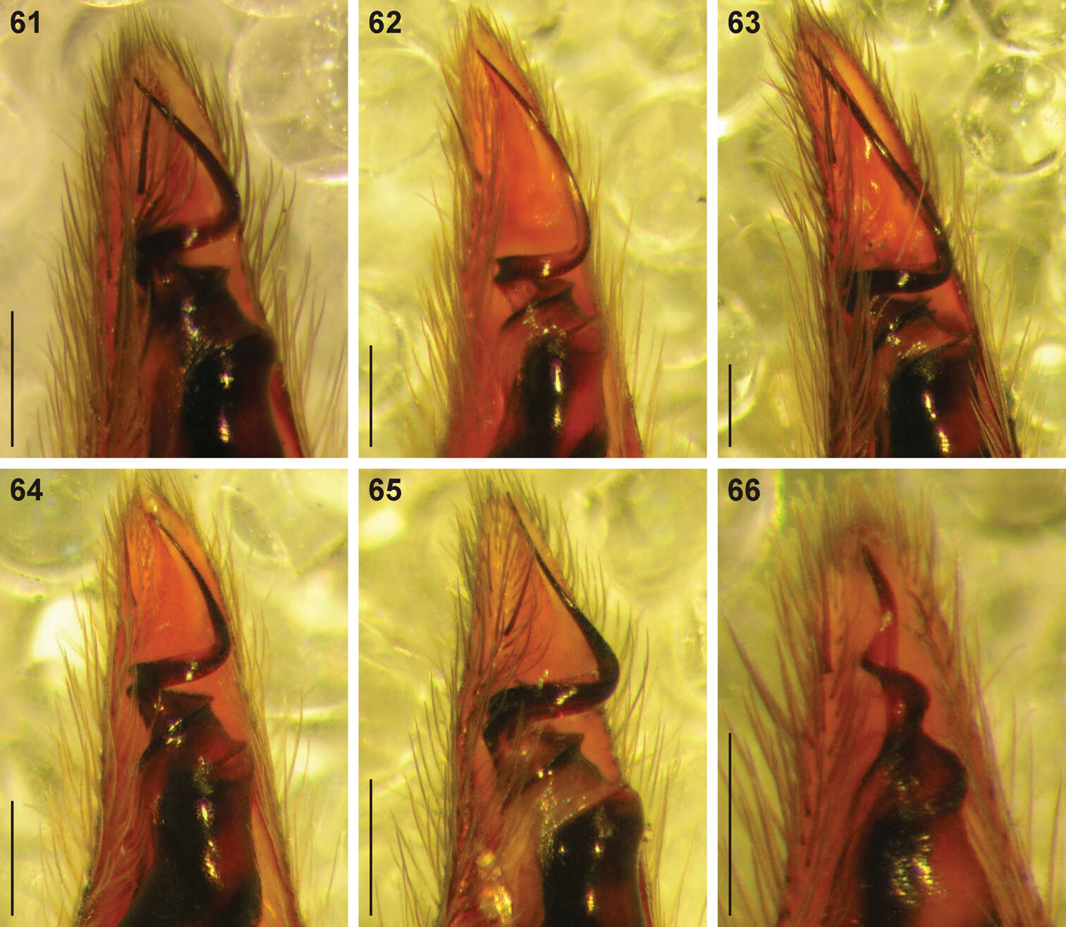
Figures 61–66.Digital microscope photographs of emboli of Copa species in ventral view: 61–65 Copa flavoplumosa Simon, 1885 from D.R. Congo (61), Cameroon (62), Tanzania (63), Botswana (64) and South Africa (65) 66 Copa kei sp. n. from South Africa. Scale bars = 0.1 mm.
-
Figures 50–56.Digital microscope photographs of emboli of Afrotropical Cambalida species in ventral view: 50 Cambalida compressa sp. n. 51 Cambalida coriacea Simon, 1909 52 Cambalida deminuta (Simon, 1909) 53 Cambalida dippenaarae sp. n. 54 Cambalida fulvipes (Simon, 1896) 55 Cambalida griswoldi sp. n. 56 Cambalida loricifera (Simon, 1885). Scale bars = 0.1mm.
-
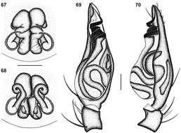
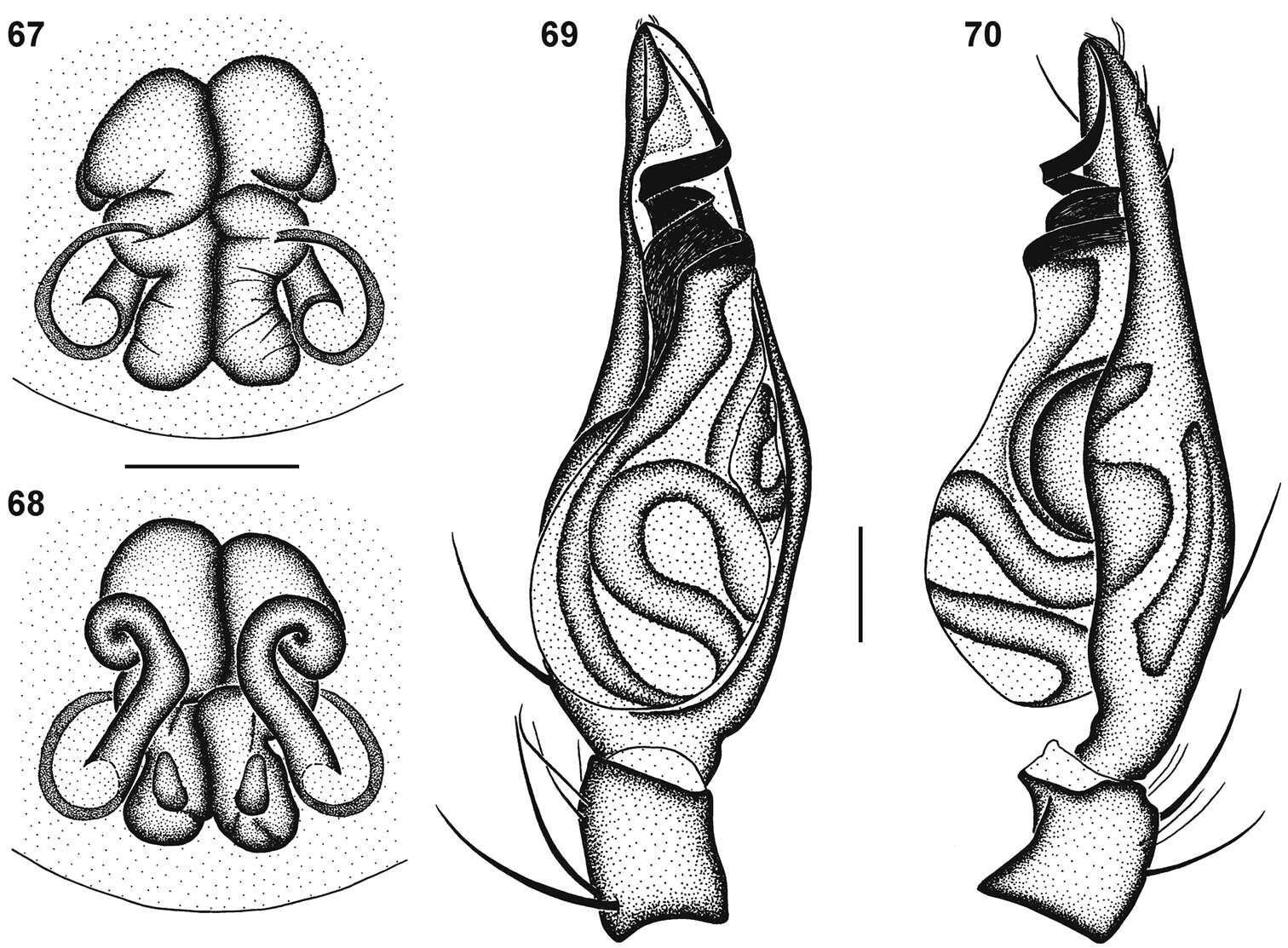
Figures 67–70.Genitalic morphology of Copa flavoplumosa Simon, 1885: 67 female epigyne, ventral view 68 same, dorsal view 69 male palp, ventral view 70 same, retrolateral view. Scale bars = 0.25 mm.
-
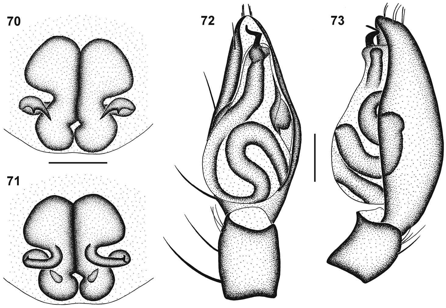
Figures 70–73.Genitalic morphology of Cambalida dippenaarae sp. n.: 70 female epigyne, ventral view 71 same, dorsal view 72 male palp, ventral view 73 same, retrolateral view. Scale bars = 0.25mm.
-
Figures 1–6.General habitus photographs of Copa flavoplumosa Simon, 1885 (1–4) and Copa kei sp. n. (5, 6): 1 female from Lesideng Research Camp, Botswana 2 female from Livingtone, Zambia 3 male and 4 female from Wildlives Game Farm, Zambia 5 female from Hogsback, South Africa 6 male from Cwebe Nature Reserve, South Africa.
-
Figures 50–56.Digital microscope photographs of emboli of Afrotropical Cambalida species in ventral view: 50 Cambalida compressa sp. n. 51 Cambalida coriacea Simon, 1909 52 Cambalida deminuta (Simon, 1909) 53 Cambalida dippenaarae sp. n. 54 Cambalida fulvipes (Simon, 1896) 55 Cambalida griswoldi sp. n. 56 Cambalida loricifera (Simon, 1885). Scale bars = 0.1mm.
-
Figures 7–12.Digital microscope photographs of Copa flavoplumosa Simon, 1885 from D.R. Congo (7–9) and Copa kei sp. n.from South Africa (10–12): 7, 10 female, dorsal habitus 8, 11 male, dorsal habitus 9, 12 sternum of female in ventral view. Scale bars = 1.0 mm.