Andrea L. Crowther, Daphne G. Fautin, Carden C. Wallace
Zookeys
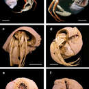
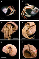
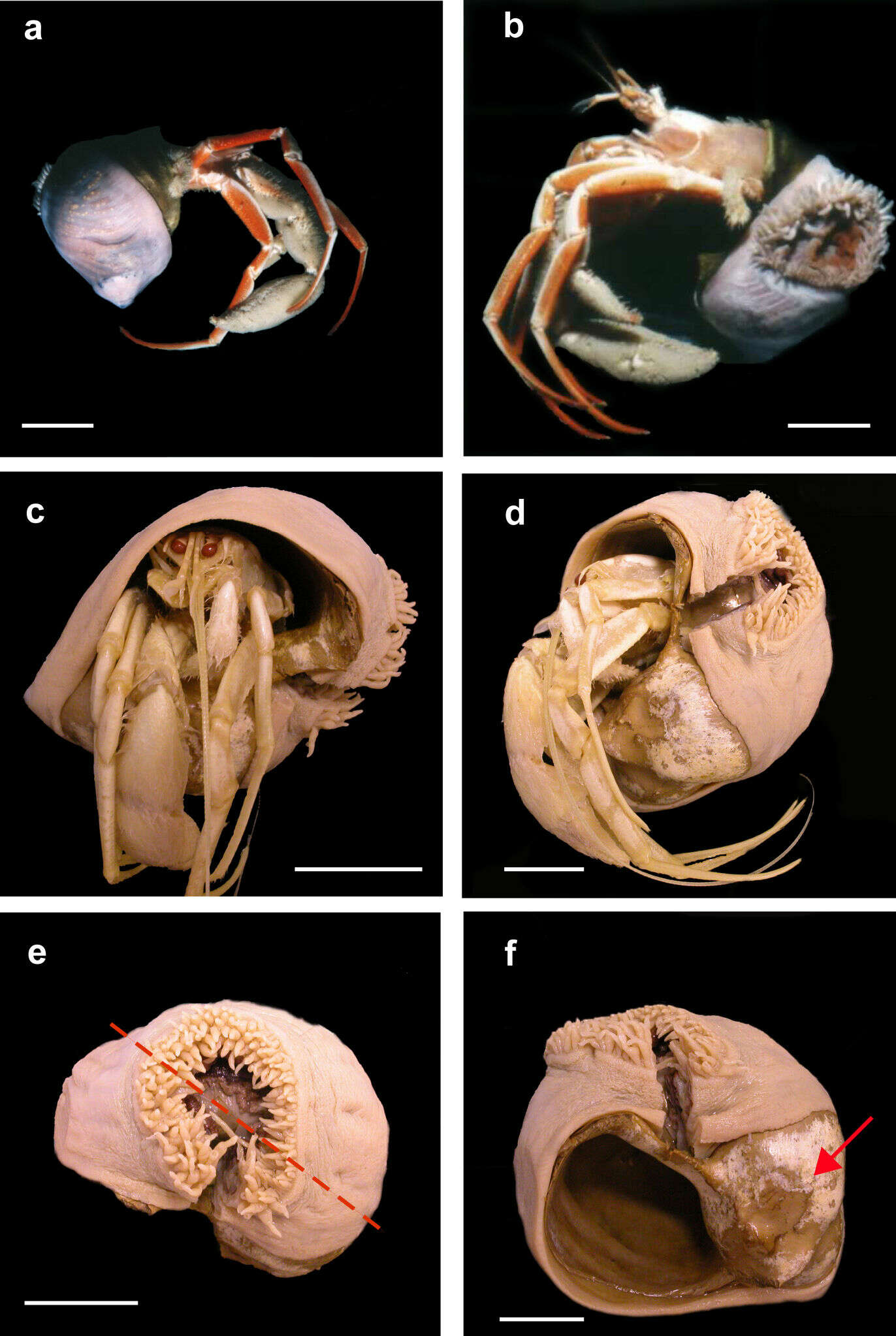
Figure 1.Stylobates birtlesi sp. n. holotype MTQ G57579 a, b soon after collection (photo: RA Birtles) c, d preserved specimen with Sympagurus trispinosus showing position of oral disc of anemone e preserved specimen: shortest tentacles beside longest ones (on right side of oral disc in this view); tentacles grade in length between longest and shortest around other side of oral disc (dashed line indicates directive axis) f preserved specimen without hermit crab showing aperture and part of carcinoecium not covered by anemone (arrow). Scale bars 20 mm.
Andrea L. Crowther, Daphne G. Fautin, Carden C. Wallace
Zookeys
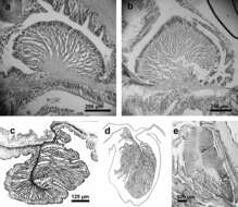
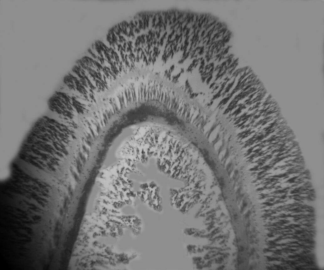
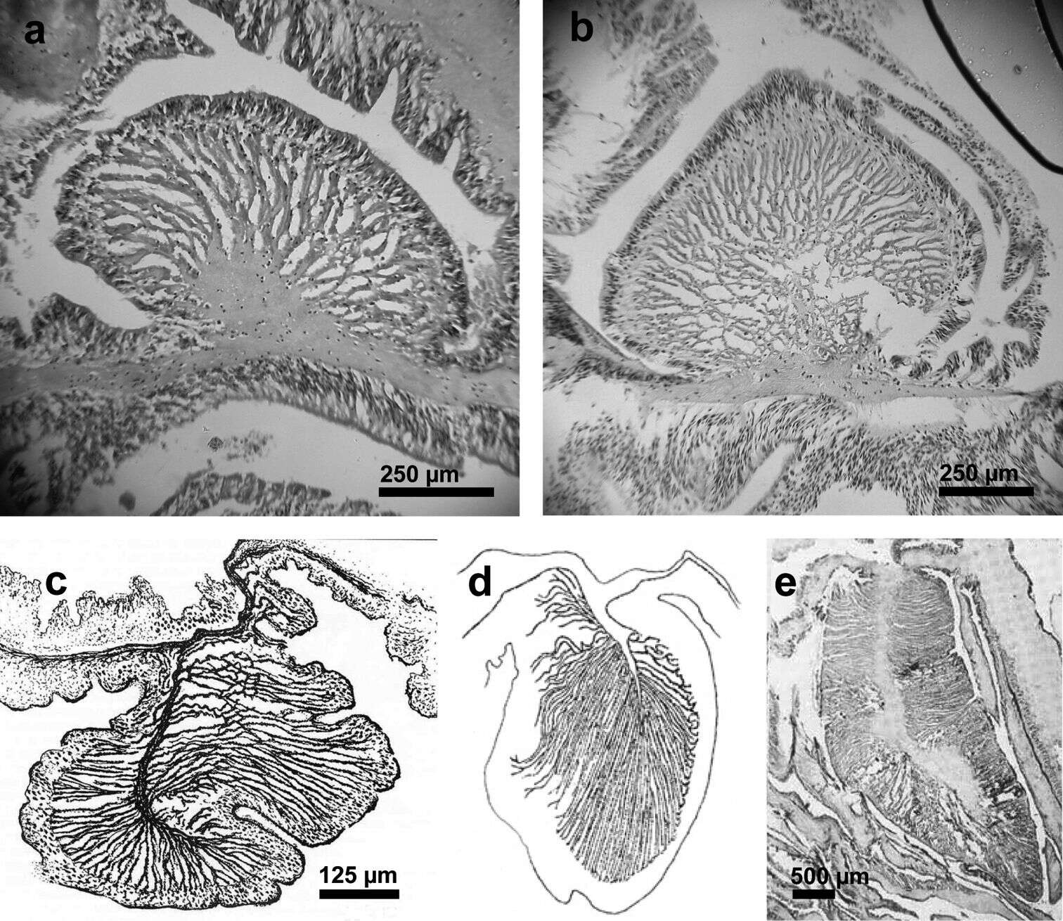
Figure 3.Endodermal circumscribed marginal sphincter muscles of Stylobates spp. a, b Palmate marginal sphincter muscle of Stylobates birtlesi sp. n. a paratype MTQ G57581 b paratype KUDIZ 003352 c-e Pinnate marginal sphincter muscles. c Stylobates aeneus (from Dunn et al. 1981) d Stylobates cancrisocia (from Carlgren 1928a) e Stylobates loisetteae (from Fautin 1987).
Andrea L. Crowther, Daphne G. Fautin, Carden C. Wallace
Zookeys
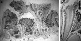
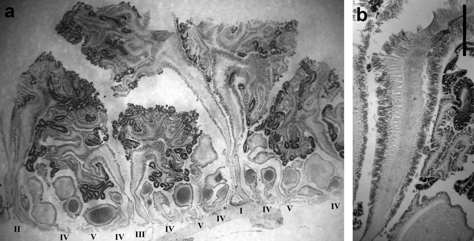
Figure 4.Mesenterial musculature and fertility of Stylobates birtlesi sp. n. holotype MTQ G57579 a mesenterial arrangement, orders indicated with Roman numerals; column wall at base of image b diffuse retractor muscle; column wall at base of image. Scale bar 1 mm.