-
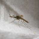
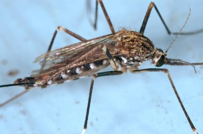
The mosquito pictured here in this 2005 photograph, was until recently known as Aedes japonicus, and is now labeled Ochlerotatus japonicus. This particular specimen was a member of the Notre Dame colony. Oc. japonicus was initially collected in the United States in New York and New Jersey, in 1998.Created: 2005
-
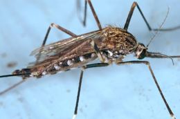
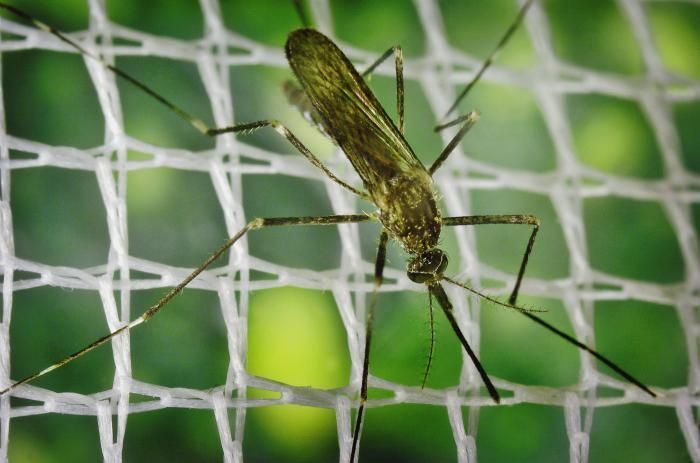
The mosquito pictured here in this 2005 photograph, was until recently known as Aedes japonicus, and is now labeled Ochlerotatus japonicus. This particular specimen was a member of the Notre Dame colony. Oc. japonicus was initially collected in the United States in New York and New Jersey, in 1998.Created: 2005
-
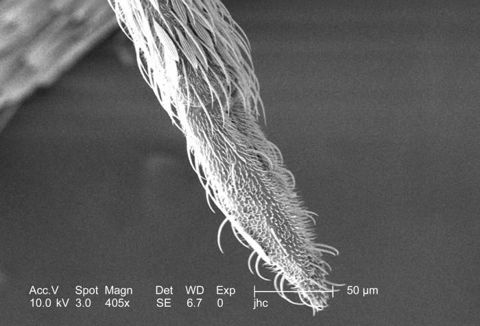
Magnified 405x, this scanning electron micrograph (SEM) revealed the ornately festooned tip of an Anopheles gambiae mosquito's proboscis. Seen in the field of view is actually the sheath that covered the pair of the needle-sharp "stylets", which together are known as the "fascicle". The larger of the two stylets, known as the "labrum", when viewed in cross-section, takes on the shape of a "V", and acts as a gutter, which directs the ingested host blood towards the insect's mouth.Created: 2006
-
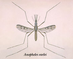
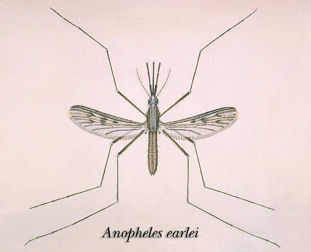
An illustration of an Anopheles earlei mosquito.Created:
-
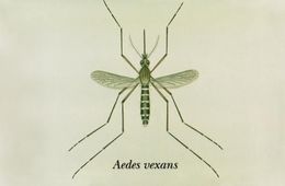
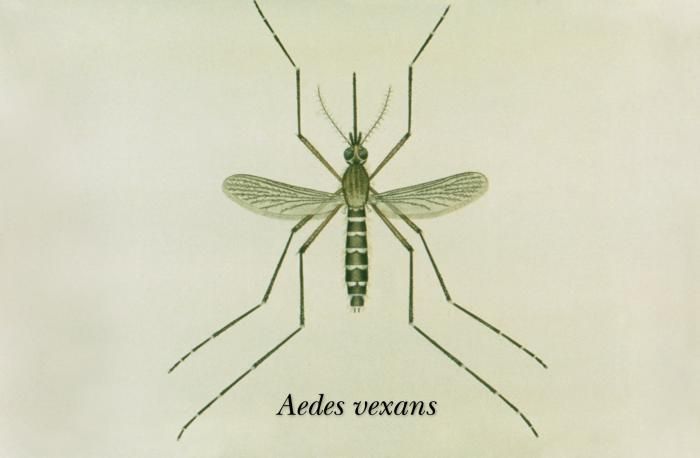
This is an illustration of an adult Aedes vexans mosquito.Created: 1976
-
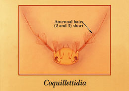
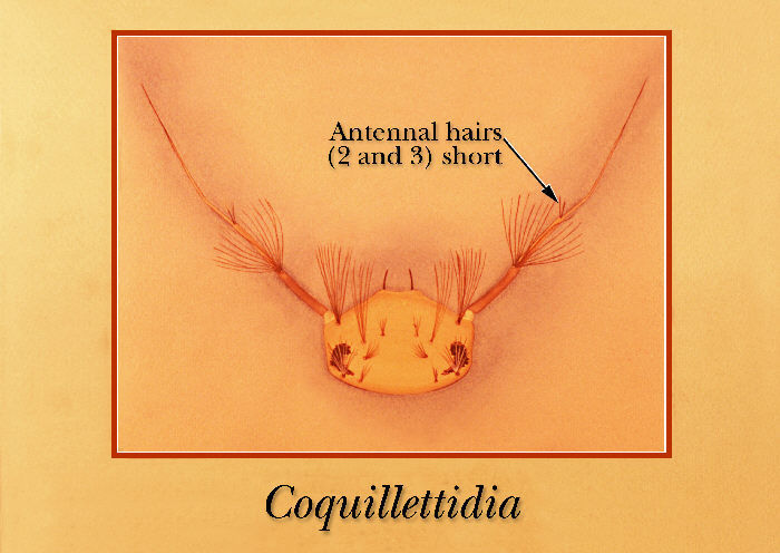
Drawing identifying the antennal structures on the head region of a Coquillettidia mosquito larva.Created: 1975
-
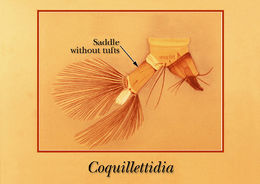
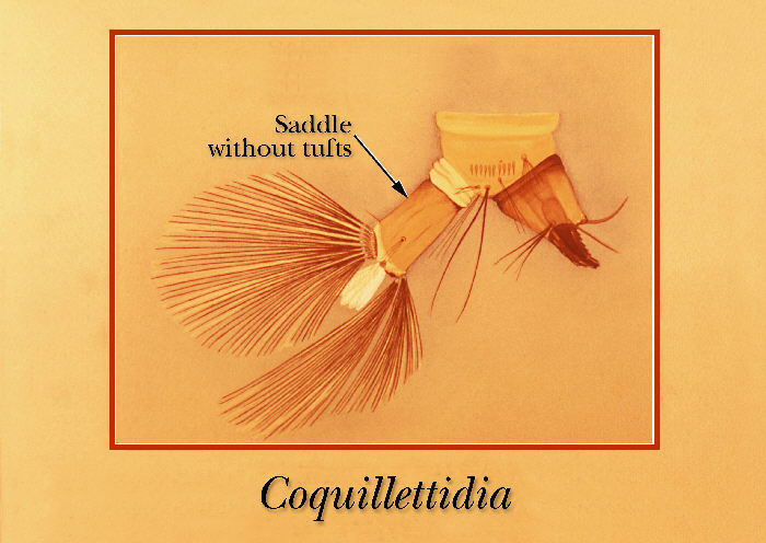
An illustration of a Coquillettidia mosquito larva identifying the terminal abdominal segments.Created: 1975
-
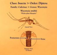
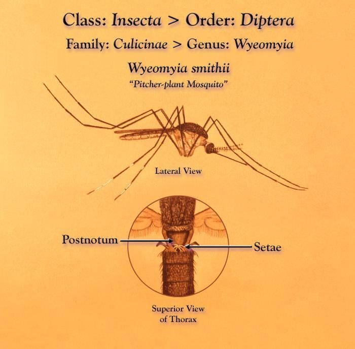
This illustration depicts a pitcher-plant, Wyeomyia smithii mosquito from a lateral view, as well as an enlarged inset highlighting its mesonotum, and its postnotal tuft of seta.Created: 1975
-
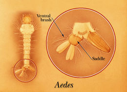
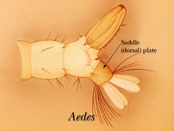
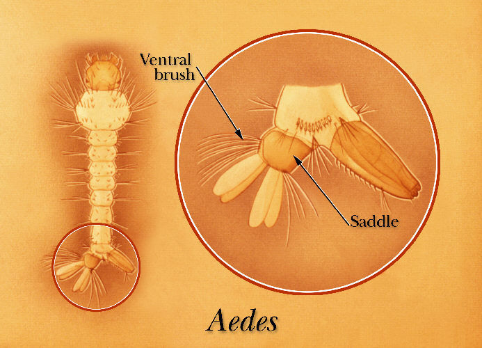
An illustration of Aedes mosquito larva with ventral brush and dorsal plate identified.Created: 1975
-
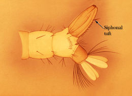
An illustration identifying the siphonal tuft of the Aedes, Uranotaenia or Psorophora mosquito larvae.Created: 1975
-
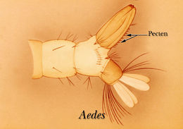
An illustration identifying the pecten on the terminal abdominal segment of an Aedes mosquito larva.Created: 1975
-
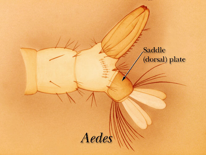
An illustration identifying the dorsal plate on the terminal segment of an Aedes mosquito larvaCreated: 1975
-
An illustration identifying the pecten on the terminal segments of an Aedes aegypti mosquito larva.Created: 1975
-
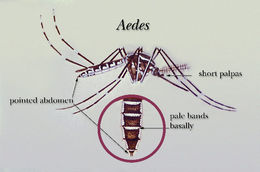
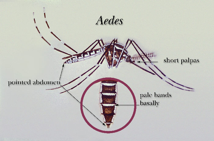
An illustration of an Aedes mosquito.Created: 1985
-
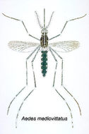
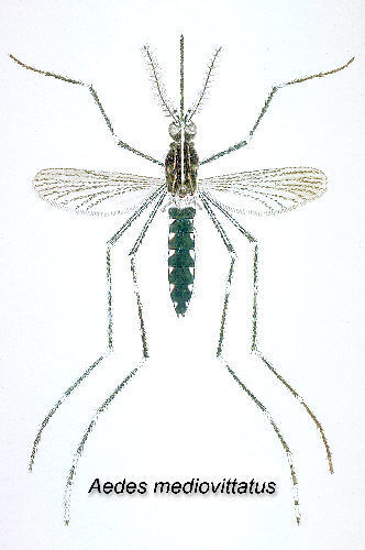
The Aedes mediovittatus mosquito has been shown to be a vector in the transmission of Dengue Fever. (Illustration)Created: 1964
-
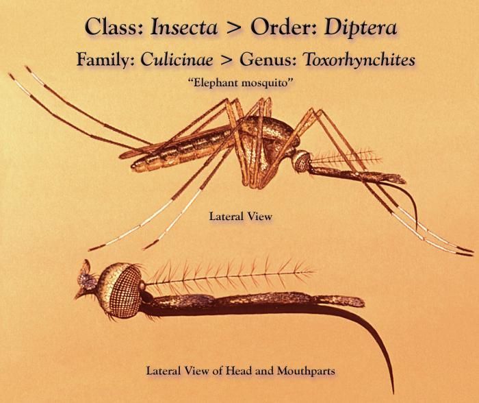
This illustration depicts a Toxorhynchites sp., Elephant mosquito from a lateral view, and an inset of its head region revealing the morphologic details of its mouthparts.Created: 1975
-
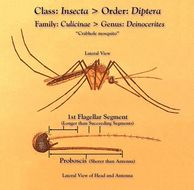
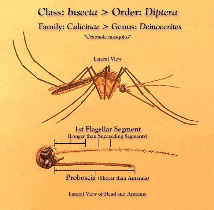
This illustration depicts a Deinocerites sp., Crabhole mosquito from a lateral view, and an inset of its head region revealing the morphologic details of its antennae.Created: 1975
-
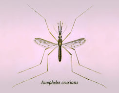
An illustration of an Anopheles crucians mosquito.Created:
-
This 2005 photograph depicted a female Anopheles albimanus mosquito while she was feeding on a human host, thereby, becoming engorged with blood.Created: 2005
-
This 2005 photograph depicted a female Anopheles albimanus mosquito while she was feeding on a human host, thereby, becoming engorged with blood.Created: 2005
-
This 2005 photograph depicted a female Anopheles albimanus mosquito while she was feeding on a human host, thereby, becoming engorged with blood.Created: 2005
-
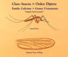
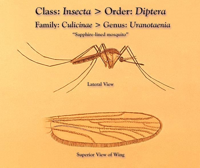
This illustration depicts a Uranotaenia sapphirina mosquito from a lateral view, and an inset of one of its wings from a superior perspective detailing its venation pattern.Created: 1975
-
This illustration depicts a Uranotaenia sapphirina mosquito from a lateral view, and an inset of one of its wings from a superior perspective detailing its venation pattern.Created: 1975
-
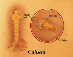
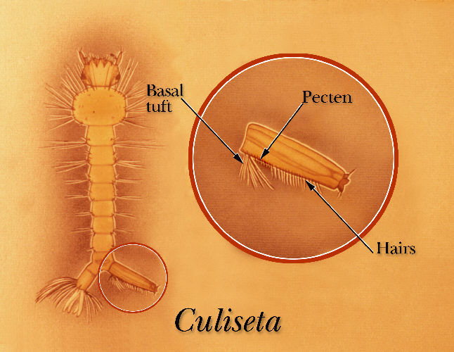
An illustration identifying the basal tuft, pecten and rows of hairs of a Culiseta mosquito larva.Created: 1975