-
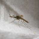
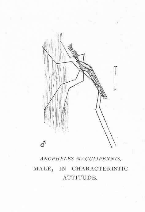
Anopheles maculipennis. Male, in characteristic attitude.
-
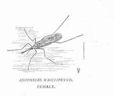
Anopheles maculipennis. Female.
-
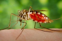
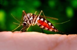
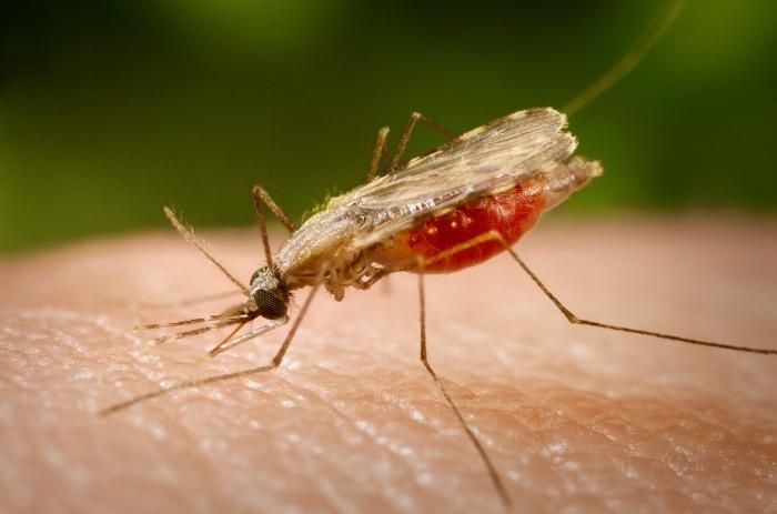
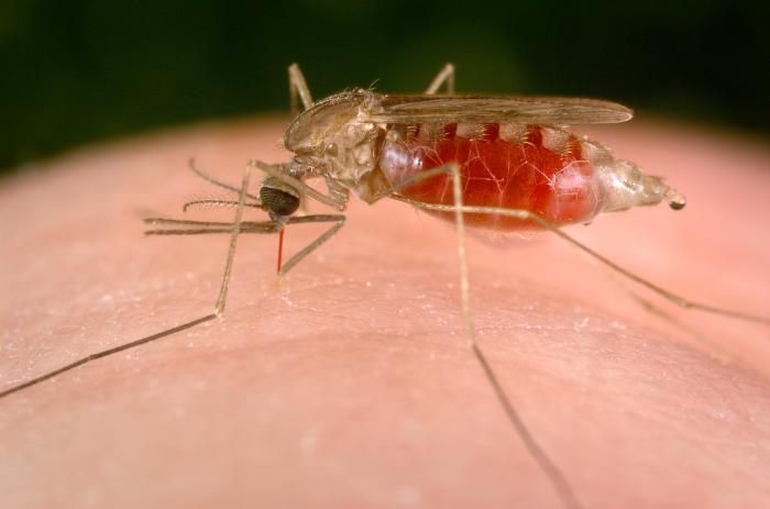
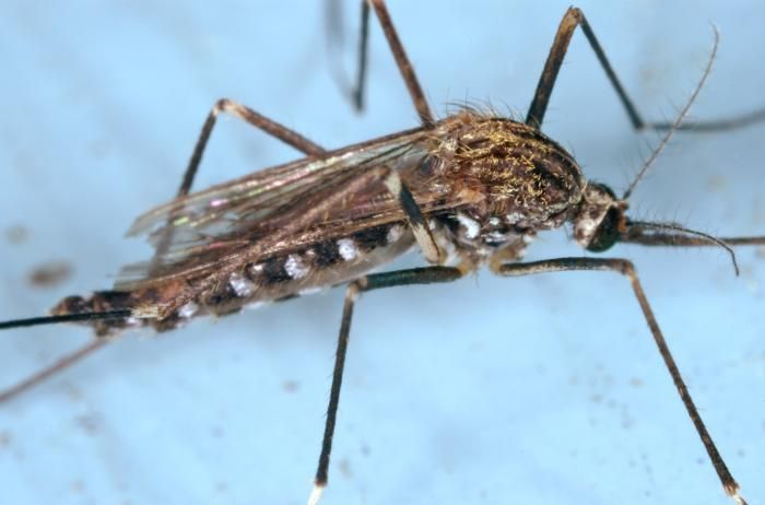
This close-up photograph shows an Anopheles minimus mosquito, a malaria vector of the Orient, as she was feeding on a human host. Note the blood meal that this mosquito had ingested, as it collected inside its stomach within its abdominal segment, extracting it from the host though its proboscis, which it had used to penetrate the skin, much like a straw.Created: 2005
-
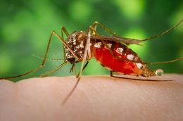
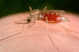
This close-up 2005 photograph shows an Anopheles minimus, a malaria vector of the Orient mosquito, from a lateral perspective as she was feeding on a human host. Note the blood meal that this mosquito had ingested, as it collected inside its stomach within its abdominal segment, extracting it from the host though its proboscis, which it had used to penetrate the skin, much like a straw.Created: 2005
-
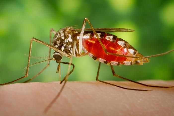
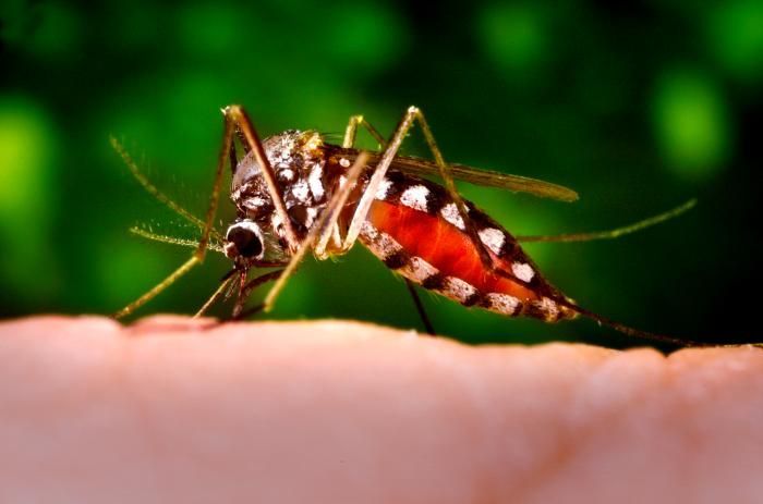
An Armigeres subalbatus mosquito of the Nagasaki colony was depicted in this 2005 photograph, as she was ingesting a blood meal after having lighted on a human finger. Note the pooling of the blood inside the mosquitos abdomen as it fills its stomach. The blood was being suctioned through the insects proboscis, which is its straw-like mouth that is used to penetrate the hosts skin much like a syringe.Created: 2005
-
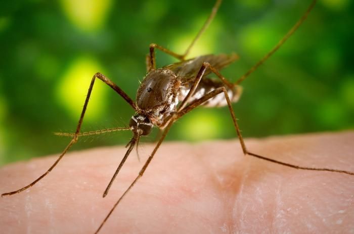
This mosquito, an Armigeres subalbatus, was found in the Nagasaki colony, and has a very broad pattern of distribution throughout Asia. This particular specimen had just lighted on a human finger, and was about to pierce the skin of its host in order to begin the process of ingesting a blood meal.Created: 2005
-
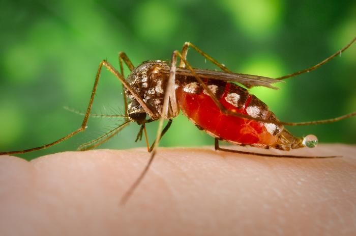
This 2005 photograph depicted an Armigeres subalbatus mosquito of the Nagasaki colony, as she was ingesting a blood meal after having lighted on a human finger. Note the pooling of the blood inside the mosquitos abdomen as it filled its stomach. The blood was being suctioned through the insects proboscis, which is its straw-like mouth that is used to penetrate the hosts skin much like a syringe.Created: 2005
-
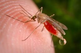
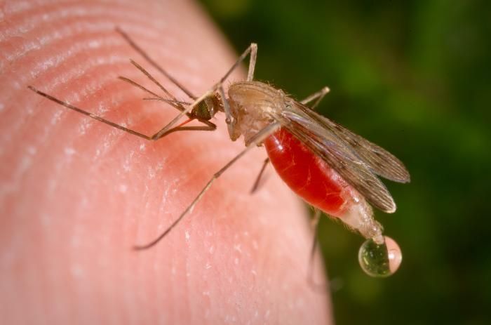
This female Anopheles freeborni is taking a blood meal from a human host by pumping the ingested blood through her labrum, which is visible here as a thin red, needle-like structure between the mosquitos head and the hosts skin.Created: 2004
-
This female Anopheles freeborni is taking a blood meal from a human host by pumping the ingested blood through her labrum, which is visible here as a thin red, needle-like structure between the mosquitos head and the hosts skin.Created: 2004
-
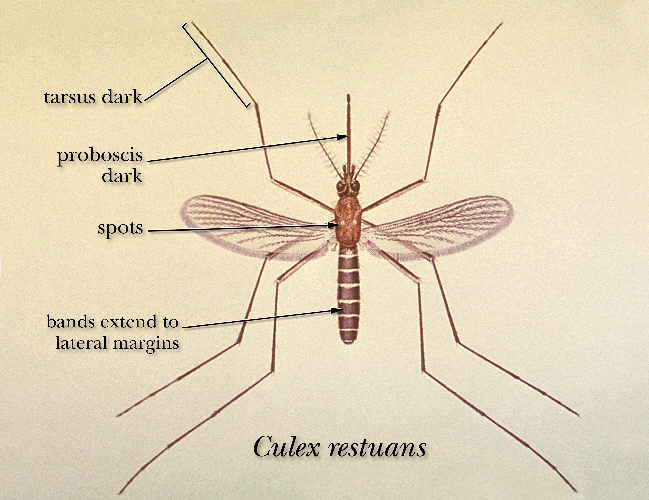
An illustration of a Culex restuans mosquito.Created:
-
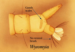
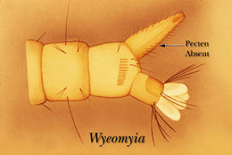
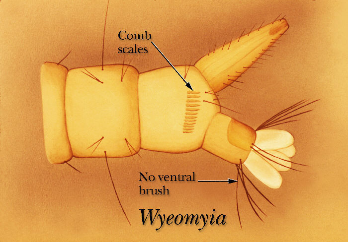
An illustration of the ventral surface of the terminal segment of a Wyeomyia mosquito larva.Created: 1975
-
A drawing showing that the Wyeomyia mosquito larva has no pectin row on the ventral surface of its siphon tube.Created: 1975
-
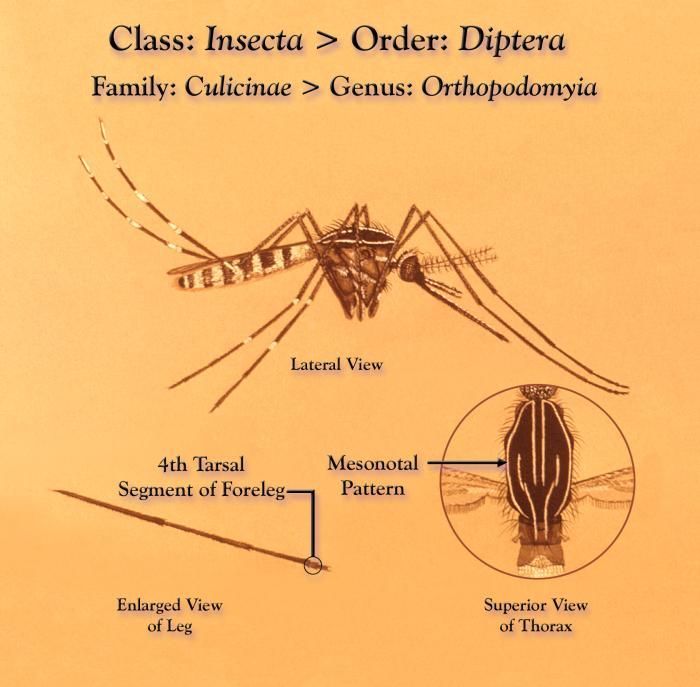
This illustration depicts a Orthopodomyia sp. mosquito from a lateral view, as well as two insets; an enlarged view of its foreleg (Lt), and a superior view of the insects thoracic region revealing the linear details of its mesonotal pattern.Created: 1975
-
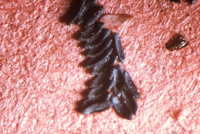
This photograph depicts a group of mosquito eggs deposited by a female Aedes triseriatus mosquito upon velour paper; Mag. 250x.Created:
-
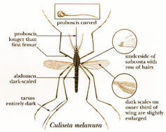
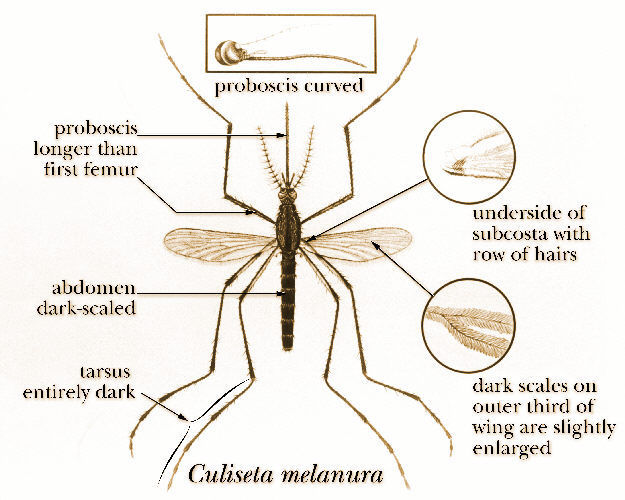
An illustration of a Culiseta melanura mosquito.Created:
-
An illustration of a Culiseta melanura mosquito.Created:
-
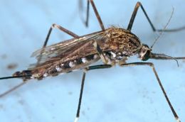
The mosquito pictured here in this 2005 photograph, was until recently known as Aedes japonicus, and is now labeled Ochlerotatus japonicus. This particular specimen was a member of the Notre Dame colony. Oc. japonicus was initially collected in the United States in New York and New Jersey, in 1998.Created: 2005
-
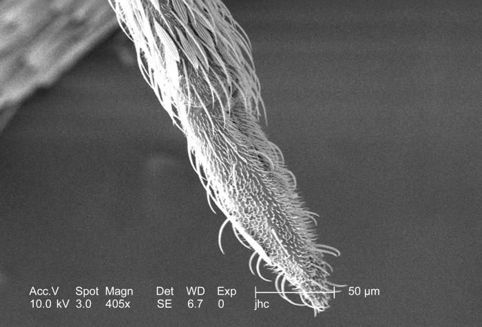
Magnified 405x, this scanning electron micrograph (SEM) revealed the ornately festooned tip of an Anopheles gambiae mosquito's proboscis. Seen in the field of view is actually the sheath that covered the pair of the needle-sharp "stylets", which together are known as the "fascicle". The larger of the two stylets, known as the "labrum", when viewed in cross-section, takes on the shape of a "V", and acts as a gutter, which directs the ingested host blood towards the insect's mouth.Created: 2006
-
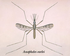
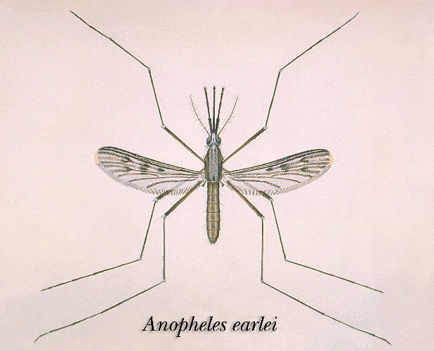
An illustration of an Anopheles earlei mosquito.Created:
-
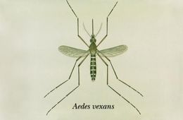
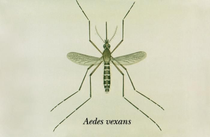
This is an illustration of an adult Aedes vexans mosquito.Created: 1976
-
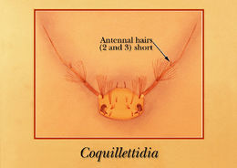
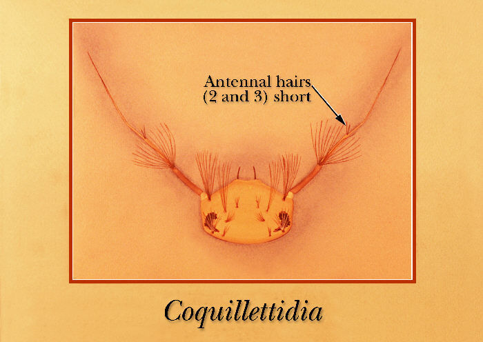
Drawing identifying the antennal structures on the head region of a Coquillettidia mosquito larva.Created: 1975
-
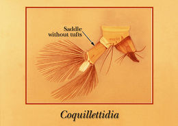
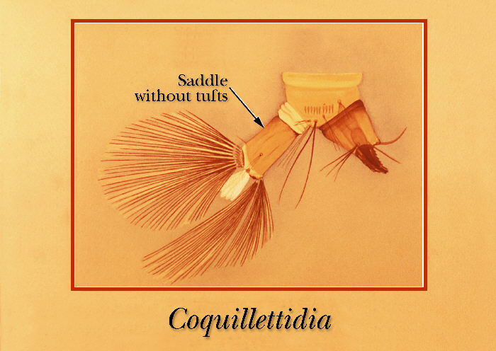
An illustration of a Coquillettidia mosquito larva identifying the terminal abdominal segments.Created: 1975