-
All Biocode files are based on field identifications to the best of the researcher’s ability at the time.
-
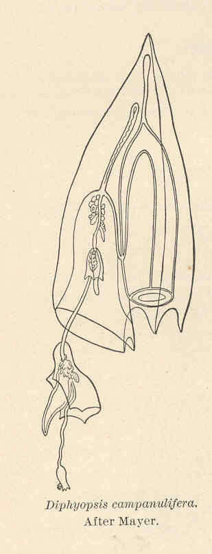
Diphyopsis campanulifera. After Mayer.
-
-
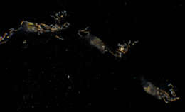
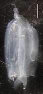
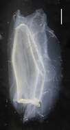
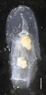
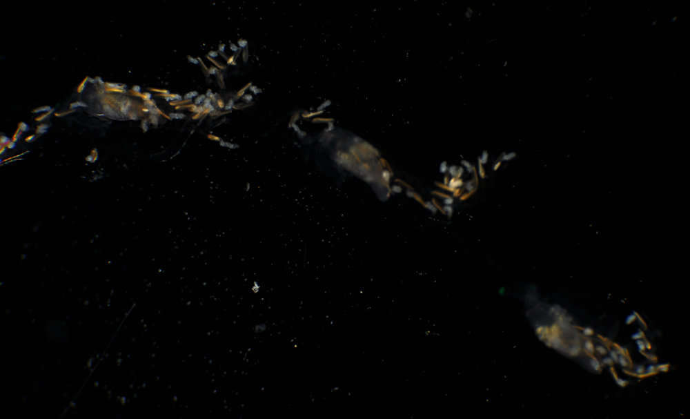
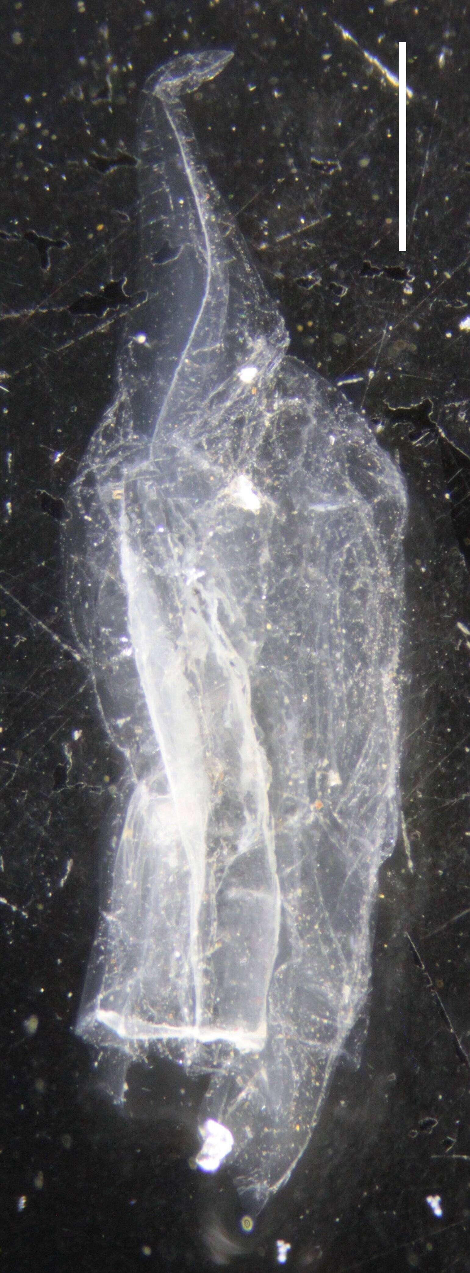
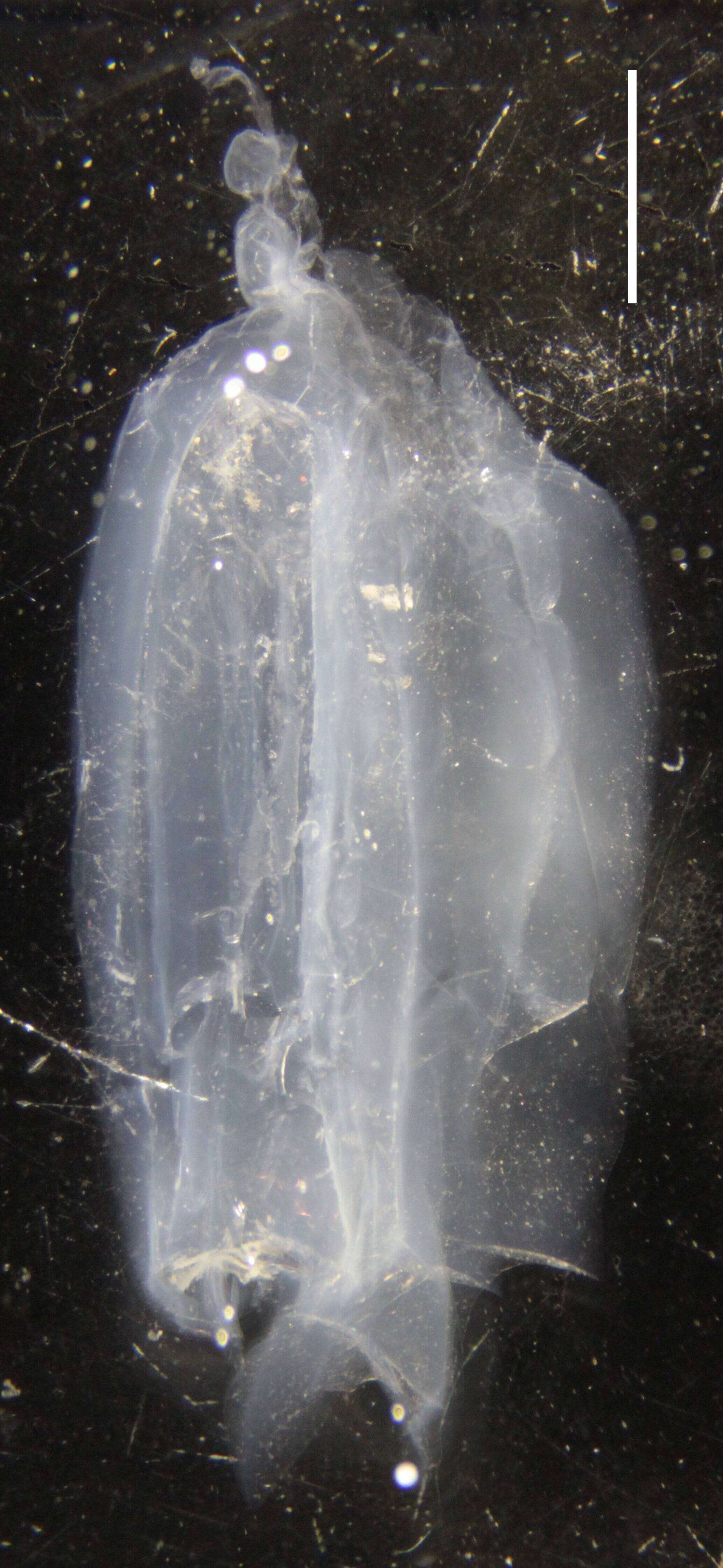
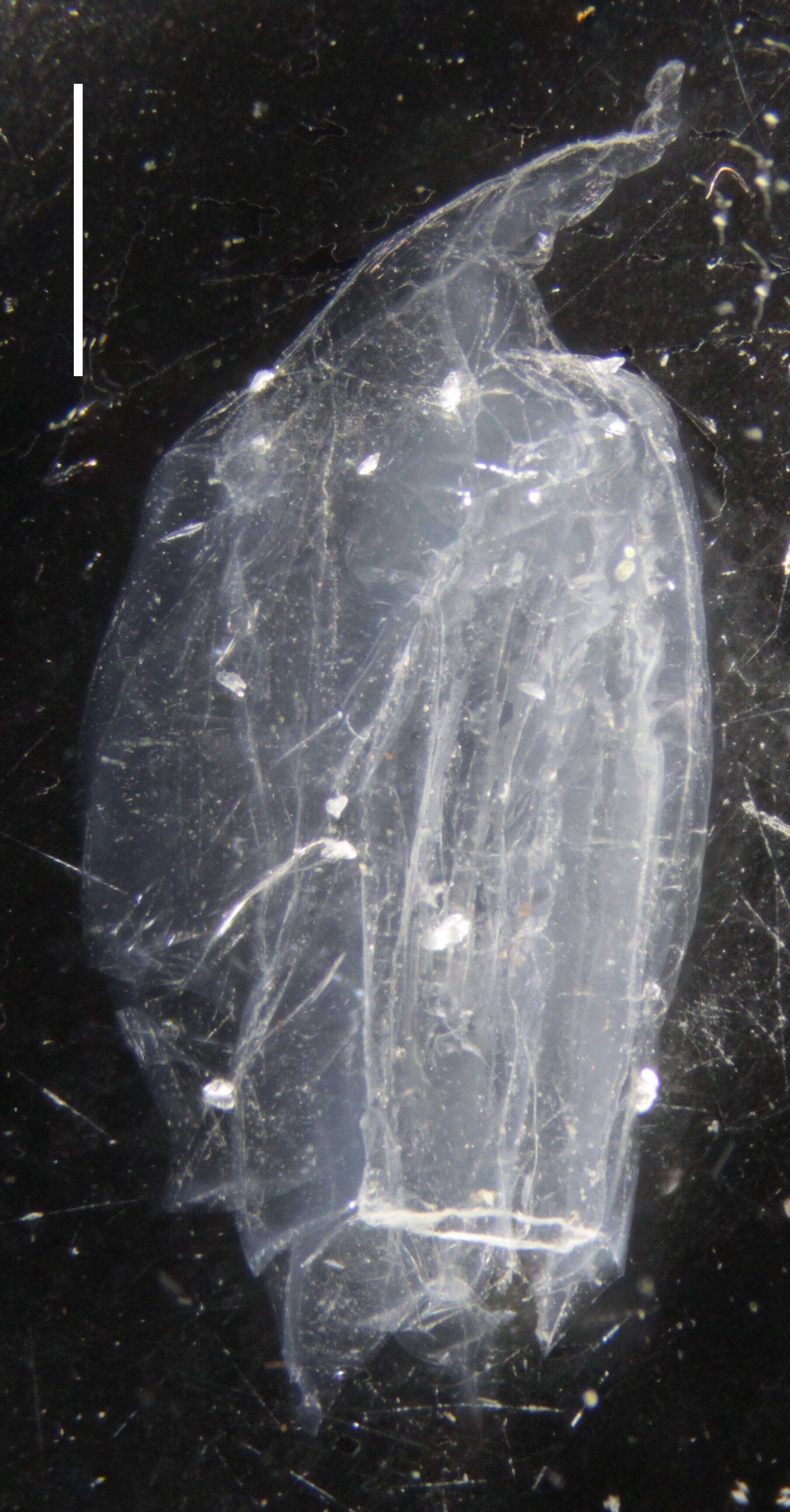
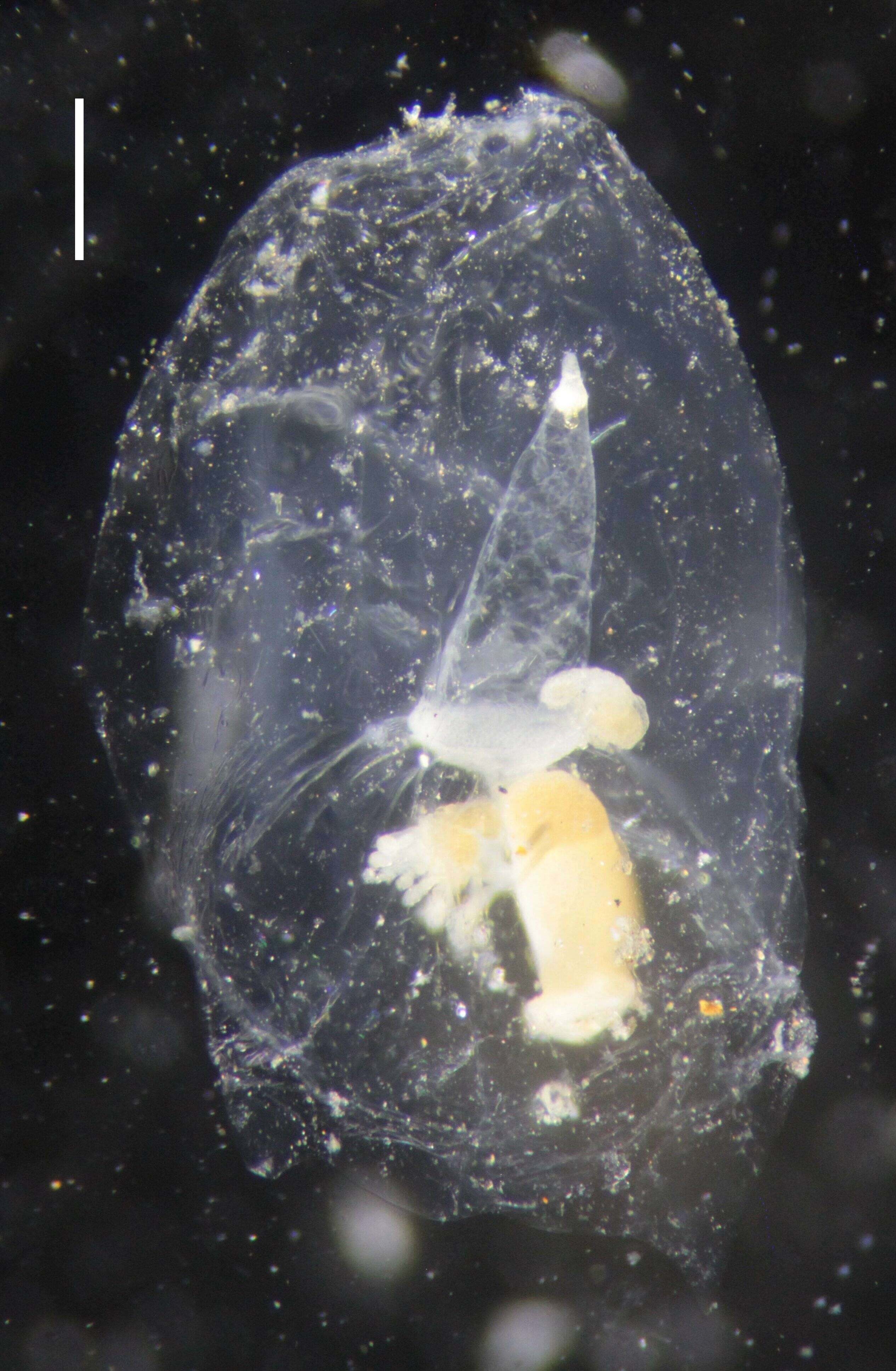
Microsope specimen image of IZ COE 28527; Anterior Nectophore; image 20; Scale Bar = 3 mm
-
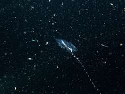
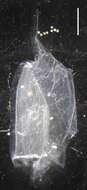
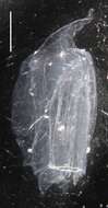
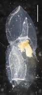
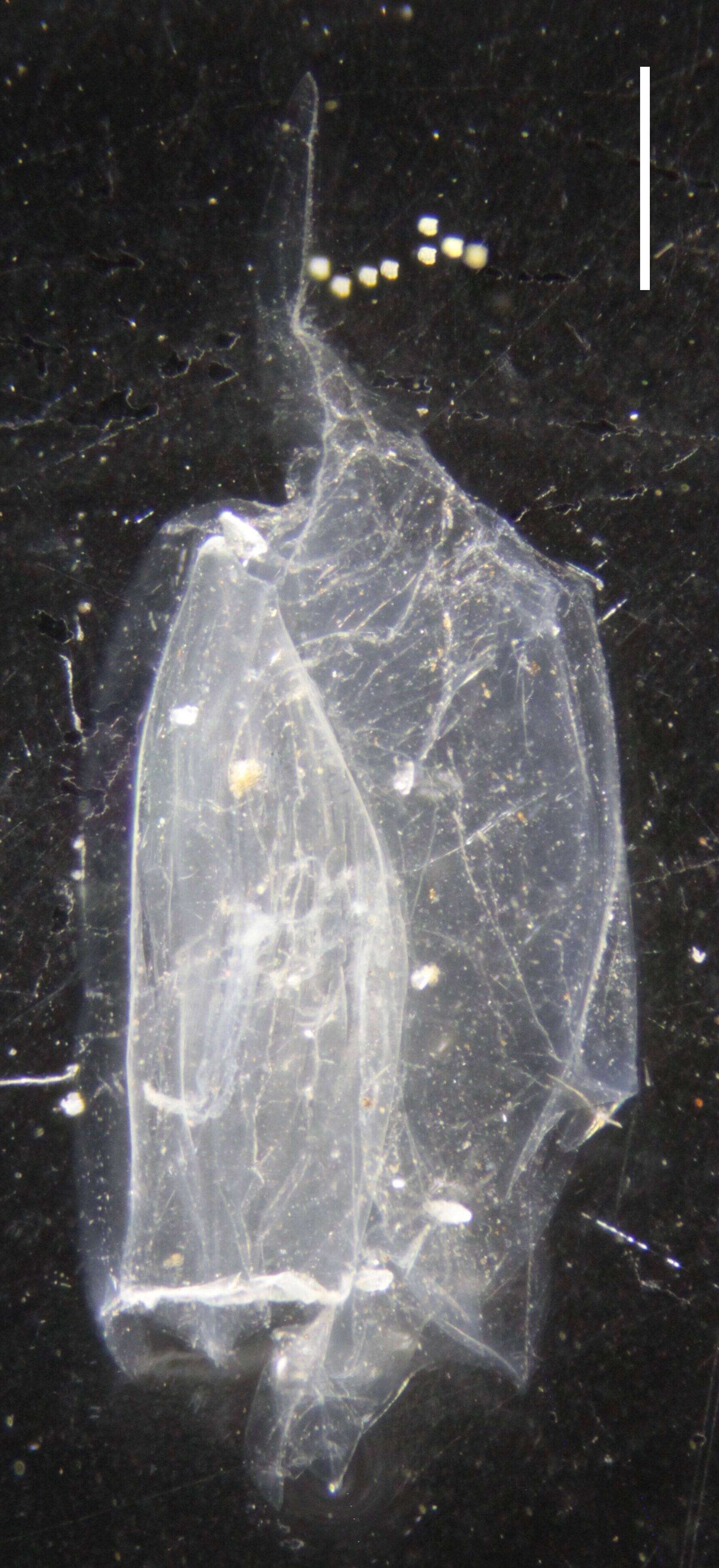
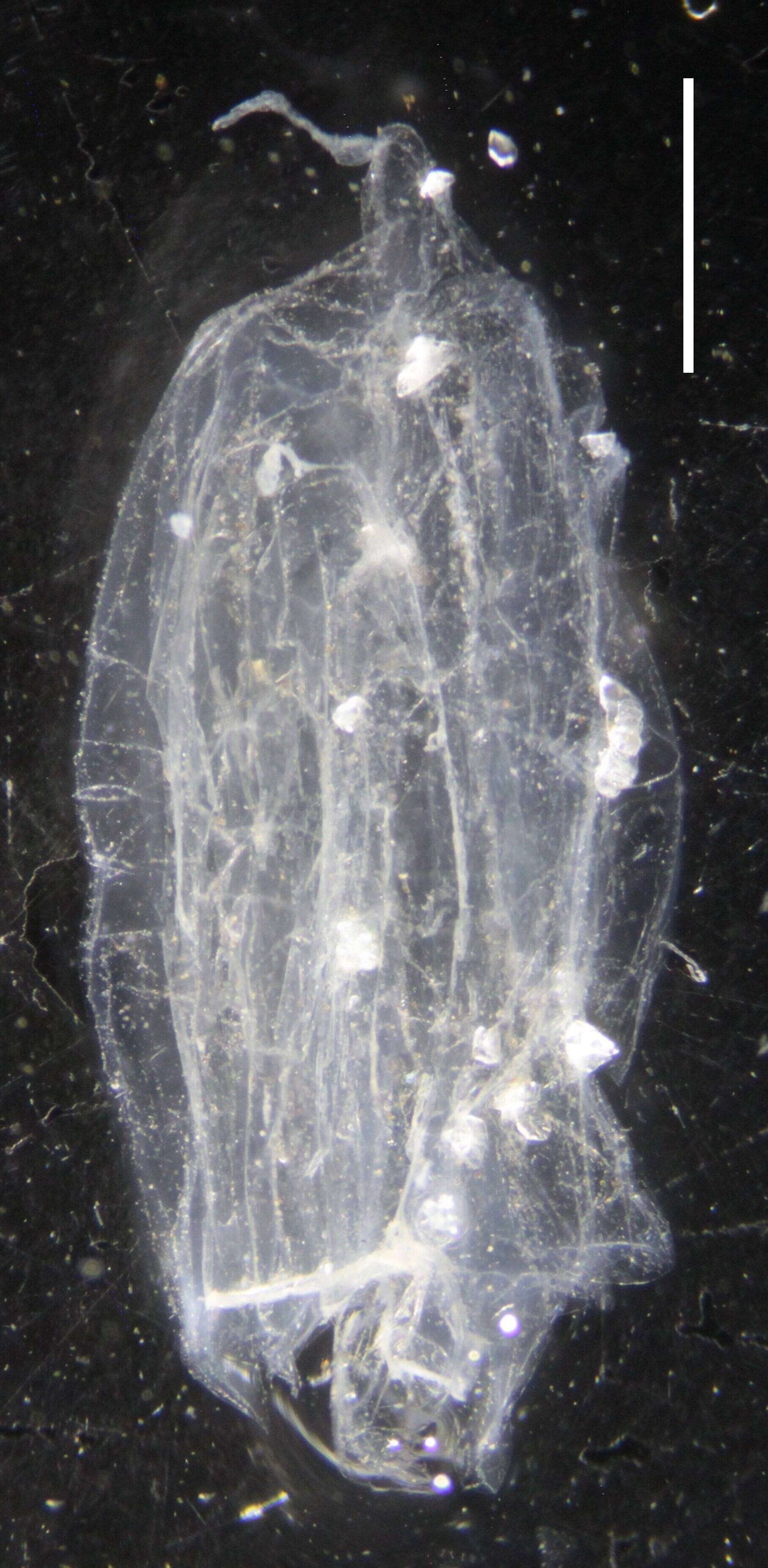
Microsope specimen image of IZ COE 28529; Posterior Nectophore; image 10; Scale Bar = 2 mm
-
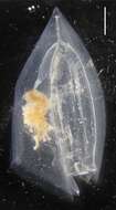
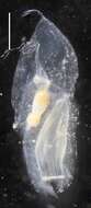
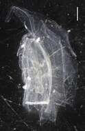
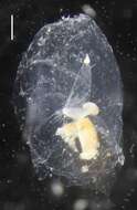
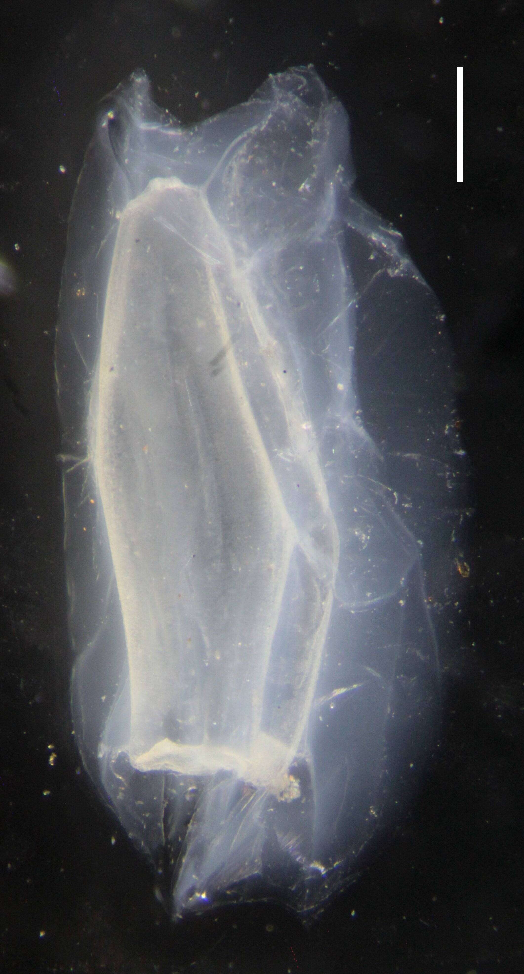
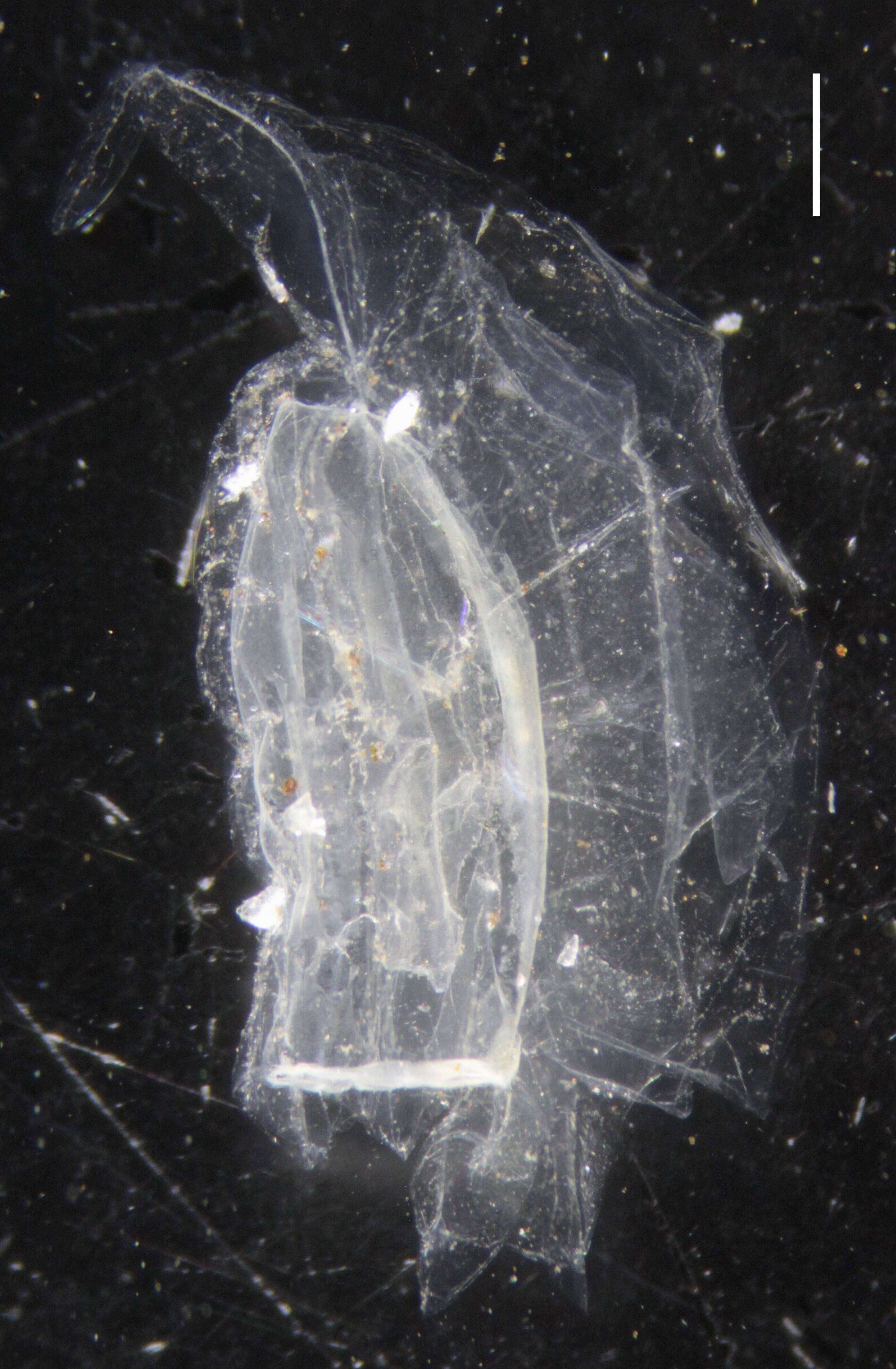
Microsope specimen image of IZ COE 28517; Posterior Nectophore; image 19; Scale Bar = 3 mm
-
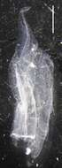
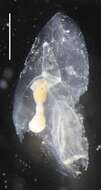
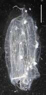
Microsope specimen image of IZ COE 28529; Posterior Nectophore; image 8; Scale Bar = 2 mm
-
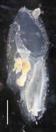
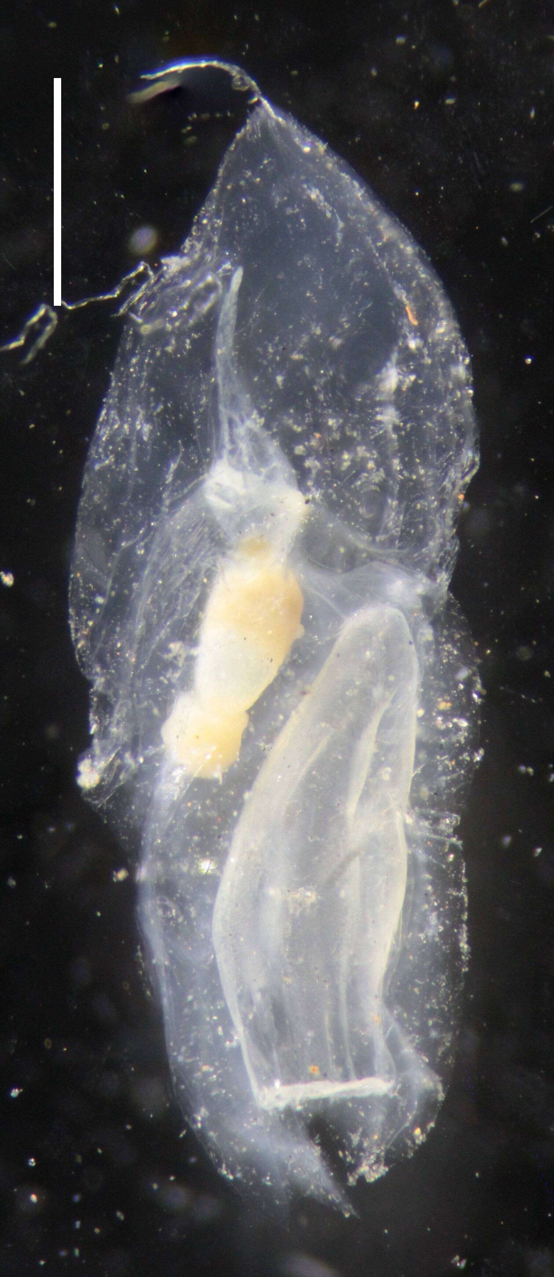
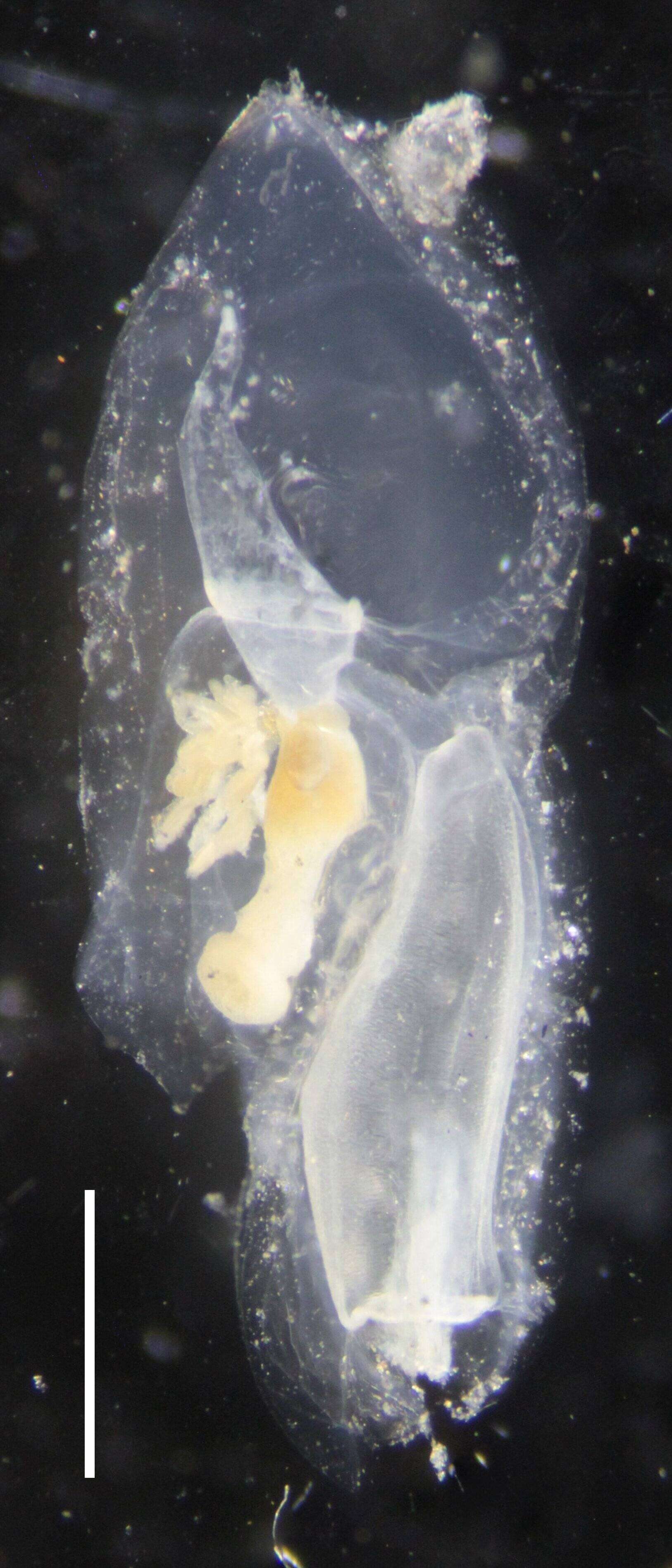
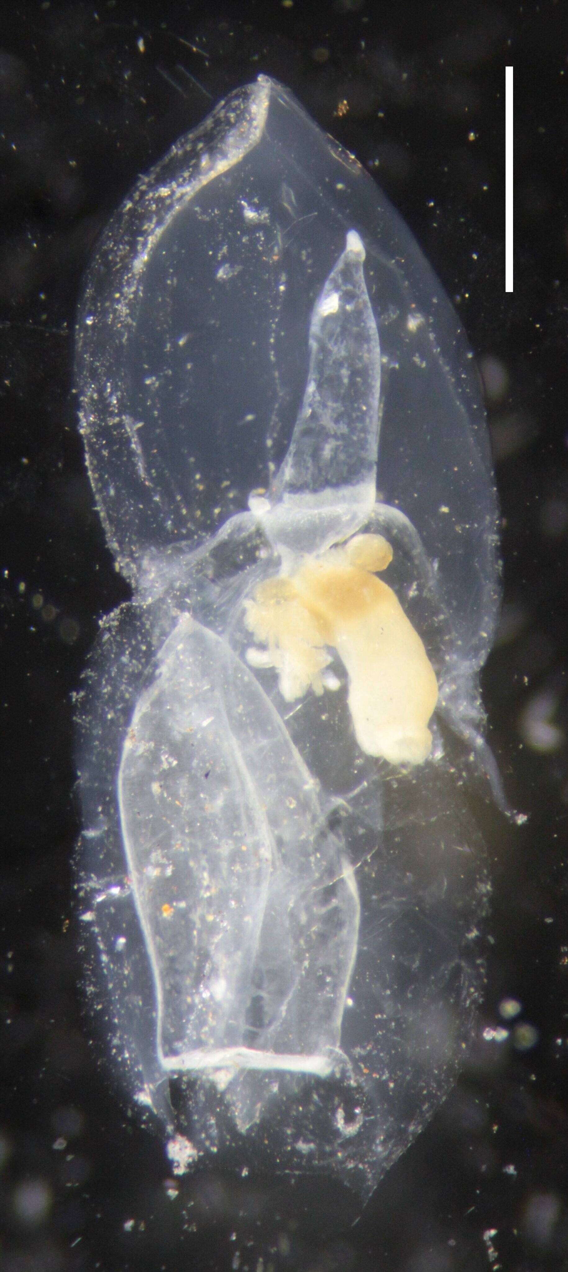
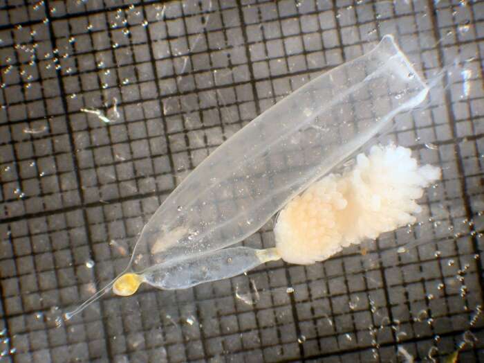
Microsope specimen image of IZ COE 28510; Eudoxid; image 13; Scale Bar = 1 mm
-
Microsope specimen image of IZ COE 28510; Eudoxid; image 16; Scale Bar = 1 mm
-
Microsope specimen image of IZ COE 28510; Gonophore; image 18; Scale Bar = 0.5 mm
-
Microsope specimen image of IZ COE 28510; Eudoxid; image 14; Scale Bar = 1 mm
-
Microsope specimen image of IZ COE 28529; Posterior Nectophore; image 7; Scale Bar = 3 mm
-
Microsope specimen image of IZ COE 28529; Posterior Nectophore; image 9; Scale Bar = 1 mm
-
Microsope specimen image of IZ COE 28510; Eudoxid; image 15; Scale Bar = 0.5 mm
-
Microsope specimen image of IZ COE 28510; Eudoxid; image 12; Scale Bar = 1 mm
-
Microsope specimen image of IZ COE 28510; Bract; image 17; Scale Bar = 0.5 mm
-
Microsope specimen image of IZ COE 28529; Posterior Nectophore; image 11; Scale Bar = 2 mm
-
anterior