-
-
-
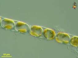
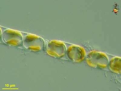
Melosira (mell-o-sigh-ra) is a centric diatom. The cells are like old-style hat boxes, or old-style pill boxes, or like petri-dishes. In Melosira, many cells are joined end to end to create a filament. The less substantial rings on the lower image are where the two halves of the frustule are joined together by the girdle bands, the more visible connections are where two cells are joined together. Each cell has a radial symmetry. As with other diatoms, plastids have chlorophylls a and c and so have a yellow brown colour. The lower picture reveals the individual disc-shaped plastids. Phase contrast.
-
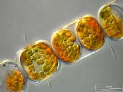
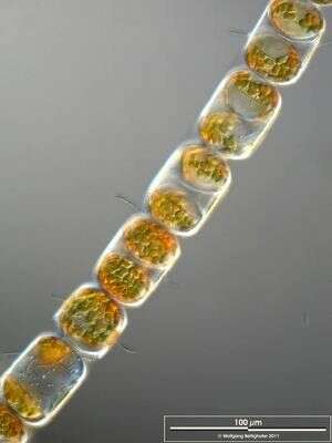
Melosira (mellow-sire-a) nummuloides, filament forming centric diatom, with multiple small plastids within the cell. Dark ground illumination. Leptosiropsis (leapt-owe-sire-op-sis) torulosa, green alga with organic wall that is produced in layers. Phase contrast microscopy.
data on this strain.
-
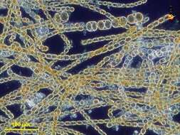
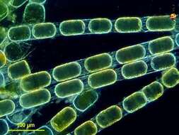
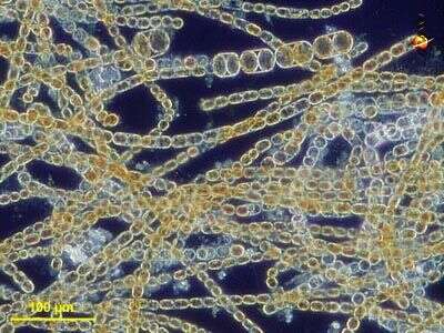
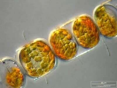
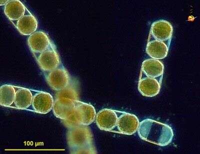
Colony with epibiotic bacteria chains. Scale bar indicates 100 µm. The image was built up using several photomicrographic frames with manual stacking technique. Sample from North Sea near Heligoland (spring diatom bloom). Images were taken using Zeiss Universal with Olympus C7070 CCD camera.
-
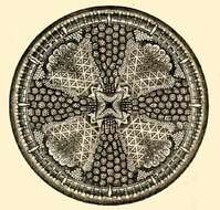
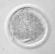
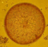
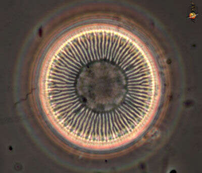
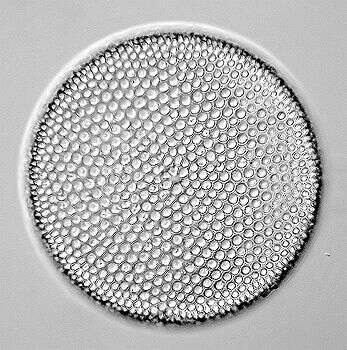
Actinoptychus heliopelta.
-
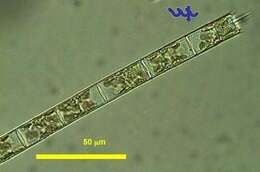
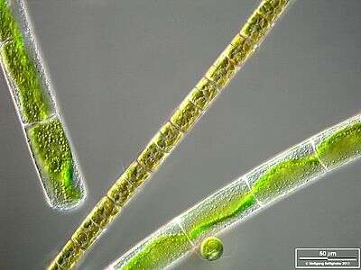
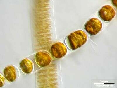
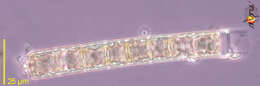
Melosira varians together with Mougeotia and Chlamydomonas. The scale bar indicates 50 µm. The specimen was gathered in the wetlands of Oderbruch (Oder valley 100 km north east of Berlin). The image was built up using several photomicrographic frames with manual stacking technique. Images were taken using Zeiss Universal with Olympus C7070 CCD camera.Image under Creative Commons License V 3.0 (CC BY-NC-SA).
-
Melosira (mell-o-sire-a) is a centric diatom. The cells are like old-style hat boxes, or old-style pill boxes, or like petri-dishes. In Melosira, many cells are joined end to end to create a filament. The less substantial rings on the lower image are where the two halves of the frustule are joined together by the girdle bands, the more visible connections are where two cells are joined together. Each cell has a radial symmetry. As with other diatoms, plastids have chlorophylls a and c and so have a yellow brown colour. Differential interference contrast.
-
Melosira (mellow-sire-a) nummuloides, filament forming centric diatom, with multiple small plastids within the cell clearly shown in this micrograph. Differential interference microscopy.
data on this strain.
-
Colony with epibiotic bacteria chains. Scale bar indicates 25 µm. The image was built up using several photomicrographic frames with manual stacking technique. Sample from North Sea near Heligoland (spring diatom bloom). Images were taken using Zeiss Universal with Olympus C7070 CCD camera.
-
-
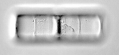
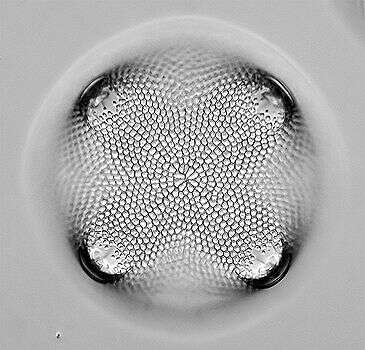
The surface of the siliceous valve of a Melosira cell. Phase contrast microscopy.
-
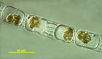
Stephanopyxis (steff-ann-o-pick-sus) palmeriana, a filament-forming centric diatom in which the cells are linked together by processes arising from the valves (the parts of the shell or frustule at the ends of each cell). The golden colour from plastids which contain chlorophylls a and c, but more significantly with carotenoids such as fucoxanthin which provide the distinctive colour. Dark ground illumination.
data on this strain.
-
Melosira moniliformis accompanied by Fragilaria islandica. Scale bar indicates 100 µm. The image was built up using several photomicrographic frames with manual stacking technique. Sample from North Sea near Heligoland (spring diatom bloom). Images were taken using Zeiss Universal with Olympus C7070 CCD camera.
-
-
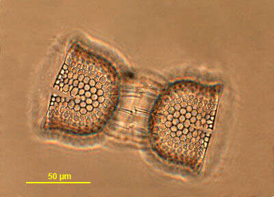
Stephanopyxis sp. isolated from a plankton net sample taken from the waters near Martha's Vineyard. Phase contrast image taken by Andrew Schurko.
-
Stephanopyxis (steff-ann-o-pick-sus) palmeriana, a filament-forming centric diatom in which the cells are linked together by processes arising from the valves (the parts of the shell or frustule at the ends of each cell). The golden colour from plastids which contain chlorophylls a and c, but more significantly with carotenoids such as fucoxanthin which provide the distinctive colour. The plastids are evident in this photograph. Differential interference microscopy.
data on this strain.
-
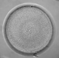
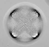
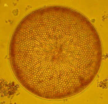
C. radiatus is one of the smaller Coscinodiscus species. It is box shaped in girdle view and the valves are very flat. The areolae form disctinct rows radiating from the valve centre. C. radiatus is a cosmopolitan species.
-
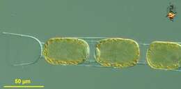
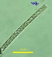
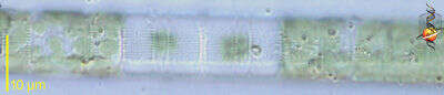
This is a typical filament of Aulacoseira (formerly Melosira) granulata (Bacillariophyta, Centrales)that was resuspended from the sediments not long before sampling. Its linking spines are of different lengths, possibly indicative of the filament having been broken at this place. In contrast, spines of equal length as in the other photo of this species are indicative of active growth. The chloroplasts here are partly compacted and do not fill the entire cell volume, indicative of the origin of this particular filament from the sediments. The other photo of this species shows actively growing, planktonic cells with chloroplasts filling the entire cell. This specimen was sampled from the shore of the lake in June 2006.
-
Phase contrast image of Stephanopyxis sp. (after being cleared with bleach) isolated from the waters near Martha's Vineyard . Photo courtesy of Andrew Schurko.
-
Stephanopyxis (steff-ann-o-pick-sus) palmeriana, a filament-forming centric diatom in which the cells are linked together by processes arising from the valves. The valves are the parts of the shell or frustule at the ends of each cell, and are more sculpted than the girdle bands, and so refract more light appearing brighter in this image, which includes many resting cells. The golden colour from plastids which contain chlorophylls a and c, but more significantly with carotenoids such as fucoxanthin which provide the distinctive colour. Dark ground illumination.
data on this strain.
-
-

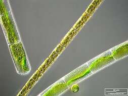
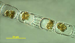
Aulacoseira (formerly Melosira) granulata (Bacillariophyta, Centrales) is one of the most common diatoms of Lake Kinneret, and certainly the major phytoplankton biomass contributor in winter (Dec â Feb), when the water column is homothermal. Typically, it occupies the entire 43 m water column. At the end of the winter bloom in early March the filaments sink and remain in the sediments in a dormant form with compressed chloroplasts till they are resuspended when the lake destratifies the following fall. It is a relatively large filamentous diatom, with cell diameter of 9 â 15 μm (median: 12.4 μm), cell height of 27-37 μm (median: 31 μm), and mean cell volume of 3700 μm3. The Kinneret Aulacoseira granulata filaments are straight, typically with 8 - 24 cells per filament. The picture shows the typical equal length marginal spines at the perimeter of the end-cell, these are âlinking spinesâ which hold adjacent cells together. The chloroplasts fill the entire cells. This specimen was sampled from the shore of the lake in June 2006. Aulacoseira granulata is a widespread centric diatom in the phytoplankton of lakes, reservoirs and rivers world-wide but particularly in African lakes and rivers, including the Nile River, L Naivasha, Kenya. It is typical of carbonate-rich, moderately eutrophic to eutrophic waters. It is used as indicator species of water with relatively low concentrations of salts, pH less than 9, and high silica concentrations.
-
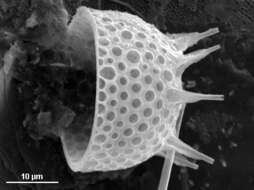
SEM image of a Stephanopyxis sp. valve. Original sample isolated from the waters near Martha's Vineyard as part of the 2005 ATOL Protistology Workshop. Image courtesy of Shauna Murray and Andrew Schurko.