-
Miranda do Douro Municipality, Braganca, Portugal
-
-
Prejano, La Rioja, Spain
-
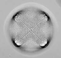
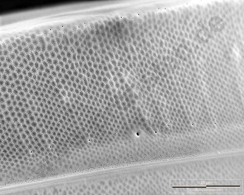
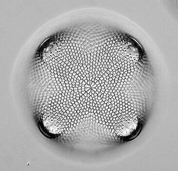
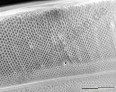
Coscinodiscus wailesii.Closeup of the lateral side of the valve. Scale bar indicates 25 m. Sample from North Sea near Heligoland (spring diatom bloom). The image was built up using several photomicrographic frames with manual stacking technique. Use of SEM equipment courtesy of Lab Dr. Karl-Heinz Schffner, Solingen, Germany. For more look at
www.protisten.de/english/gallery_main/gallery_main.htmlFor high-resolution images please ask postmaster@protisten.de.
-
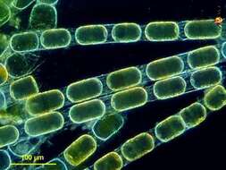
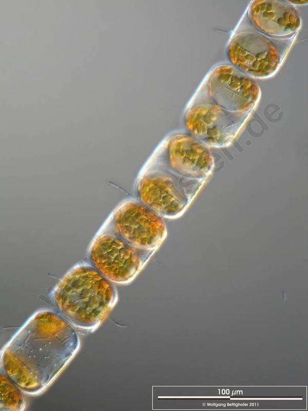
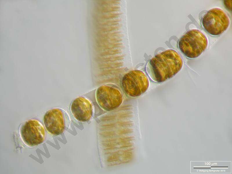
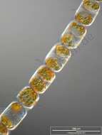
Melosira moniliformis.Colony with epibiotic bacteria chains. Scale bar indicates 100 m. The image was built up using several photomicrographic frames with manual stacking technique. Sample from North Sea near Heligoland (spring diatom bloom). Images were taken using Zeiss Universal with Olympus C7070 CCD camera.For more look at
www.protisten.de/english/gallery_main/gallery_main.htmlFor high-resolution images please ask postmaster@protisten.de.
-
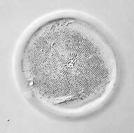
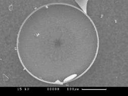
Fig 5: Scanned electron micrograph image of C.wailesii in the valve view.
-
-
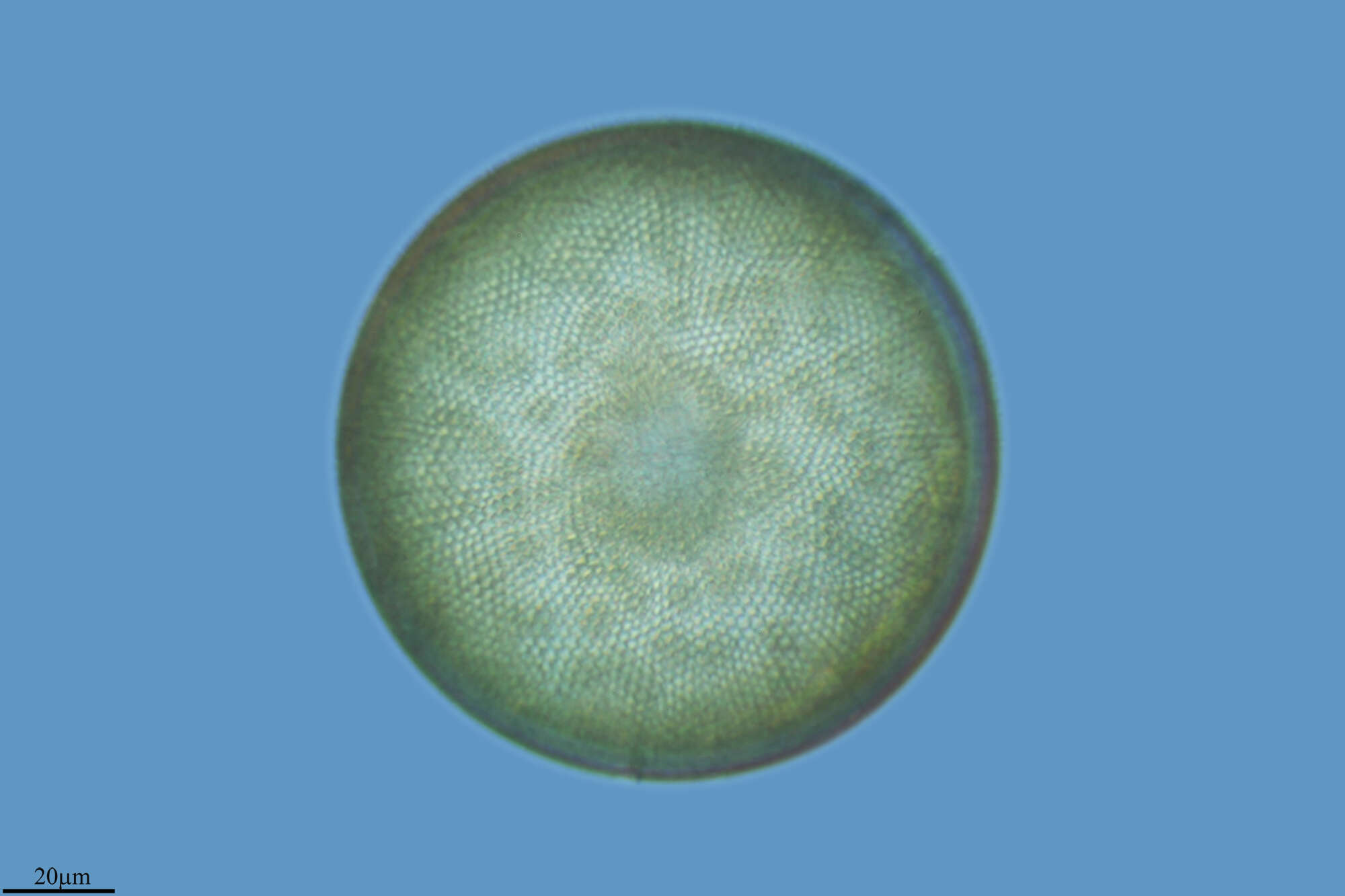
The surface of the siliceous valve of a Melosira cell. Phase contrast microscopy.
-
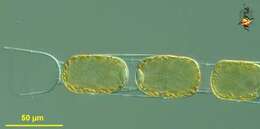
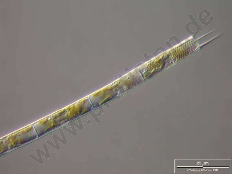
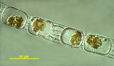
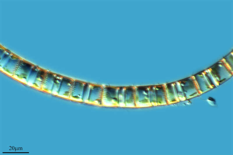
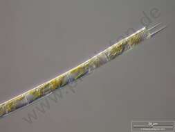
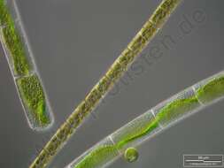
Stephanopyxis (steff-ann-o-pick-sus) palmeriana, a filament-forming centric diatom in which the cells are linked together by processes arising from the valves (the parts of the shell or frustule at the ends of each cell). The golden colour from plastids which contain chlorophylls a and c, but more significantly with carotenoids such as fucoxanthin which provide the distinctive colour. Dark ground illumination.
data on this strain.
-
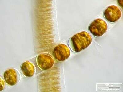
Melosira moniliformis accompanied by Fragilaria islandica. Scale bar indicates 100 µm. The image was built up using several photomicrographic frames with manual stacking technique. Sample from North Sea near Heligoland (spring diatom bloom). Images were taken using Zeiss Universal with Olympus C7070 CCD camera.
-
Grove, O, Galicia, Spain
-
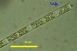
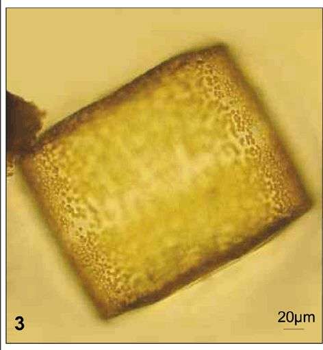
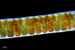
Aulacoseira granulata.Scale bar indicates 25 m. The specimen was gathered in the wetlands of Oderbruch (Oder valley 100 km north east of Berlin). The image was built up using several photomicrographic frames with manual stacking technique. Images were taken using Zeiss Universal with Olympus C7070 CCD camera.For more look at
www.protisten.de/english/gallery_main/gallery_main.htmlFor high-resolution images please ask postmaster@protisten.de.
-
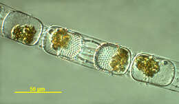
Melosira varians together with Mougeotia and Chlamydomonas. The scale bar indicates 50 m. The specimen was gathered in the wetlands of Oderbruch (Oder valley 100 km north east of Berlin). The image was built up using several photomicrographic frames with manual stacking technique. Images were taken using Zeiss Universal with Olympus C7070 CCD camera.For more look at
www.protisten.de/english/gallery_main/gallery_main.htmlFor high-resolution images please ask postmaster@protisten.de..
-
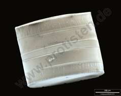
Coscinodiscus wailesii.SEM of girdle view. The ligulae which fit in the open girdle bands are weakly visible. Scale bar indicates 100 m. Sample from North Sea near Heligoland (spring diatom bloom). The image was built up using several photomicrographic frames with manual stacking technique. Use of SEM equipment courtesy of Lab Dr. Karl-Heinz Schffner, Solingen, Germany. For more look at
www.protisten.de/english/gallery_main/gallery_main.htmlFor high-resolution images please ask postmaster@protisten.de.
-
Melosira moniliformis accompanied by Fragilaria islandica. Scale bar indicates 100 m. The image was built up using several photomicrographic frames with manual stacking technique. Sample from North Sea near Heligoland (spring diatom bloom). Images were taken using Zeiss Universal with Olympus C7070 CCD camera.For more look at
www.protisten.de/english/gallery_main/gallery_main.htmlFor high-resolution images please ask postmaster@protisten.de.
-
Fig 3: Coscinodiscus wailesii Light micrograph of a Lugol's preserved cell in girdle view
-
-
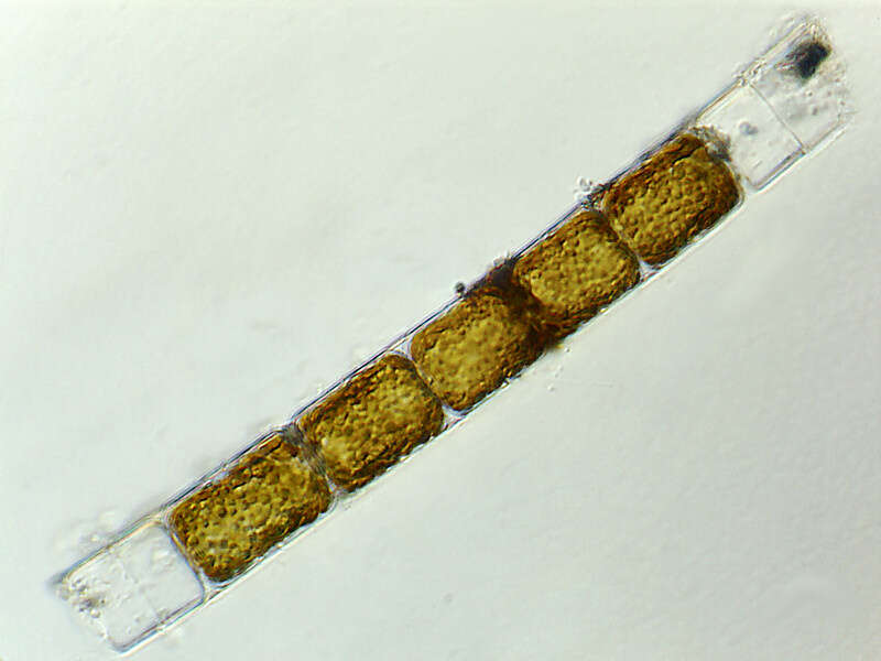
Stephanopyxis sp. isolated from a plankton net sample taken from the waters near Martha's Vineyard. Phase contrast image taken by Andrew Schurko.
-
Stephanopyxis (steff-ann-o-pick-sus) palmeriana, a filament-forming centric diatom in which the cells are linked together by processes arising from the valves (the parts of the shell or frustule at the ends of each cell). The golden colour from plastids which contain chlorophylls a and c, but more significantly with carotenoids such as fucoxanthin which provide the distinctive colour. The plastids are evident in this photograph. Differential interference microscopy.
data on this strain.
-
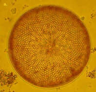
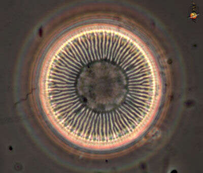
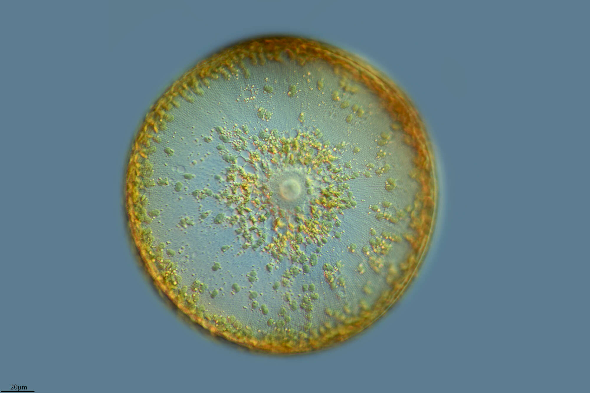
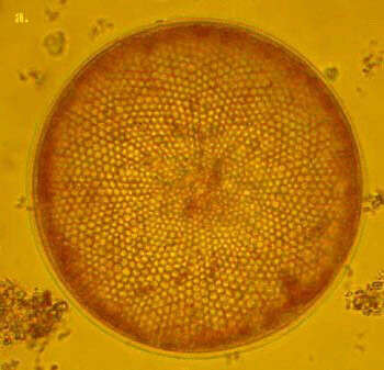
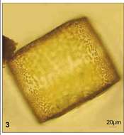
C. radiatus is one of the smaller Coscinodiscus species. It is box shaped in girdle view and the valves are very flat. The areolae form disctinct rows radiating from the valve centre. C. radiatus is a cosmopolitan species.
-
Grove, O, Galicia, Spain
-
Ribadelago, Castille and Leon, Spain
-
Grvalos, La Rioja, Espaa
-
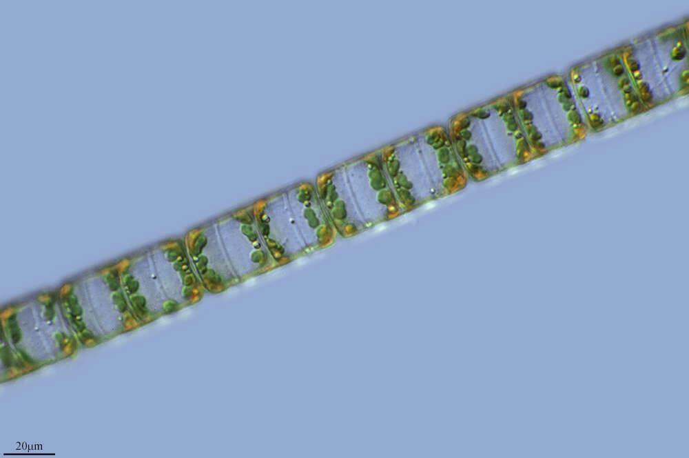
This is a typical filament of Aulacoseira (formerly Melosira) granulata (Bacillariophyta, Centrales)that was resuspended from the sediments not long before sampling. Its linking spines are of different lengths, possibly indicative of the filament having been broken at this place. In contrast, spines of equal length as in the other photo of this species are indicative of active growth. The chloroplasts here are partly compacted and do not fill the entire cell volume, indicative of the origin of this particular filament from the sediments. The other photo of this species shows actively growing, planktonic cells with chloroplasts filling the entire cell. This specimen was sampled from the shore of the lake in June 2006.