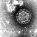
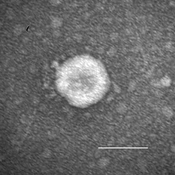
Description: English: Porcine epidemic diarrhea virus particles seen by negative-stain electron microscopy of fecal samples. Negative staining with 1% phosphotungstic acid. Scale bar indicates 100 nm. Date: 1 March 2015. Source: https://wwwnc.cdc.gov/eid/article/21/3/14-1165_article. Author: Dennis Hanke, Maria Jenckel, Anja Petrov, Mathias Ritzmann, Julia Stadler, Valerij Akimkin, Sandra Blome, Anne Pohlmann, Horst Schirrmeier, Martin Beer, and Dirk Höper.
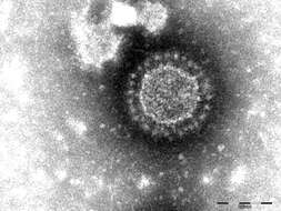
Description: English: Porcine deltacoronavirus (OH-FD22) particle detected in intestinal contents from a gnotobiotic pig. The sample was negatively stained with 3% phosphotungstic acid. Scale bar indicates 100 nm. Date: 1 April 2015. Source: https://wwwnc.cdc.gov/eid/article/21/4/14-1859_article. Author: Kwonil Jung, Hui Hu, Bryan Eyerly, Zhongyan Lu, Juliet Chepngeno, and Linda J. Saif.
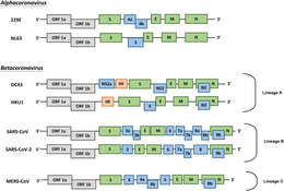
Sin-Yee Fung, Kit-San Yuen, Zi-Wei Ye, Chi-Ping Chan, and Dong-Yan Jin
Wikimedia Commons
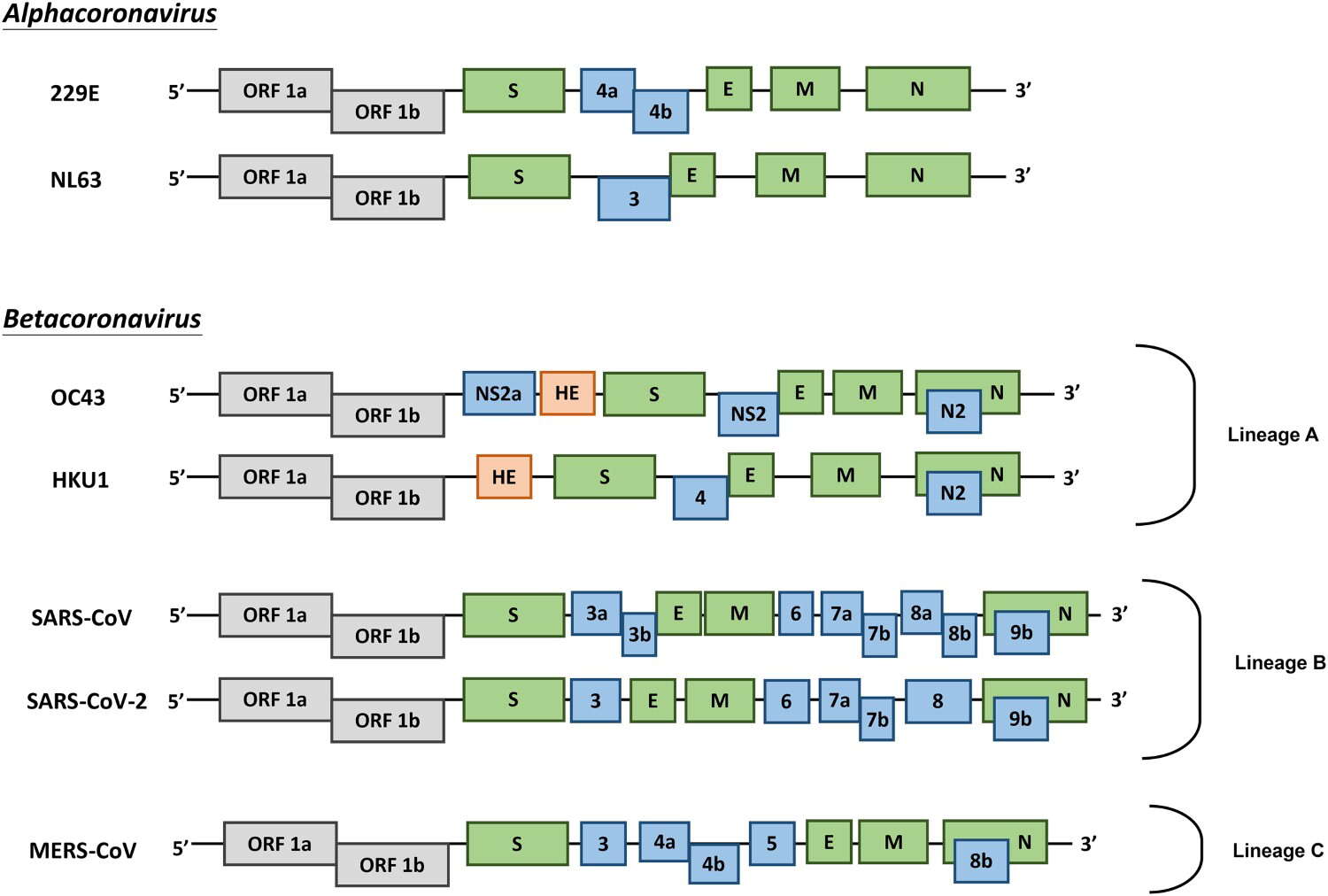
Description: English: Genome organization of HCoVs. Schematic diagram of seven known HCoVs is shown (not in scale). The genes encoding structural proteins spike (S), envelope (E), membrane (M), and nucleocapsid (N) are in green. The gene encoding haemagglutinin-esterase (HE) in lineage A of betacoronaviruses is in orange. The genes encoding accessory proteins are in blue. Date: 14 March 2020. Source: https://www.tandfonline.com/doi/full/10.1080/22221751.2020.1736644. Author: Sin-Yee Fung, Kit-San Yuen, Zi-Wei Ye, Chi-Ping Chan, and Dong-Yan Jin.
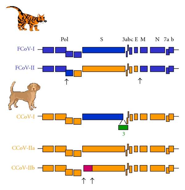
Description: English: Genetic relationships between the different feline and canine coronaviruses genotypes (FCoV and CCoV). The feline sequences are coloured in blue, the canine sequences in orange, and the porcine sequences in purple. Arrows indicate the putative sites of recombinations. The genes encoding for the polymerase polyprotein (pol), the structural spike (S), the envelope (E), the membrane (M), and the nucleocapsid (N) proteins are indicated. The genes encoding the accessory proteins are designated by numerals. Date: 31 July 2011. Source: https://www.hindawi.com/journals/av/2011/609465/. Author: Sophie Le Poder.
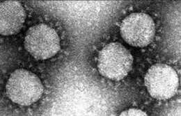
Description: English: Electron microscopy images of thin sections and partially purified virions from cells infected with coronavirus. Electron microscopy images of concentrated Transmissible gastroenteritis coronavirus (TGEV). Date: 1 August 2002. Source: https://wwwnc.cdc.gov/eid/article/8/8/02-0042_article. Author: Cristina Riquelme, David Escors, Javier Ortego, Carlos M. Sanchez, Branislava Uzelac-Keserovic, Konstantin Apostolov, and Luis Enjuanes.