Cimex的圖片
描述:
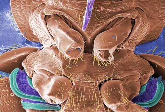
This digitally-colorized scanning electron micrograph (SEM) revealed some of the ultrastructural morphology displayed on the ventral surface of a bedbug, Cimex lectularius. From this view, at the top, you can see the insects skin piercing mouthparts it uses to obtain its blood meal, as well as a number of its disarticulated six jointed legs. Youll also notice a beautiful diaphanous structure at the bottom of the image. It is speculated that this wondrous ultrastructural organ is most probably a scent gland, or related to the dissemination of scent, which may be pheromonal in nature. A further dissection of this, and the adjacent mesothoracic region, could possibly reveal an internalized aspect of this organ, which would be glandular in nature, and actually involved in the production of the aromatic chemical. See PHIL 11742, 11743, and 11744 for successively greater magnifications of this marvelous structure.
Created: 2009
包含在以下頁面:
- Life
- Cellular
- Eukaryota (真核生物)
- Opisthokonta
- Metazoa
- Bilateria
- Protostomia
- Ecdysozoa (蜕皮动物总门)
- Arthropoda (節肢動物)
- Pancrustacea
- Hexapoda (六足亞門)
- Insecta (昆蟲)
- Pterygota (有翅亞綱)
- Neoptera (新翅下纲)
- Paraneoptera
- Hemiptera (半翅目)
- Heteroptera (異翅亞目)
- Cimicomorpha (臭蟲總科)
- Cimicoidea
- Cimicidae (臭虫科)
- Cimex
- Cimex lectularius (溫帶床蝨)
- Panarthropoda
此圖片未被精選在任何收藏裡。
來源資訊
- 攝影者
- Janice Haney Carr
- 提供者
- Public Health Image Library
- 原始內容
- 原始媒體檔案
- 參訪來源
- 合作夥伴網站
- Public Health Image Library
- ID