323022 1 En 5 Fig3lr HTML

描述:
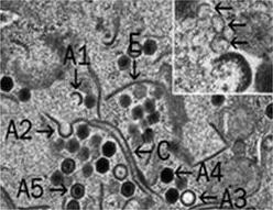
Description: English: Transmission electron micrographs of FHM cells infected by Frog virus 3 (FV3). Enlargement of a viral assembly site showing virions in various stages of assembly. Full (A4 and A5) and empty (A3) viral particles are shown as well as two possible intermediates (A1 and A2) and two aberrant forms (C and E). The inset indicates membranes (arrows), possible originating from the ER, that play a role in virion morphogenesis. Date: 2015. Source: Fig. 3 lower right at https://link.springer.com/chapter/10.1007/978-3-319-13755-1_5 Ranavirus Replication: Molecular, Cellular, and Immunological Events. In: Ranavirus Replication: Molecular, Cellular, and Immunological Events. In: Gray M., Chinchar V. (eds) Ranaviruses. Springer, Cham. doi:10.1007/978-3-319-13755-1_5 . Author: Jancovich J.K., Qin Q., Zhang QY., Chinchar V.G; Gray M., Chinchar V. (eds). Other versions: .
包含在以下頁面:
此圖片未被精選在任何收藏裡。
來源資訊
- 許可
- cc-by-sa-3.0
- 版權
- Jancovich J.K., Qin Q., Zhang QY., Chinchar V.G; Gray M., Chinchar V. (eds)
- 建立者
- Jancovich J.K., Qin Q., Zhang QY., Chinchar V.G; Gray M., Chinchar V. (eds)
- 來源
- Fig. 3 lower right at https://link.springer.com/chapter/10.1007/978-3-319-13755-1_5 Ranavirus Replication: Molecular, Cellular, and Immunological Events. In: Ranavirus Replication: Molecular, Cellular, and Immunological Events. In: Gray M., Chinchar V. (eds) Ranaviruses. Springer, Cham. doi:10.1007/978-3-319-13755-1_5
- 原始內容
- 原始媒體檔案
- 參訪來源
- 合作夥伴網站
- Wikimedia Commons
- ID