Stenhelia Boeck 1865的圖片
描述:
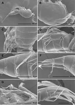
Figure 1.Stenhelia pubescens Chislenko, 1978, scanning electron micrographs, female 1: A habitus, lateral B cephalothorax, lateral C free thoracic somites, lateral D fifth pedigerous somite and genital double-somite, lateral, with one spermatophore attached on ventral side E fourth and fifth urosomites, lateral F anal somite and caudal rami, lateral G first legs and proximal part of second and third legs, lateral H distal part of right antennula, dorsal.