松骨海綿目的圖片
描述:
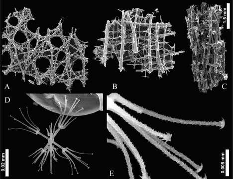
Figure 4. Scaning Electron Microscopy of Indeilla gen. n. ridgenensis sp. n. Frameowrk and spicules of the holotypes A dermal layer B atrial layer C lateral view D discohexaster E secondary ray tuft of discohexaster
Figure 4. Scaning Electron Microscopy of Indeilla gen. n. ridgenensis sp. n. Frameowrk and spicules of the holotypes A dermal layer B atrial layer C lateral view D discohexaster E secondary ray tuft of discohexaster