Cimex resmi
Açıklama:
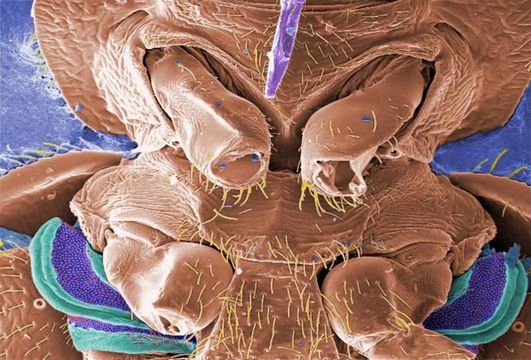
This digitally-colorized scanning electron micrograph (SEM) revealed some of the ultrastructural morphology displayed on the ventral surface of a bedbug, Cimex lectularius. From this view, at the top, you can see the insects skin piercing mouthparts it uses to obtain its blood meal, as well as a number of its disarticulated six jointed legs. Youll also notice a beautiful diaphanous structure at the bottom of the image. It is speculated that this wondrous ultrastructural organ is most probably a scent gland, or related to the dissemination of scent, which may be pheromonal in nature. A further dissection of this, and the adjacent mesothoracic region, could possibly reveal an internalized aspect of this organ, which would be glandular in nature, and actually involved in the production of the aromatic chemical. See PHIL 11742, 11743, and 11744 for successively greater magnifications of this marvelous structure.
Created: 2009
Aşağıdaki Sayfalarda Bulunmaktadır:
- Life
- Cellular
- Eukaryota (Ökaryot)
- Opisthokonta
- Metazoa
- Bilateria
- Protostomia
- Ecdysozoa
- Arthropoda (Eklem bacaklılar)
- Pancrustacea
- Hexapoda (Altı bacaklılar)
- Insecta (Böcek)
- Pterygota (Kanatlı böcekler)
- Neoptera
- Paraneoptera
- Hemiptera (Yarım kanatlılar)
- Heteroptera (Yarım kanatlılar)
- Cimicomorpha
- Cimicoidea
- Cimicidae
- Cimex
- Cimex lectularius (Tahtakurusu)
- Panarthropoda
Bu resim hiçbir koleksiyonda yer almıyor.
Kaynak Bilgileri
- lisans
- cc-publicdomain
- fotoğrafçı
- Janice Haney Carr
- sağlayıcı
- Public Health Image Library
- orijinal
- orijinal medya dosyası
- kaynağı ziyaret et
- ortak site
- Public Health Image Library
- ID