Comprehensive Description
provided by Smithsonian Contributions to Zoology
Eustenogaster eximia (Bingham)
Members of the Stenogastrinae are unique among the Vespidae in the facility with which they hover in the air like a dragonfly, a capability that led Carpenter (1988) to christen them “hover wasps.” The female uses this ability to hover in front of a spider web, and delicately pick off small insects trapped in the outer strands, or small commensal spiders on the periphery of the web of a larger host spider.
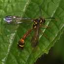
Eustenogaster eximia is the only member of the Stenogastrinae that occurs in Sri Lanka. The female is a relatively large, slender wasp (Figure 11), 19–21 mm long, superficially resembling a large eumenid such as Delta flavopictum (Blanchard) (Figure 10).
The species was described as Ischnogaster eximius Bingham (1890) from a male reared by E.E. Green from a nest obtained near Galle, Sri Lanka. Bingham also included a few notes and a figure of the nest which had been furnished by Green. Stenogaster eximioides Dover and Rao from southern India was synonymized as only a color variant of eximia by Dover (1925). It is, however, currently recognized as a subspecies of eximia by Das and Gupta (1983), who also synonymized Paravespa eva Bell (1936) under eximioides. There is a slight difference in coloration between eximia and eximioides, a character that J. van der Vecht (personal communication) thinks is probably not constant. J. van der Vecht, and later, C.K. Starr, confirmed that my specimens from Sri Lanka are all eximia. The reported occurrence of eximia in Thailand and Malaya (Dover, 1931) is questionable and needs confirmation. Pending a revisionary study of Eustenogaster, it seems preferable to recognize eximia as a single taxon of disjunct distribution restricted to quite humid areas of southwestern Sri Lanka and southern India.
Within Sri Lanka eximia occurs in densely shaded areas of lowland rain forest. It was collected at the following localities by personnel associated with the Smithsonian’s Ceylon Insect Project, 1972–1980.
Kalutara District: Morapitiya
Kegalla District: Kitulgala
Ratnapura District: Gilimale and Weddagala, Sinharaja Jungle
Monaragala District: Wellawaya
Galle District: Kottawa and Kanneliya, Sinharaja Jungle
These localities have elevations that range from 65 to 610 m and an average annual rainfall of 2600–5000 mm. There are specimens from Kandy in the Colombo Museum collected in 1909 and 1917, but I did not find it during my frequent visits to Kandy, although I collected in Udawattakele Sanctuary and Kandy Reservoir jungles. We obtained most of our data and nests in the Kanneliya section of the Sinharaja Jungle, and lesser numbers in Kottawa, Kitulgala, and Wellawaya (Figures 16–30). These localities are all in the Wet Zone except for Wellawaya in the Intermediate Zone, where we found a single nest in a wooded ravine beneath an overhanging stream bank.
NEST
ARCHITECTURE.—Nests are constructed in sheltered spots in the rain forest, usually beneath earth or rock overhangs along a stream (Figures 12–15) where they are protected from rain. The nest is suspended from a slender exposed rootlet or stem of a fern frond at some distance from the tip. The top of the comb is attached firmly to the substrate which does not penetrate into the cells.
The distinctive completed nest is a more or less pear-shaped structure constructed from masticated bits of rotten vegetative matter that dry to form a thin, brittle carton that is normally 0.24–0.36 mm thick on the exterior surface.
L.R. Batra* analyzed the composition of carton in fresh nests collected by P.B. Karunaratne in Morapitiya, June 1990. Using three samples, about 1 mm3, from different sites of the pseudenvelope, he found that microscopically they contained about 10% by volume of masticated fungal hyphae of a species of Polyporaceae or Thelephoraceae (Basidiomycetes, Aphyllophorales). Members of those families cause wood rots which make the wood soft, friable, and workable by many species of insects that feed upon it, bore into it, or use it in nest-making. The fungi may remain viable in the wood for several years. He identified the fungus from the clamp connections of brown vegetative hyphae.
He found two additional kinds of hyphae, skeletal and binding, which may or may not belong to the same basidiomycetous species.
The skeletal hyphae were thick walled, usually branched, unseptate, and straight except for being slighdy bent or flexuous with the thin apices. He stated that these hyphae develop from the vegetative cum generative hyphae. They are called skeletal hyphae because they are characteristic of genera and species whose fruiting bodies are hard and tough, as contrasted with soft and fleshy fruiting bodies of commercial mushrooms such as Agaricus.
The binding hyphae were similar to the skeletal hyphae but had much thicker walls, and, thus, narrower lumens. He found them intertwined and mixed with the vegetative and skeletal hyphae. The binding hyphae in the Basidiomycetes further strengthen the fruiting body. The vegetal hyphae of many Basidiomycetes, such as the entire genus Termitomyces Heim, are known to act as a bonding agent for the rotting wood fibers that constitute the bulk of the carton, i.e., material that has passed through worker guts, in nests of fungus-growing subterranean termites such as Odontotermes obesus (Rambur) and O. gurdaspurensis (Holmgren and Holmgren) in India (Batra and Batra, 1966).
Batra stated that the relatively large volume of fungal matter in the carton of the wasp nest was intriguing, and that the wasp was certainly using wood that was rotted by fungi. He said that rotting wood has only vegetative or generative hyphae in its interior. However, toward the exterior, where the wasp would have obtained the wood, skeletal hyphae or even binding hyphae would be found as the fruiting body primordia appeared. Thus, it is quite possible that the wasp incorporated into the carton pieces of fruiting bodies of the Aphyllophorales that grew on the surface of the wood. He stated that the fungus did not form a regular mass which would have indicated that it developed after the nest carton was assembled.
He took three additional samples of carton of the same size, and cultured them on 2% water agar, and potato dextrose agar. He found two common soil-inhabiting saprophytes in each sample—Cunninghamella elegans Matruchot (Phycomycetes, Mucorales), and a species of Cephalosporium (Hyphomycetes, Moniliales). These saprophytic fungi are presumably chance contaminants, and have no essential role in carton formation.
The pseudenvelope is frequently covered by numerous, short erect hyphae (Figures 43, 44, 52, 53) that occur also on the inner cell walls (Figures 54, 55). Batra identified this fungus as a species of Fusarium (Hyphomycetes, Moniliales, Tuberculariaceae) by finding its spores on the carton of these recently collected nests.
The nest shape is rather variable (Figures 16–23) and depends basically on the number and arrangement of cells, which range from 4 to 26 in eximia with an average of 13.3 cells per nest (n = 34). The peripheral cells are rounded on the outer surface, so that the upper 10 to 15 mm of the nest has distinct longitudinal grooves between adjacent cells (Figure 19, g). Below this area of peripheral cells the pseudenvelope narrows toward the terminal spout. There are often thin, sometimes denticulate ridges on the carton, extending downward from the peripheral cells (Figure 17, r, and 35). Sometimes there are thin elongate wings or teeth on top of the nest, and the peripheral cells are not so clearly demarcated (Figure 17, w, t). Occasionally there are one or more aborted cells up to 12 mm long on the outer surface of the pseudenvelope which the female did not include within the completed nest (Figure 23).
The nest narrows below the cells to form a slender spout with the nest entrance on the surface toward the bank (Figure 17). The wall of the spout surrounding the entrance is perforated by a lacy network of carton through which the wasp may peer while remaining hidden. The perforated area is margined by a narrow flange of carton giving the spout a lanceolate appearance (Figure 18, f). The spout is extremely fragile because of the perforations. The perforations are made from larger masses of masticated material (Figures 31–34), 0.6–1.0 mm thick. One wasp included in the perforated area a large chunk of unworked vegetative matter 2.4 mm long and 0.4–0.8 mm thick (Figure 34). The spout must frequently be broken during storms as the nest is buffeted by the wind or driving rain. Many of the active nests that we found in the field had the spout broken off. The spout apparently is not replaced if it is damaged.
We found a single anomalous nest at Kanneliya in 1979. The pseudenvelope divided into two spouts with entrance holes at the bottom, each giving access to the comb. The older spout had the tip broken off, the other was complete. The single female escaped from the nest. Pagden (1958, fig. 7a) described a similar nest of an unidentified species of Eustenogaster (his Group I of Stenogaster in Malaya). These two nests may possibly be rare examples of spout replacement following supersedure of one female by another.
Bell (1936) found nests of eximia in areas of high rainfall in the Western Ghats near Bombay. His nests were located in the same kinds of habitats and were as variable in shape as those I found in Sri Lanka.
The profound variation of nest architecture in eximia nests creates doubt about the validity of the elaborate scheme of nest architecture proposed by Ohgushi and his colleagues (1983) for four species of Sumatran Eustenogaster which they designated as E1–4. Nests of types E1–3 are all found in eximia. The nest types which they designated as E4, and later as E5 (Ohgushi et al., 1986), could be variation within calyptodoma. Critical taxonomic study of wasps associated with nest variations is required to substantiate any system of nest architecture. It seems probable that there may be considerable plasticity in nest architecture within a single species of Eustenogaster.
Some species of Eustenogaster, including eximia, do not construct an ant guard on the support above the nest. However, Pagden (1958, fig. 8) found an unidentified species of this genus in Malaya that provided such a guard, and C.K. Starr (personal communication) also found ant guards in several species of Eustenogaster in the Philippines.
MATERIALS USED.—I did not observe eximia gathering decaying vegetative material for nest construction, but scanning electron micrographs (SEM) of fragments from about a dozen nests provide some knowledge. Some sections were made principally of decayed xylem fragments with some intermixed amorphous vegetative matter (Figure 36). I also found occasional pieces of decayed hardwood with profuse vessel-pitting as seen from inside of a vessel element (the many tiny perforations) (Figure 37), mixed in with xylem fragments. Small particles of mud occurred rarely (Figures 38–39, m).
Some of my nests (Figures 16, 17, 19, 23, 24) are mostly unicolorous, the pulp apparently coming from a single source. Other nests are prettily patterned (Figures 18, 20, 21, 27) with alternating bands in two colors, undoubtedly made from pulp from different sources. SEMs of banded sections of carton showed no appreciable difference in composition. Still other nests (Figure 22) are irregularly blotched, suggesting that a damaged nest might have been patched with pulp from a different source.
Green (1924) erred in stating that the nest of eximia in Sri Lanka is “built of earthy matter.” Bell (1936) mentioned the extreme fragility of the Indian nests, but he, too, was mistaken in stating that they were built of mud mixed with saliva. Nestor nest fragments from both Bell and Green in the British Museum (Natural History) are of carton (personal commnunication, M.C. Day).
Williams (1919) published a brief account of the Philippine luzonensis (Rohwer). He observed females gathering nest material from the trunk of a fallen tree and reported that the “moist, and well-decayed wood [was] chewed into a pulp and formed into a delicate paper which is not rainproof.” He made no notes on application of the pulp to the nest, but mentioned that the pulp-gathering trips were made at intervals of about 15 minutes. The presumption is that the actual processing of a load of pulp and its application to the nest is a reasonably prolonged process.
Pagden (1958) stated that several unidentified species of Eustenogaster (his Group I of Stenogaster) in Malaya constructed their nests of “triturated vegetable matter.”
Hansell (1981) analyzed pieces of nest material from three nests of another stenogastrine in Thailand, Parischnogaster mellyi (Saussure). He found the carton composed principally of plant fragments with very little intermixed soil. The plant material was of two types, woody xylem fragments possibly from rotten stems, and pieces of cuticular cell layer of plants, some of which bore discernible hairs.
Ohgushi et al. (1983) found a small amount of mud mixed with the carton in some of the nests made by several unidentified species of Eustenogaster in Sumatera Barat, Indonesia. Neither Williams nor Pagden mentioned any admixture of mud in their nests, and Hansell and I found it infrequently. I believe that any inclusion of mud in Eustenogaster nests is accidental. It probably occurs when the wasp collects leaf fragments or pulp from decaying wood lying on the forest floor.
CONSTRUCTION.—The wasp initiates the nest by constructing a comb of shallow cells beneath the supporting substrate (Figure 26). The cells are about 6.5 mm wide, and hexagonal in cross section except for those on the periphery which have the outer surface rounded (Figures 29, 30). When the cell walls reach 5–10 mm in length, she may begin to lay eggs, one in each cell beginning at the center of the comb. We found a 25-celled comb with cell walls about 10 mm long in which eggs or tiny larvae were present in five of the central cells. Oviposition and brood rearing apparently proceed at a leisurely pace. During this period the cell walls are extended downward to their full length of 13–15 mm.
Occasionally, though, the entire pseudenvelope including spout may be completed before eggs are laid, or very soon after the first eggs are deposited. I found one such nest with completed pseudenvelope in which the 17 cells were very short, and six of the central cells, 5–8 mm long, each contained an egg or tiny larva.
The interior cell walls are 0.13–0.28 mm (n = 13) thick, while the pseudenvelope is 0.24–0.36 mm (n = 12) thick. If ribs are present on the lower exterior of the pseudenvelope, that wall and the rib are 0.69–0.75 mm (n = 6) thick.
There is no true envelope of carton separated from the comb in most species of Eustenogaster. The single known exception is calyptodoma (Sakagami and Yoshikawa) from Borneo and Malaya. In this unusual species the cells are attached directly to a flat surface such as a slanting rock or piece of wood, and the envelope is separated from the comb by as much as 5 mm. Exceptionally the envelope may be partially in contact with the comb. The nest has a terminal spout as in other species of the genus.
BROOD DEVELOPMENT
EGG.—The egg of eximia is short, sausage-shaped, 1.8–2.1 mm long and 0.8–0.9 mm wide (n = 6) (Figure 40). The wasp begins to lay eggs in the central comb cells first, placing each at the inner (i.e., upper) end of the cell. All eggs and young larvae that I observed were immersed in a globular mass of thick, sticky, milky-white substance. This is consistent with conditions reported in nests of other species of Stenogastrinae.
I did not observe oviposition in eximia, but Turillazzi (1985a) studied it in several species of Parischnogaster. He noted that the female secreted a drop of viscid, milky-white substance from the tip of her abdomen, collected it in her mouthparts, touched the drop to the egg as it was extruded from the abdomen, and then deposited the egg and substance in the cell. Additional droplets of substance were placed in the cell after the larva hatched. Hansell (1982) suggested that the drops were secreted from Dufour’s gland. Carpenter (1988) commented that the oviposition sequence of the Stenogastrinae is unique in the Vespidae.
Turillazzi also noted that the abdominal substance, possibly mixed with oral secretions, was used to form the ringlike ant guard placed on the rootlet a short distance above the nest of Parischnogaster.
LARVAL FOOD.—I examined the gut contents of eight larvae from Kanncliya preserved in alcohol in 1975. The larvae ranged from about a third grown to fully mature. The results were as shown below.
I also obtained some data from the cell contents of several nests at Kanneliya in 1975 and 1980. The contents from one cell in each of three nests in 1975 were negative for larval food; each contained only the globule of abdominal substance and an egg or newly hatched larva. One cell of another nest from 1975 contained the following: the head and abdominal integument of a worker minor of a small myrmicine ant, possibly Oligomyrmex Mayr, the modified pedipalp of a male spider, 0.25 mm long, and the head of a small coleopterous larva, all of these parts having been fed upon by the wasp larva. There was also a whole, tiny collembolan, 0.44 mm long; this will be discussed in the section, Nest Associates. Another cell from the same nest contained only a globule of the abdominal substance and an egg.
Each of two cells in the 1980 nest contained a small larva and some food. I recovered the following food items from these cells. (1) Two workers of a myrmicine ant, 2.4 mm long, a species of Crematogaster Lund possibly belonging to the biroi group. One ant had been malaxated, possibly by the foundress, and partially embedded in a small mass of abdominal substance, and one ant had the thorax and abdomen hollowed out, presumably by the larva of eximia. (2) A small fragment of chitin, 0.25 mm long, from an unidentified arthropod. The lower pseudenvelope of this nest was damaged, thus allowing easy access by casual visitors. In one of these two cells there was an adult ephydrid fly, 2 mm long, attracted to some of the larval food, a small dipterous maggot, 1.8 mm long, a worker termite, 2.5 mm long, and an adult mite, 0.25 mm long; these arthropods will be discussed in the section, Nest Associates.
It was not surprising to find identifiable parts of spiders in many of the guts and in one cell. Williams (1928) figured a female off. depressigaster (Rohwer) hovering before a spider web suspended over a stream. She picked small midges from a strand of the web, using her mandibles and legs. Pagden (1958) reported that unidentified species of Eustenogaster (his Group I of Stenogaster) in Malaya hovered in front of spider webs and plucked small flies from the web with the forelegs. He noted that the spider was not disturbed by the wasp’s activities, and suggested that the flies were too small to attract the spider. This curious habit presumably is widespread among Stenogastrinae.
However, I was surprised to discover that most of the identifiable fragments of insects were of Hymenoptera rather than Diptera. Iwata (1967) reported finding unidentified chitinized fragments on the body of a larva of P. mellyi in Thailand, and also found a wasp larva feeding on a minute lepidopterous larva. He also recovered chitinized arthropod fragments from a food mass of a species of Eustenogaster near micans (Saussure). Spradbery (1975) found no identifiable fragments in the spherical food mass provided for first and second instar larvae of S. concinna. He noted that the food mass for maturing larvae in the fourth and fifth instars contained fragments of legs, integument, and wings of small Diptera that he presumed to be Cecidomyiidae. It is probable that this larval food also was obtained from spider webs.
The developing eximia larva remains curled at the upper end of the cell, instead of extending lengthwise along the cell axis as is normal in other vespids. As the larva becomes full grown, the female gradually begins to narrow the cell mouth (Figures 29, 30) to a diameter of 2 to 5 mm; it is never entirely closed.
COCOON.—The mature larva of eximia constructs an unusual cocoon. It secretes a substance, presumably from the salivary glands, that dries to form a glistening, delicate film, 2.9–9.0 µm in thickness. The secretion is applied to the entire interior of what is to become the pupal cell but it does not seal the narrowed opening of the cell (Figures 42, 44–47, 51). Individual strands of silk are not apparent when examined with a binocular microscope, just the glistening surface.
I preserved some combs of eximia in ethyl alcohol. Careful maceration of sections of pupal cells freed bits of the film from the vegetative matrix of the cell. Subsequent staining of these cocoon sections with acid fuchsin revealed that there are, indeed, strands of fine silk underlying the glistening film that now appears transparent (Figures 49, 50).
The source of the glistening film overlying the strands of silk is a puzzle. Perhaps it also is silk from the salivary glands, but is applied broadly, as if painted on, rather than narrowly as are the fine strands. The film and underlying strands of silk dissolve in hot KOH solution. The North American crabronid wasp Crossocerus (Blepharipus) stictochilos Pate spins a fusiform cocoon that has the same basic structure. A scanning electron micrograph (SEM) of part of the cocoon wall of this wasp (Figure 48) shows that it is composed of a mesh of fine silken strands 3.6–7.0 µm wide overlying a thin, glistening film. The entire cocoon of stictochilos also dissolves in hot KOH solution.
Using scanning electron microscopy, I discovered that the cocoon of eximia often covers hyphae of a species of Fusarium that is frequently found on both the exterior of the pseudenvelope and the interior cell walls. Heating sections of dried pupal cell walls in KOH solution dissolved the cocoon, revealing hyphae and fragments of vegetative matter forming the wall (Figure 54). SEMs (Figure 42, 43) of the outer and inner surfaces of a section of pseudenvelope from a pupal cell on the nest periphery show a dense growth of hyphae on the exterior, and the smooth film lining the inside of the cell. Note in Figure 51 that the cocoon (c) walls off the pupa that it contains, leaving a narrow space (f) containing hyphae and the cell wall (w) formed from vegetative material. It seems probable that the cocoon may serve a vital function by protecting the inert pupa from infestation and possible death from the Fusarium.
Other workers, e.g., Williams (1919), van der Vecht (1972), and Spradbery (1989), noted that cells of several genera of Stenogastrinae containing pupae, or cells from which adults had emerged, were coated with a glistening material, but they did not interpret this as a cocoon. Sakagami and Yoshikawa (1968) stated that the base of the pupal cell of calyptodoma was covered by a spun fibrous film, but that the pupa was not enclosed in a cocoon. Hansell (personal communication) stated that in his experience calyptodoma did not line the base of the cell with such a fibrous layer, but that the walls were coated with a thin film of secretion. My experience with eximia, detailed above and in Figure 55, suggests that this fibrous layer might have been a mass of hyphae.
It seems probable that this type of cocoon occurs throughout the Stenogastrinae. It is known to occur in all genera except Metischnogaster van der Vecht. In his notes on nests of the two known species of that genus, cilipennis (Smith) and drewseni (Saussure), Pagden (1958, 1962) did not mention whether the pupal cells were lined with a glistening film.
PUPATION.—The mature larva lies in a curled position at the upper end of the cell until the pupal eye disks are visible through the integument. It then assumes the characteristic stenogastrine pupal position (Figure 41). The scutum, with the posterior pair of spines, is at the outer (lower) end of the cell. The scutal spines are presumed to anchor the pupa against the narrowed lower end of the cell. The thorax posterior to the scutum and the long abdominal petiole are aligned upward along the length of the cell on one side. The remainder of the abdomen is flexed downward along the opposite side of the cell with its tip against the apex of the upturned mouthparts beneath the head.
I obtained no direct data on the duration of early brood stages. However, emergence dates of adults from a nest collected at Kanneliya on 11 January 1975 suggest a pupal period of about 3 weeks in eximia. The 21-celled nest had 11 cells with narrowed lower ends, 3 with either nearly full grown or mature larvae, and 8 with pupae in various stages toward eclosion as adults. The nest was kept in a sealed plastic sack and examined daily. One mature larva pupated by the 12th, but the other larvae and one older pupa died. Adults eclosed from the eight viable pupae as follows: 18 Jan; 23 Jan; 24 Jan; 26 Jan; 28 Jan; 29 Jan; 31 Jan; and 1 Feb. It is reasonable to assume that this last female is from the larva that pupated on 12 January, thus giving a pupal period of about 21 days.
Hansell (1987) estimated an average duration of 42.7 days from oviposition to pupation in E. calyptodoma, and an average duration of 21.4 days for the pupal period. Spradbery (1975) stated that the pupal period was more than 24 days in Stenogaster concinna. Turillazzi (1985b) noted a mean period of 44.5 days from egg to adult in Parischnogaster nigricans serrei (du Buysson), and a mean pupal period of 17.1 days. Samuel (1987) found a mean egg-to-adult time of 103 days in Liostenogaster flavolineata (Cameron), and a mean pupal period of 33 days.
The rather prolonged periods of pupal duration in stenogastrines contrast markedly with the shorter such periods of 9–18 days in many North American eumenine wasps (Krombein, 1967).
I did not find a larval meconium in the cells from which I recovered eximia pupae. This is consistent with observations by Turillazzi and Pardi (1982) and Sakagami and Yamane (1983), who found that females of two species of Parischnogaster removed the larval fecal pellets.
NEST ASSOCIATES
NATURAL ENEMIES.—In my experience eximia is remarkably free of parasites and predators. I preserved a few larvae of various stages from nests at Kanneliya. One of the mature wasp larvae had seven small chalcidoid larvae, 0.6 mm long, attached to the inner surface of the abdominal integument. They were identified as Eulophidae, possibly a species of Melittobia Westwood. Members of this genus commonly infest nests of solitary wasps and bees (Krombein, 1967). I reared two species in Sri Lanka from nests of the eumenid wasp, Paraleptomenes mephitis (Cameron) (Krombein, 1978a). There is also a possibility that the eulophid might have been a species of Nesolynx Ashmead, which Spradbery reared from a tachinid parasite of a stenogastrine in New Guinea.
Ants are frequent predators of other wasp nests, raiding them for brood or for prey provided for the brood. The only ants in my nests were three small myrmicine workers belonging to species of Crematogaster and possibly Oligomyrmex Mayr. They were definitely part of the larval food. They might, of course, have entered the nest to raid its contents, but were converted by the foundress into larval food.
Williams (1928) mentioned that larvae of species of Stenogaster, the generic name he used for species of Eustenogaster and Parischnogaster in the Philippines, were heavily parasitized by the ichneumonid, Theronia Holmgren. H.K. Townes advised me that P. timida (Williams) was probably the species from which T. pyramida williamsi Gupta was reared. Williams also recorded Vespa deusta Lepeletier as raiding the nest of P. depressigaster (Rohwer) to pillage the brood. Spradbery (1975) found an unidentified tachinid in four nests of S. concinna in Papua New Guinea, three puparia and one larva that had devoured half of a wasp larva. The three puparia were hyperparasitized by eulophid wasps; he found adult eulophids in two other nests, and assumed that they were searching for tachinids because they had not parasitized any of the brood. Spradbery (1989) reared the tachinid, Euvespivora decipiens (Walker) from nests of Anischnogaster iridipennis (Smith) in Papua New Guinea, and a eulophid wasp, a species of Nesolynx, from puparia of the tachinid.
Iwata (1967) recorded the eulophid, Syntomosphyrum species, as attacking the larva of P. striatula (Buysson) in Thailand.
COMMENSAL ORGANISMS.—I presume that the termite and collembolan recovered from nests in 1975 and 1980 were just casual visitors. They had not been malaxated by the female wasp, nor had the larva fed on them.
The adult dipteran was an ephydrid, a species of Rhyncopsilota near magnicornis Hendel. W.N. Mathis advised me that, so far as is known, species of this genus are attracted to injured Crematogaster ants, and feed on their body fluids. The dipterous larva from the same nest was a first instar larva of Ephydroidea, possibly a species of Drosophilidae.
The mite was an adult oribatid, a family whose members are frequently associated with decaying vegetative matter. It may have been introduced into the nest with some of the nesting material.
In the preceding section on Brood Development I mentioned a nest from which I reared a sequence of adult wasps. I also reared a small lyonetiid moth, Opogona praecincta Meyrick, from one cell. This nest had been kept in a sealed plastic sack, so this was not a chance infestation. The larva may have developed on some of the organic debris in the nest.
Spradbery (1975) recorded that a newly emerged male of S. concinna had 53 phoretic mites attached to its wings.
ADULT
BEHAVIOR.—Both sexes of eximia visit flowers for nectar. One shrub visited is wild olive, known in Sinhalese as gal veralu, one of several species of Elaeocarpus Linnaeus. I netted several females returning to their nest, each of which exuded a droplet of liquid, presumably nectar, between my fingers. Apparently this is the first flower visiting record for a stenogastrine.
Turrillazzi (1985a) noted that “drops of transparent liquid are also regurgitated by the wasps onto the eggs and larvae” and that the drops can be sucked up by the adults and serve as a readily available food reserve. In view of my notes on flower visits by adults, it is possible that this transparent liquid noted by Turillazzi is actually nectar.
We noted occasional female behavior unlike anything reported previously for Stenogastrinae. Usually the occupant(s) emerged quickly and flew off, when we disturbed a nest. Rarely, however, one or more individuals stayed inside the nest, and made a tapping sound on the pseudenvelope. This is reminiscent of the behavior described by Rau (1933) for the Panamanian social vespid wasp, Synoeca septentrionalis Richards (reported as S. surinama var. cyanea (Fabricius)). These wasps build a large paper nest against a tree trunk. When disturbed, they may warn off an intruder by beating their wings against the inner surface of the envelope.
I did not observe patrolling by males or associations of males such as has been reported for a few species of Stenogastrinae (summarized by Carpenter, 1988).
EUSOCIALITY.—There is evidence that eximia may rarely exhibit primitive eusociality. Most of the nests that I observed or recovered in the field contained only a single female. The occupants were counted in 35 nests from Kanneliya and Kottawa in the Sinharaja rain forest in 1972, 1975, and 1980. Twenty-three nests had only a single female occupant, presumably the foundress. A large nest from Kottawa contained four wasps, two of each sex; these specimens are not now available for examination. Eleven nests had two occupants, but both wasps escaped from five of these nests and could not be sexed. Three of the remaining six nests contained a female and a male; one male was a teneral specimen, and the other two were presumed to be progeny of the respective female.
The three remaining nests contained two females, one nest in each of the three years. The two females from the 1972 nest were killed and pinned, but the two females from the 1975 and 1980 nests were preserved in fluid. Subsequent examination of the abdominal contents demonstrated that the females from the latter two nests had the ovaries more or less well developed with oocytes in several stages of development. One female in each nest had mature ovaries with at least one egg ready for oviposition and others less developed. The other female in each nest had marginally mature ovaries with only one egg about two-thirds mature and the others less developed. The females with mature eggs were undoubtedly the foundresses, and the second female in each nest was presumably one of the daughters.
The 1975 nest was quite large and had 21 cells. It undoubtedly had been active for a lengthy period, because 11 of the cells were narrowed at the outer end and contained 8 pupae in various stages of coloring, and 3 nearly mature or fully grown larvae. Four of the remaining cells had larvae about half grown, and 6 were either empty or contained an egg or tiny larva beneath the globular mass of abdominal secretion.
When I found the 1980 nest, the pseudenvelope had been damaged and the lower portion was missing. There were 12 cells in the comb, one with the lower end narrowed, and three with an egg or tiny larva beneath the usual globule of abdominal substance; the other cells were empty.
Hansell (1987) found a greater degree of eusociality in calyptodoma nests in the Malay Peninsula. He kept 26 nests under observation without disturbing their occupants. When these control colonies were collected, they comprised 47 females and 5 males. There were about the same number of nests with one or two females, only five contained three or four females, and males were present in only five nests.
Hansell termed other nests experimental. He made holes in the envelope to observe behavior, captured the occupants, marked them with paint, and released them. He examined the ovaries of females from 24 experimental and control nests with more than one female. Only four of those nests had more than one female with mature ovaries, and in three of these nests the maturity of ovaries of the second female was marginal.
I obtained no information on division of labor within pairs of females. Hansell found that in calyptodoma the older female guarded the nest entrance while the younger female did most of the foraging for food. He also noted that a young female usually left the nest in a week, either to found her own nest or to usurp the nest and brood of another female.
I believe that limited eusociality is more likely to occur in the nest of a species such as calyptodoma rather than in a species such as eximia. In the former the envelope is separate from the comb, allowing space for more individuals to function within the nest. The pseudenvelope of most Eustenogaster species leaves very little room at the bottom of the comb for more than one individual to operate. C.K. Starr (personal communication) advised me that his observations of Eustenogaster in the Philippines gave “indirect evidence that luzonensis and an undescribed species are at least usually solitary.”
- bibliographic citation
- Krombein, Karl V. 1991. "Biosystematic Studies of Ceylonese Wasps, XIX: Natural History Notes in Several Families (Hymenoptera: Eumenidae, Vespidae, Pompilidae and Crabronidae)." Smithsonian Contributions to Zoology. 1-41. https://doi.org/10.5479/si.00810282.515