Description of Coscinodiscus wailesii
provided by BioPedia
Cells are often as high as wide. Cells are rectangular in outline or with an undulating margin, depending on the focal plane. In girdle view a prominent hyaline area is visible at the centre of the valve with wide interstriae radiating from it, resulting in an irregular fasciculation, 5-6- valve areolae are present in 10µm. Areolae are small but cribra are still visible with the light microscope. there are two marginal rings of processes. The outer ring contains two larger rings of processes that are 120-180 degrees apart. Chloroplast shape is irregular.
Biology
provided by Diatom LifeDesk
C. wailesii is a large solitary diatom found in coastal and oceanic waters, primarily in the Pacific and Atlantic Oceans. Because of its size, distinctive morphology, and relative ease of cultivation, more information is available for this species than any other species of Coscinodiscus.
- license
- cc-by-nc
- copyright
- Hargraves, Paul
Cytology
provided by Diatom LifeDesk
Cytological events associated with cell wall morphogenesis, and the internal organelle structure, have been studied by Schmid & Volcani (1983), and subsequently expanded by Schmid (1984, 1988, 1990). As with many planktonic diatoms, the greater portion of the cell volume (as much as 90%) consists of a vacuole, in this case with an interior of extremely acidic pH (Kesseler 1967). Chemical constituents of C. wailesii natural populations have been reported by Ono et al., 2008.
- license
- cc-by-nc
- copyright
- Hargraves, Paul
Diagnostic Description
provided by Diatom LifeDesk
The cells are cylindrical or tympaniform, with the pervalvar axis varying from about ½ to the whole length of the diameter. The transition from valve face to valve mantle is abrupt, forming nearly a 90 degree angle. The valve center is irregularly hyaline due to a variable termination of the radial rows of areolae, which number 5-6 in 10µm. At the small end of its size spectrum, the valve structure is significantly altered, and a central rosette replaces the hyaline area (Schmid 1990). The valve mantle is large, usually 30-40µm in the pervalvar direction, with slightly larger areolae, 4-6 in 10µm. The cingulum of each theca consists ot a broader valvocopula (~50-60µm) and a narrower copula (20-25µm). Occasionally there are three bands in the cingulum (Schmid & Volcani 1983). There are three rings of rimoportulae (labiate processes). One ring is at the base of the mantle, 2-3 areolae in from the margin. Each rimoportula is the terminus of a hyaline line (interstriae). The rimoportulae are ~10-18µm apart. In this ring are also two larger rimoportulae (macrolabiate processes), separated by an angle of 120-180 degrees. The second ring is at the valve/mantle junction, difficult to see with light microscopy, and individual rimoportulae may be ~5-15µm apart. The third ring is irregular, and at about 1/3 the radial distance from the center. Each rimoportula is the terminus of an incomplete radial row of areolae. The broad mantle with its hyaline lines, coupled with the irregular hyaline area and irregular third rimoportula row, are distinctive features. For further discussion of frustule morphology, see Gran & Angst (1931) and Hasle & Lange (1992).
- license
- cc-by-nc
- copyright
- Hargraves, Paul
Diseases
provided by Diatom LifeDesk
Coscinodiscus wailesii is subject to parasitic infections by the nanoflagellate Pirsonia diadema, which is at least partly specific to this diatom (Kuehn 1998). Fatal infections by the bacterium Alteromonas sp. are also known (Nagai & Imai 1998). It is likely other pathogens exist.
- license
- cc-by-nc
- copyright
- Hargraves, Paul
Distribution
provided by Diatom LifeDesk
Coscinodicus wailesii has been found throughout the Pacific Ocean since its first description. It is an invasive species in the Atlantic Ocean, first observed in 1977 in the English Channel (Boalch & Harbour 1977), misidentified as Coscinodiscus nobilis, and along the U.S. East coast in the late 1970s. It is absent from a 1964-74 survey of diatoms along the U.S. East coast (Marshall 1976). It appeared in Narragansett Bay in 1978 (Hargraves unpubl.; and found in the Chesapeake Bay in 1980 (Marshall, 1982). The earlier report of the species from Chesapeake Bay (Patten et al. 1963) cannot be confirmed. During the next decade it spread to the South Atlantic, both eastern (Senn 2002) and western (Fernandes et al. 2001; Lutz et al. 2006) sides. It was not present in the Indian Ocean during the 1964-65 R/V ‘Meteor’ expedition according to Simonsen (1974); current status in the Indian Ocean could not be confirmed but it may have also invaded there. Its ability to survive long periods in darkness (Nagai 1995) suggests that transport in ballast water may have aided in its distribution. It is mostly found in coastal and shelf waters, and is apparently absent from boreal and polar environments.
- license
- cc-by-nc
- copyright
- Hargraves, Paul
General Description
provided by Diatom LifeDesk
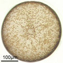
Each cell is cylindrical, with the pervalvar axis approaching or equivalent to the cell diameter, giving the appearance of a square or rectangular shape when viewed in girdle view. In valve view the cell is round, sometimes flat but often slightly concave. Chloroplasts are quite small and very numerous.
- license
- cc-by-nc
- copyright
- Hargraves, Paul
Habitat
provided by Diatom LifeDesk
Although it is primarily a coastal species, it occasionally is found beyond the continental shelf, and in estuaries. When abundant it apparently produces copious amounts of polysaccharide exudates ("slime", "mucilage") that can interfere with fisheries activities by clogging nets (Boalch & Harbour 1977; Mahoney & Steimle 1980), and is therefore considered a potential Harmful Algal Bloom species.
- license
- cc-by-nc
- copyright
- Hargraves, Paul
Life Cycle
provided by Diatom LifeDesk
Details of the sexual cycle (auxospore formation) have not appeared in detail, though spermatogenesis was examined by Jensen et al.(2003), and the asexual mitotic events were documented by Schmid & Volcani (1983). No morphologically distinct resting spore is known. Nagai (1995) reports resting cells in the sediments of Seto Inland Sea, distinguished by partly plasmolyzed cells, which responded quickly to increased illumination. About 70% of resting cells “rejuvenated”, and 80% of these divided within 48 hours. Nagai also reports dark survival of at least 15 months.
- license
- cc-by-nc
- copyright
- Hargraves, Paul
Size
provided by Diatom LifeDesk
Valve diameter in litt. of natural populations is about 160-500µ. However, cells in cultures have been found as small as 50µm diameter (Schmid, 1990) and post-auxospore cells as large as 550µm. In volume, this is one of the largest diatoms: for a cell of diameter 500µm and a pervalvar axis of 350µm, the volume is about 6.9 X 107 µm3. However, since most of the volume is vacuole, metabolic activity is mostly confined to areas adjacent to the frustule wall.
- license
- cc-by-nc
- copyright
- Hargraves, Paul
Toxicity
provided by Harmful Phytoplankton Project
No direct toxic effects have been observed but C. wailesii produces large amounts of mucilage which, at high cell densities, which can clog up fishing gear and smother the seabed.
- bibliographic citation
- Guide to UK Coastal Planktonic Ciliates © 2001 DJS Montagnes, University of Liverpool http://www.liv.ac.uk/ciliate/
- author
- David J.S. Montagnes
Brief Summary
provided by Harmful Phytoplankton Project
The shape and extent of the central hyaline area is variable. It can be very small and surrounded by a ring of larger areolae.
- Edward, M., John, AWG., Johns, DG & Reid, PC 2001. Case history and persistence of the non-indigenous diatom Coscinodiscus wailesii in the north-east Atlantic. Journal of the Marine Biological Association of the UK. 81:207-211.
- Hasle GR, Syvertsen EE. (1996) In: Identifying Marine Diatoms and Dinoflagellates. C. R. Tomas ed., Academic Press, New York, pp. 5-386.
- Tomas C ed. (1997) Identifying marine diatoms and dinoflagellates. pp 598. Academic Press Ltd. London
- bibliographic citation
- Guide to UK Coastal Planktonic Ciliates © 2001 DJS Montagnes, University of Liverpool http://www.liv.ac.uk/ciliate/
- author
- David J.S. Montagnes
Comprehensive Description
provided by Harmful Phytoplankton Project
Cells are often as high as wide, cells are rectangular in outline or with an undulating upper valve edge, depending on the focal plane. In girdle view a prominent hyaline area at the centre of the valve with wide interstrae radiating from it resulting in an irregular fasciculation. Areolae are small. Cribra visible with LM. There are two marginal rings of processes.
- bibliographic citation
- Guide to UK Coastal Planktonic Ciliates © 2001 DJS Montagnes, University of Liverpool http://www.liv.ac.uk/ciliate/
- author
- David J.S. Montagnes
Diagnostic Description
provided by Harmful Phytoplankton Project
Cells are often as high as wide, cells are rectangular in outline or with an undulating upper valve edge, depending on the focal plane. This is due to a concentric depression near the steep mantle (Tomas 1997). In valve view a prominant hyaline area at the centre of the valve is visible with wide interstrae radiating from it, resulting in an irregular fasciculation. Hyaline lines are more conspicuous on the valve mantle than the valve face. Areolae are small. Cribra are visible with LM Two rings of small processes are located in the area between valve face and mantle. The outer ring contains two larger processes that are 120-180 degrees apart (Tomas 1997). Numerous small, irregular chloroplasts are present
- bibliographic citation
- Guide to UK Coastal Planktonic Ciliates © 2001 DJS Montagnes, University of Liverpool http://www.liv.ac.uk/ciliate/
- author
- David J.S. Montagnes
Distribution
provided by Harmful Phytoplankton Project
Their distribution has been noted in European shelf seas. It is a significant member of the diatom community in the North-East Atlantic.
- bibliographic citation
- Guide to UK Coastal Planktonic Ciliates © 2001 DJS Montagnes, University of Liverpool http://www.liv.ac.uk/ciliate/
- author
- David J.S. Montagnes