-
Keratella, a rotifer (metazoa). Rotifers typically have a stiffened body wall (lorica) in segments which can telescope, with a corona of feeding cilia at the anterior end and with toes posteriorly. This genus has no foot nor toe, as an adaptation to a pelagic life. Rotifers are common members of the microbial communities of many aquatic ecosystems. Although they are multicellular animals, they may be only be 100 microns long, and so overlap in size with ciliates. They can be confused with ciliates because they use cilia to capture their food. However, they can be distinguished because they have a lorica, may have podites, and a strong muscular pharynx.
-
Centers for Disease Control/Division of Parasitic Diseases and Malaria
EOL staff
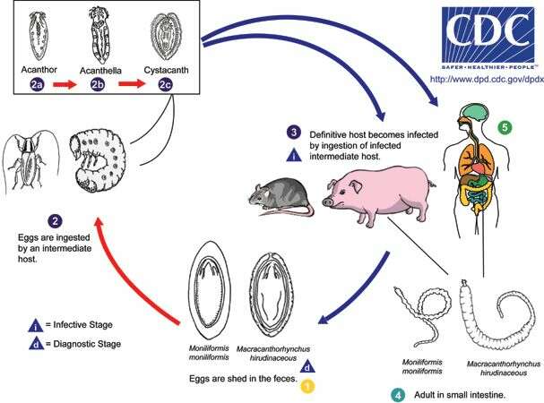
Life cycle of acanthocephalans Moniliformis moniliformis and Macracanthorhynchus hirudinaceus Eggs are shed in the feces of the definitive hosts (1), which are usually rats in the case of M. moniliformis and swine for M. hirudinaceous, although carnivores and primates, including humans, may serve as accidental hosts. The eggs contain a fully-developed acanthor when shed in feces. The eggs are ingested by an insect intermediate host (2) (usually scarabaeoid or hydrophilid beetles for M. hirudinaceus and beetles or cockroaches for M. moniliformis). Within the hemocoelom of the insect, the acanthor (2a) molts into a second larval stage, called an acanthella (2b). After 6-12 weeks, the worm reaches the infective stage called a cystacanth (2c). The definitive host becomes infected upon ingestion of intermediate hosts containing infective cystacanths (3). In the definitive host, liberated juveniles attach to the wall of the small intestine, where they mature (4) and mate in about 8-12 weeks. In humans (5), the worms seldom mature, or mature but will rarely produce eggs.From
Centers for Disease Control Parasites and Health website
-
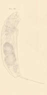
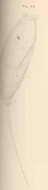
Diurella percellus Gosse, Left side of extended specimen.
-
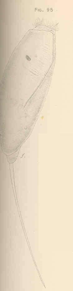
Diurella tenuior Gosse. Larger specimen, dorsal or dorso-dextral view.
-
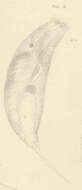
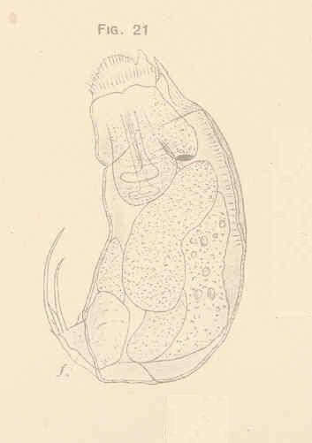
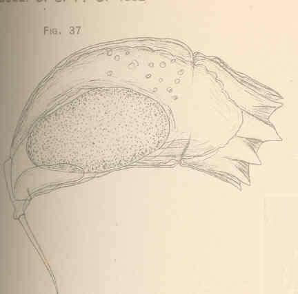
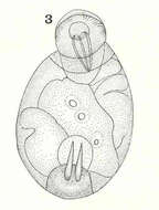
Taphrocampa annulosa Gosse, as seen from above; body curved so that the food is not visible.
-
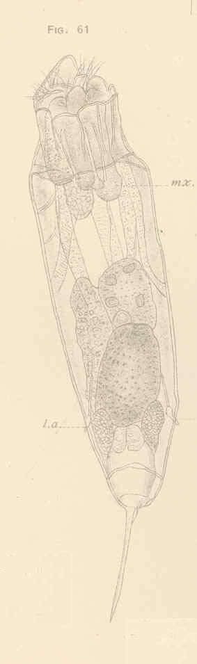
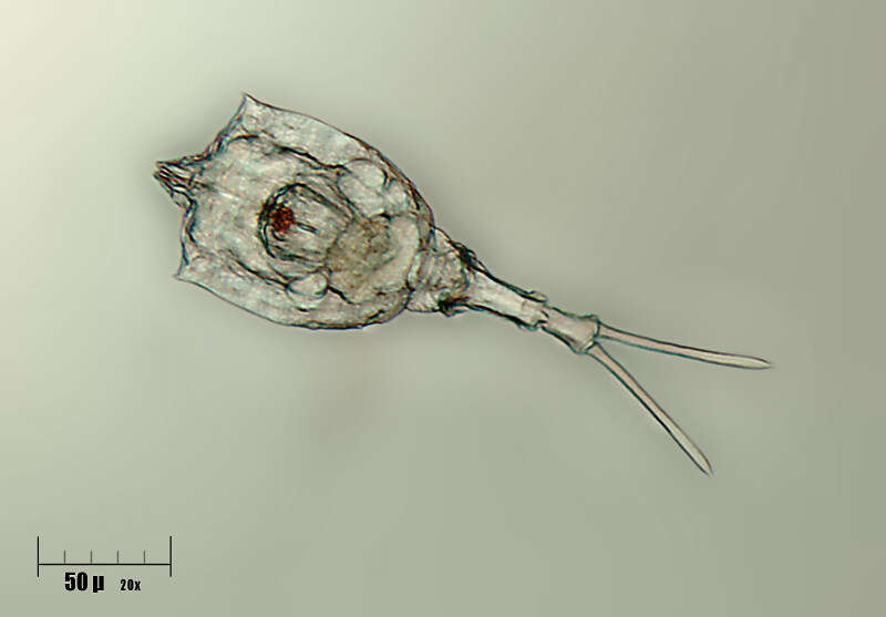
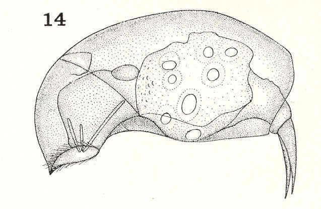
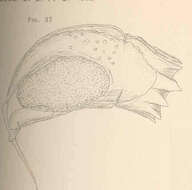
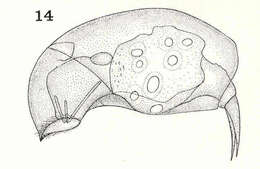
Rattulus capucinus Werz. & Zach. Ventral view. The toe extends obliquely toward the observer, so that its full length is not seen.
-
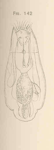
Pleurotrocha parasitica n. sp. Ventral view.
-
Phuripong Meksuwan, Pornsilp Pholpunthin, Hendrik Segers
Zookeys
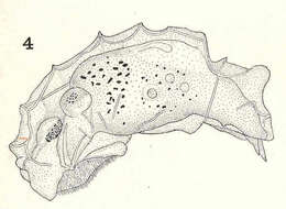
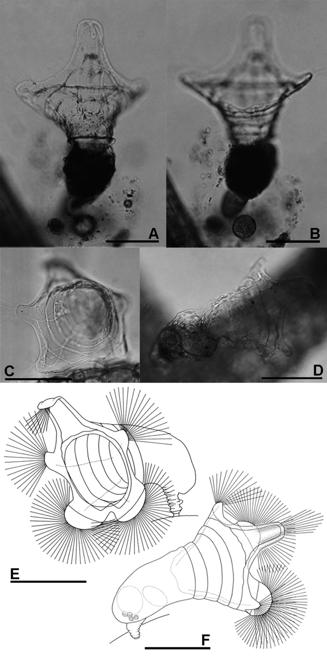
Figure 2.A, B Collotheca ferox (A dorsal view B ventral view) C–F Collotheca orchidacea sp. n. (C, E frontal D, F dorsal). Scale bars: A–F = 100 µm (A, B by Rapeepan Jaturapruek).
-
-
-
Lepadella. Rotifer observed in sandy and muddy marine sediments in the vicinity of Broome, Western Australia in September 2003. This image was taken using phase contrast optics. This work was supported by the Australian Biological Resources Study.
-
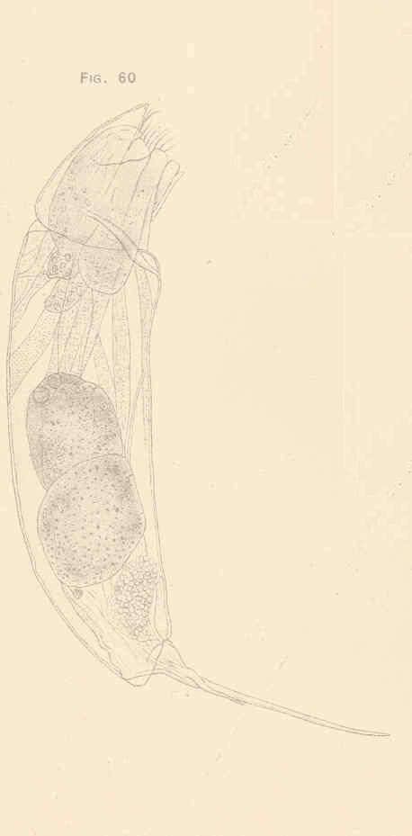
Diurella rousseleti Voigt. Side view of retracted specimen.
-
Taphrocampa annulosa Gosse. Side view.
-
Rattulus capucinus Wierz. & Zach. Right side.
-
Pleurotrocha parasitica n. sp. Side view.
-
Phuripong Meksuwan, Pornsilp Pholpunthin, Hendrik Segers
Zookeys
Figure 4.A Collotheca heptabrachiata, lateral B Collotheca ornata, ventral C Stephanoceros fimbriatus, lateral D Collotheca stephanochaeta, lateral E Collotheca ambigua, ventral F Collotheca algicola, ventral G, H Collotheca ornata f. cornuta (G dorsal H lateral) I Collotheca campanulata f. longicaudata, attachment stalk J Collotheca ferox, ventral corona margin. Scale bars: A, B, D–H, J = 50 µm, C, I = 100 µm.
-
-
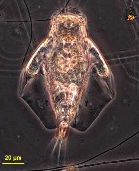
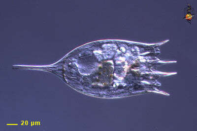
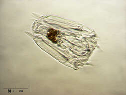
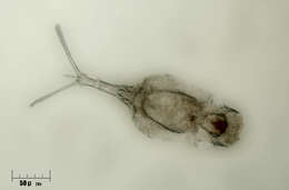
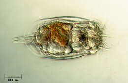
This is a bdelloid rotifer (metazoa), body with an exoskeleton usually in elements that will telescope, posterior end often with two podites (like gastrotrichs to which they are probably related). Very common in aquatic habitats especially those which are prone to dry out - like moss, soils and the contents of pitcher plants. They can resist drying because they can form cysts, but some species are known to be cryptobiotic - that is they can dry out completely, and the organisms can come back to life when wetted. Usually eat bacteria or suspended protists, using the cilia of the corona at the front end of the body to sweep particles into the front of the body where an armoured mastax or jawed pharynx helps to grind the food. Rotifers are common members of the microbial communities of many aquatic ecosystems. Although they are multiceullar animals, they may be only be 100 microns long, and so overlap in size with ciliates. They can be confused with ciliates because they use cilia to capture their food. However, they can be distinguished because they have an exoskeleton, usually two posterior toes, and a tough pharyngeal region just behind the head. Hendrik Seegers says this is probably a non-contracted Lecane because of the toes which appear to bear claws and the more or less regular lower half of the body, below the constriction. Phase contrast.
-
Diurella rousseleti Voigt. Side view of retracted specimen.
-
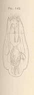
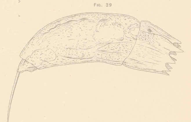
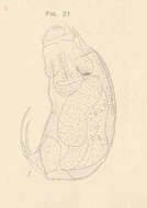
Elosa worrallii Lord, dorsal view, after Lord.
-
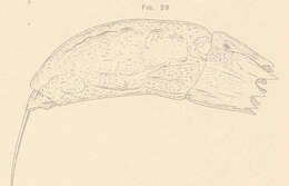
Rattulus carinatus Lamarck. Dorsal view.
-
Phuripong Meksuwan, Pornsilp Pholpunthin, Hendrik Segers
Zookeys
Figure 2.A, B Collotheca ferox (A dorsal view B ventral view) C–F Collotheca orchidacea sp. n. (C, E frontal D, F dorsal). Scale bars: A–F = 100 µm (A, B by Rapeepan Jaturapruek).
-
-
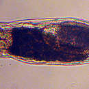
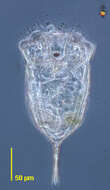
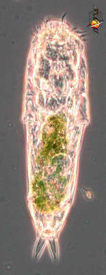
A somewhat withdrawn individual - rotifers can be like that. Differential interference contrast optics.