-
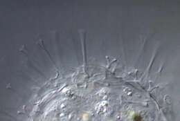
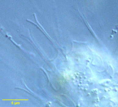
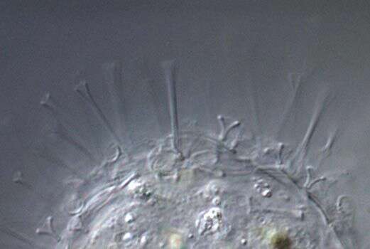
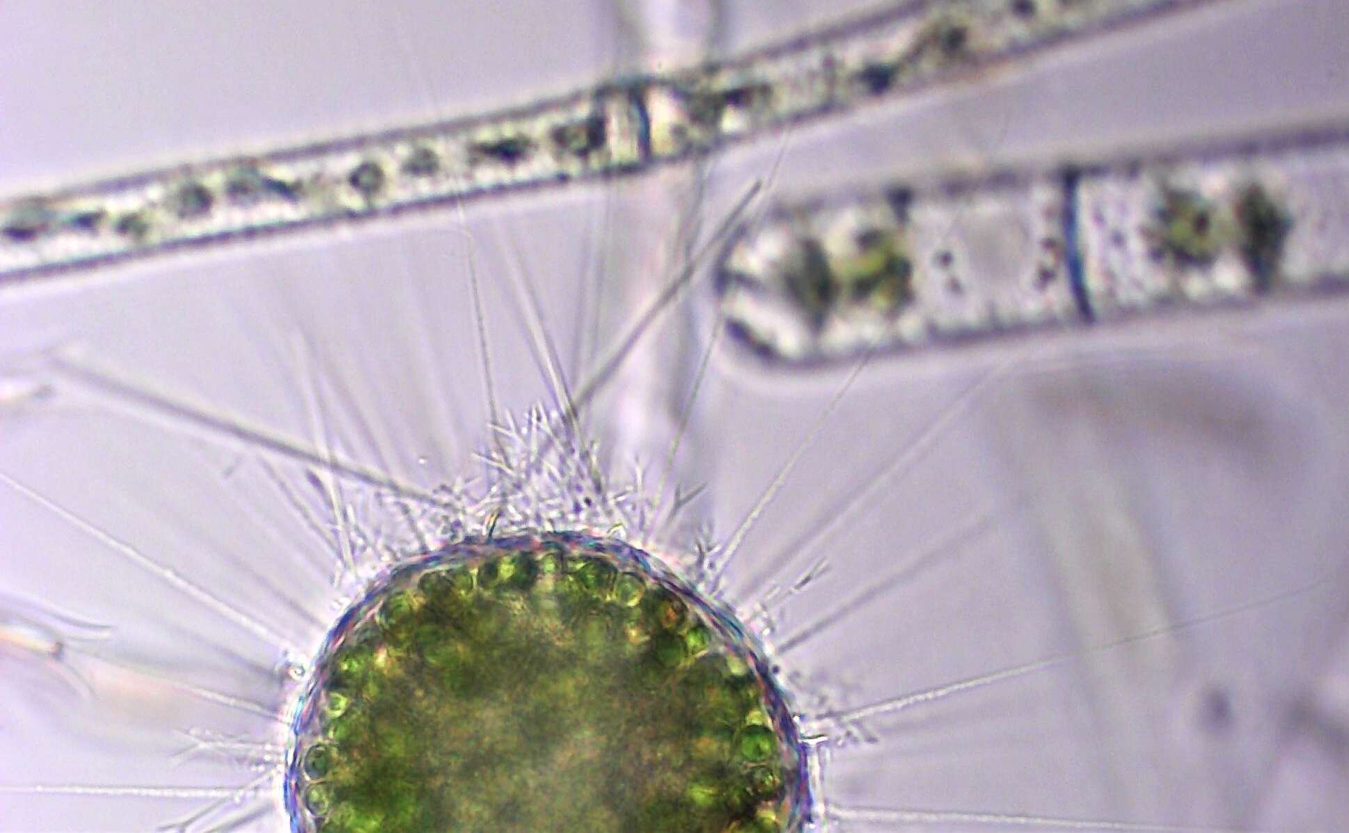
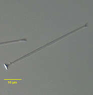
Detail of long forked radiating siliceous spine of Acanthocystis turfacea (Carter,1863), demonstrating the typical circular baseplate. From standing fresh water near Boise, Idaho.DIC.
-
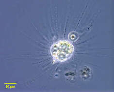
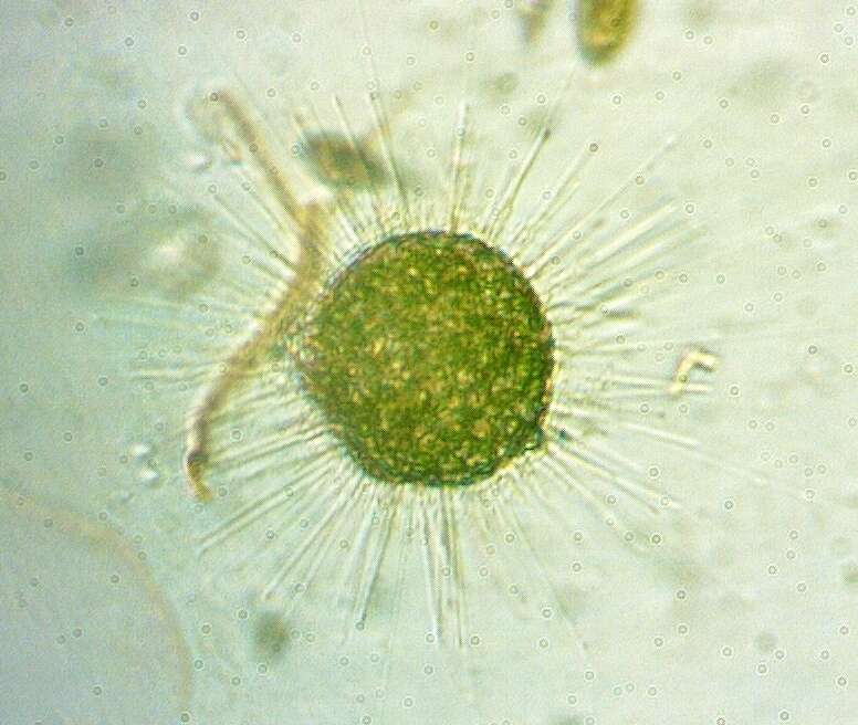
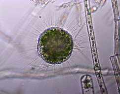
Detail of radiating forked siliceous spines of Acanthocystis turfacea (Carter,1863) which have detached from the periplast under the pressure of the coverglass. A. turfacea is a centroheliozoon with tangentially layered siliceous scales and two types of forked radial siliceous spines, one short, the other long (both seen here). The radiating axopodia contain extrusomes (not seen in this image). Endosymbiotic zoochlorellae are visible in this image. From standing fresh water near Boise, Idaho.DIC.
-
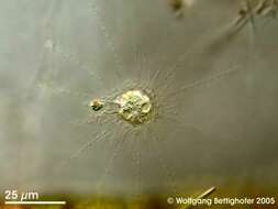
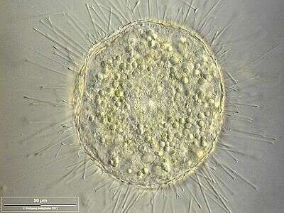
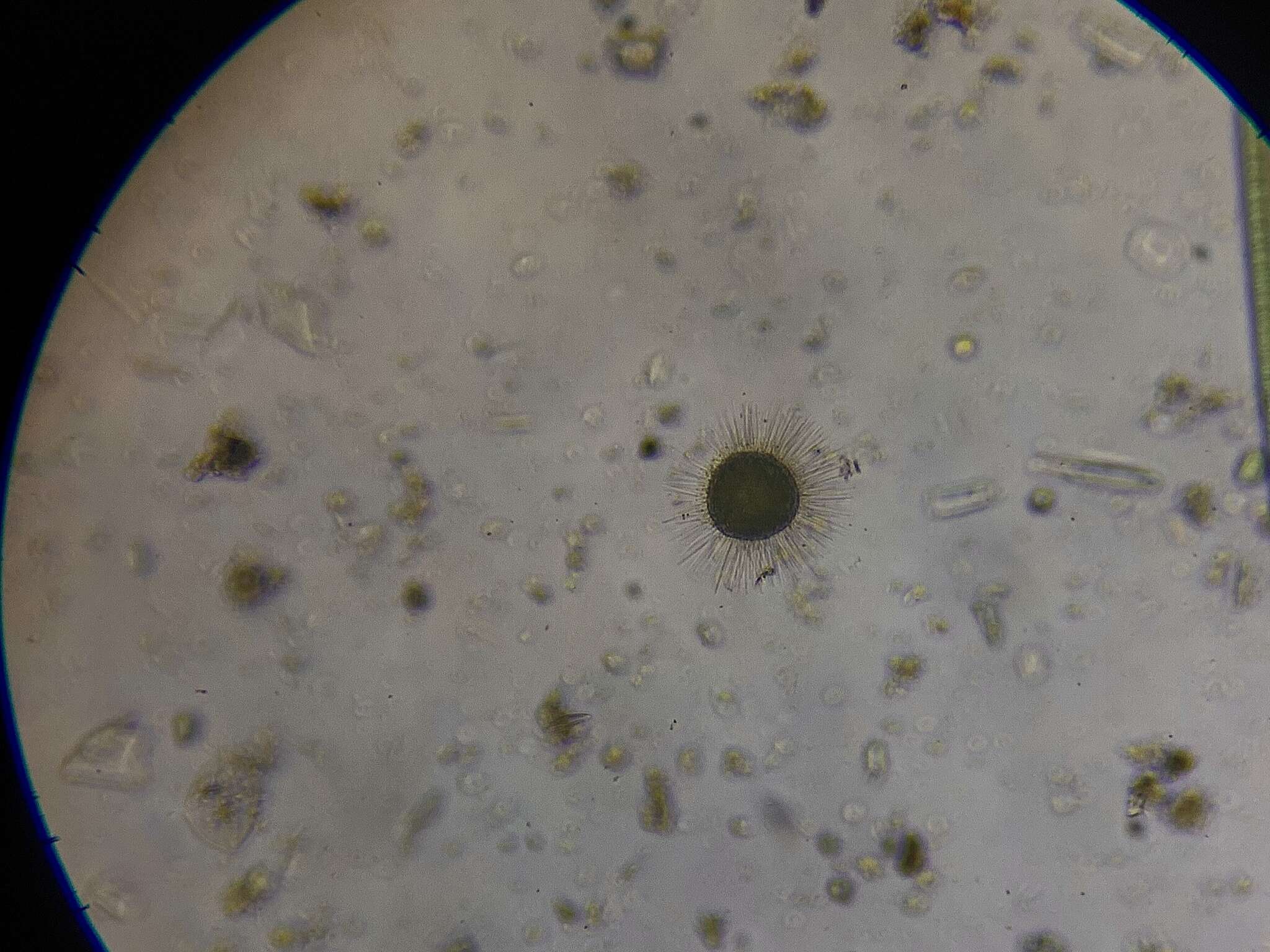
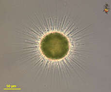
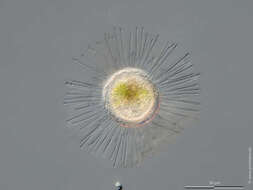
This species has spines of two lengths and that fork at their extremity. It also frequently occurs, as in this case, with symbiotic green algae. Phase contrast micrograph.
-
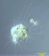
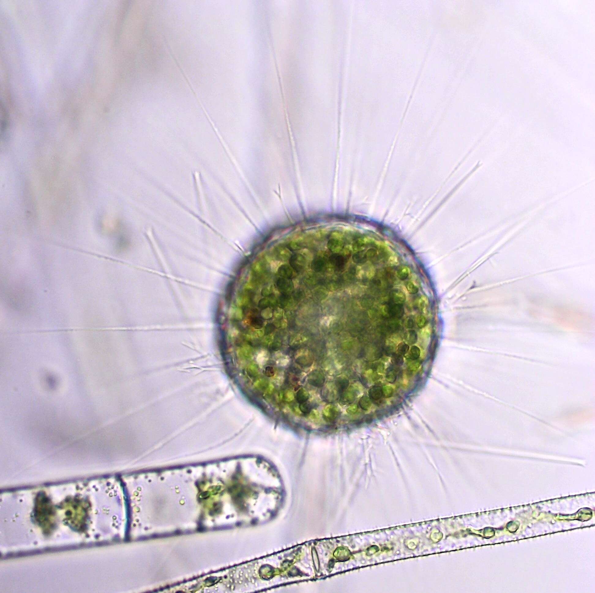
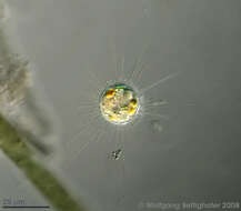
Scale bar indicates 50 µm.Sample from a wetland at the Pillersee (Tyrol, Austria). The image was built up using several photomicrographic frames with manual stacking technique. Images were taken using Zeiss Universal with Olympus C7070 CCD camera.Image under Creative Commons License V 3.0 (CC BY-NC-SA).
-
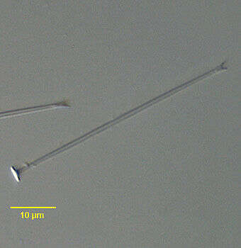
Detail of long radial trumpet-shaped siliceous spines of Raphidocystis tubifera (Penard, 1904). The periplast is composed of 3 types of siliceous elements: elliptical tangential plate scales, long radial trumpet-like scales and short, broad radial funnel-shaped scales. From slow flowing organically enriched freshwater stream near Boise, Idaho. DIC.
-
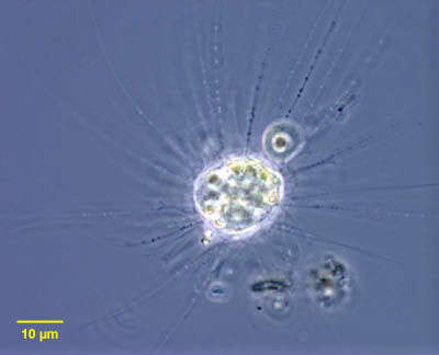
Raphidocystis tubifera (Penard, 1904), a centrohelid heliozoan. The periplast is composed of 3 types of siliceous elements: elliptical tangential plate scales, long radial trumpet-like scales and short, broad radial funnel-shaped scales. The latter two types are seen well in this image at the upper margin of the periplast. The tangential elements are difficult to see. The long axopodia and their extrusomes are visible on the viewer's right in this image. Although species identification rests on EM morphology of the tangential plate scales, the organisms in these images conform to the description of R. tubifera (Mikrjukov,K.A. Arch. Protistenkd. 147: 205-212). From slow flowing organically enriched freshwater stream near Boise, Idaho. Phase contrast.
-
Raphidocystis tubifera (Penard, 1904), a centrohelid heliozoan. The periplast is composed of 3 types of siliceous elements: elliptical tangential plate scales, long radial trumpet-like scales and short, broad radial funnel-shaped scales. The latter two types are seen well in this image at the upper margin of the periplast. The tangential elements are difficult to see. The long axopodia and their extrusomes are visible at 12 o'clock in this image. Zoochlorellae are seen in the cytoplasm. Although species identification rests on EM morphology of the tangential plate scales, the organisms in these images conform to the description of R. tubifera (Mikrjukov,K.A. Arch. Protistenkd. 147: 205-212). From slow flowing organically enriched freshwater stream near Boise, Idaho. Differential interference contrast.
-
Raphidocystis tubifera (Penard, 1904).DIC.
-
Raphidocystis tubifera (Penard, 1904). Phase contrast.
-
Raphidocystis tubifera (Penard, 1904). DIC.
-
This centrohelid heliozoan is characterized by trumpet shaped siliceous skeleton elements. This specimen shows the enormous food vacuole production (funnel-shaped pseudopod) preceding the ingestion process. Further pictures in ZIP archive show steps of ingestion. Collected from littoral region (stand of Phragmites) of a rain storage reservoir in Kiel (Schleswig-Holstein, Germany). Images were taken using Zeiss Universal with Olympus C7070 CCD camera.
-
-
-
-
-
-
-
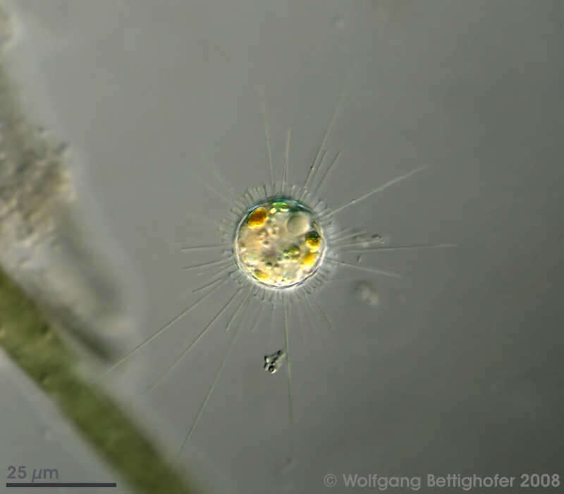
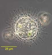
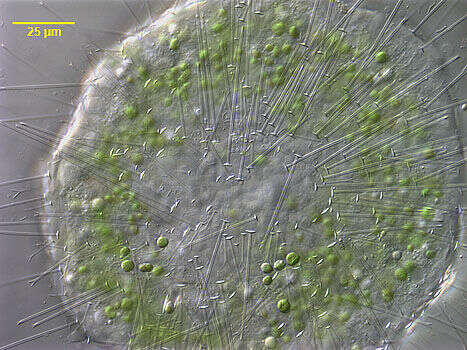
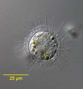
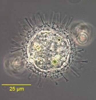
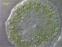
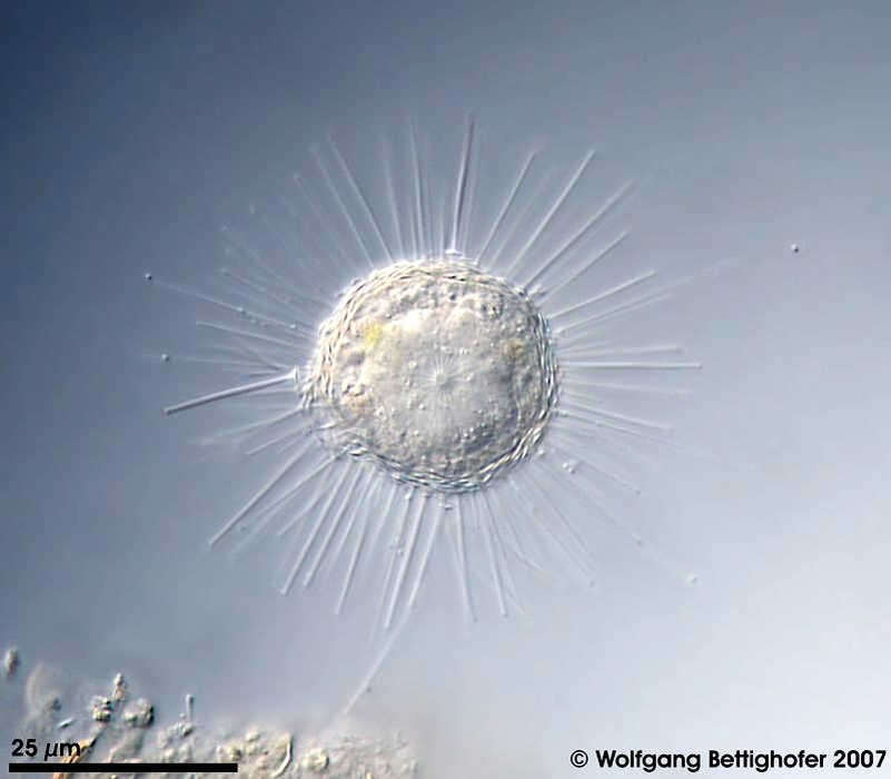
Acanthocystis pectinata The picture shows the two types of actinomorphic siliceous spines (the shorter type is forked, see inset top right) and the tangential scales. Nucleus is clearly shown together with some food vacuoles. Scale bar indicates 25 µm. Sample from sphagnum pond Dosenmoor near Neumuenster (Schleswig-Holstein. Images were taken using Zeiss Universal with Olympus C7070 CCD camera.Image under Creative Commons License V 3.0 (CC BY-NC-SA). Place name: Bog Dosenmoor near Neumuenster (Schleswig-Holstein, Germany) Latitude: 54.136219 Longitude: 10.026433 Das Bild zeigt die beiden Arten der radiären kieselsäurehaltigen Stacheln (der kürzer Typ ist gegabelt, siehe Kasten oben rechts) und die tangentialen Kieselschuppen. Der Zellkern ist deutlich zu sehen, zusammen mit einigen Nahrungsvakuolen. Der Messbalken markiert eine Länge von 25 µm. Probe aus dem Dosenmoor in der Nähe von Neumünster. Mikrotechnik: Zeiss Universal, Kamera: Olympus C7070. Creative Commons License V 3.0 (CC BY-NC-SA). For permission to use of (high-resolution) images please contact postmaster@protisten.de.
-
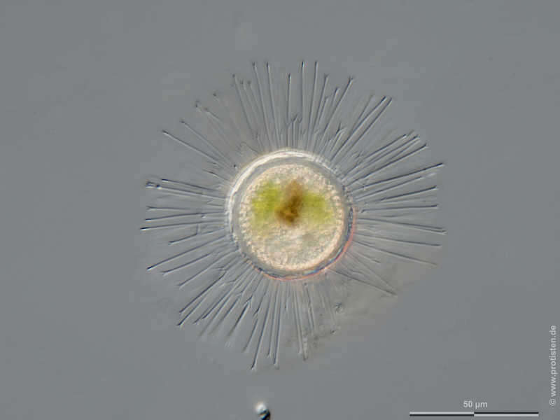
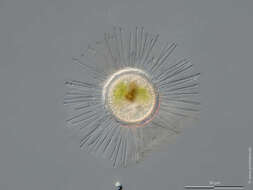
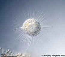
Acanthocystis turfacea Scale bar indicates 50 µm. Sample from Bog Waasenmoos Pass Thurn near Mittersil, Tyrol, Austria. Sampling date 10/2019. The image was built up using several photomicrographic frames with manual stacking technique. Images were taken using Zeiss Axioplan with Olympus OM-D M5 MKII. Image under Creative Commons License V 3.0 (CC BY-NC-SA). Place name: Bog Waasenmoos Pass Thurn near Mittersil (Tyrol, Austria) Latitude: 47.30234117 Longitude: 12.41751194 Multiebenen-Abbildung, manuell gestapelt. Der Messbalken markiert eine Länge von 50 µm. Probe aus dem Waasenmoos bei Pass Thurn Nähe Mittersil, Tirol. Datum der Aufsammlung: 10/2019. Mikrotechnik: Zeiss Axioplan, Kamera: Olympus OM-D M5 MKII. Creative Commons License V 3.0 (CC BY-NC-SA). For permission to use of (high-resolution) images please contact postmaster@protisten.de.
-
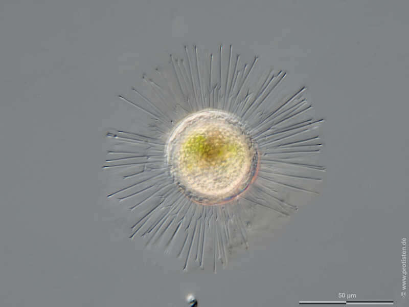
Acanthocystis turfacea Scale bar indicates 50 µm.Sample from a wetland at the Pillersee (Tyrol, Austria). The image was built up using several photomicrographic frames with manual stacking technique. Images were taken using Zeiss Universal with Olympus C7070 CCD camera.Image under Creative Commons License V 3.0 (CC BY-NC-SA). Place name: Wetland at the Pillersee (Tyrol, Austria) Latitude: 47.531785 Longitude: 12.573095 Multiebenen-Abbildung, manuell gestapelt. Der Messbalken markiert eine Länge von 50 µm. Probe aus einer Feuchtwiese beim Pillersee in Tirol. Mikrotechnik: Zeiss Universal, Kamera: Olympus C7070. Creative Commons License V 3.0 (CC BY-NC-SA). For permission to use of (high-resolution) images please contact postmaster@protisten.de.
-
Acanthocystis turfacea Scale bar indicates 50 µm. Sample from Bog Waasenmoos Pass Thurn near Mittersil, Tyrol, Austria. Sampling date 10/2019. The image was built up using several photomicrographic frames with manual stacking technique. Images were taken using Zeiss Axioplan with Olympus OM-D M5 MKII. Image under Creative Commons License V 3.0 (CC BY-NC-SA). Place name: Bog Waasenmoos Pass Thurn near Mittersil (Tyrol, Austria) Latitude: 47.30234117 Longitude: 12.41751194 Multiebenen-Abbildung, manuell gestapelt. Der Messbalken markiert eine Länge von 50 µm. Probe aus dem Waasenmoos bei Pass Thurn Nähe Mittersil, Tirol. Datum der Aufsammlung: 10/2019. Mikrotechnik: Zeiss Axioplan, Kamera: Olympus OM-D M5 MKII. Creative Commons License V 3.0 (CC BY-NC-SA). For permission to use of (high-resolution) images please contact postmaster@protisten.de.
-
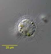
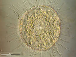
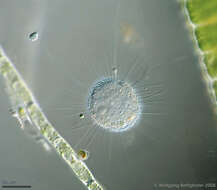
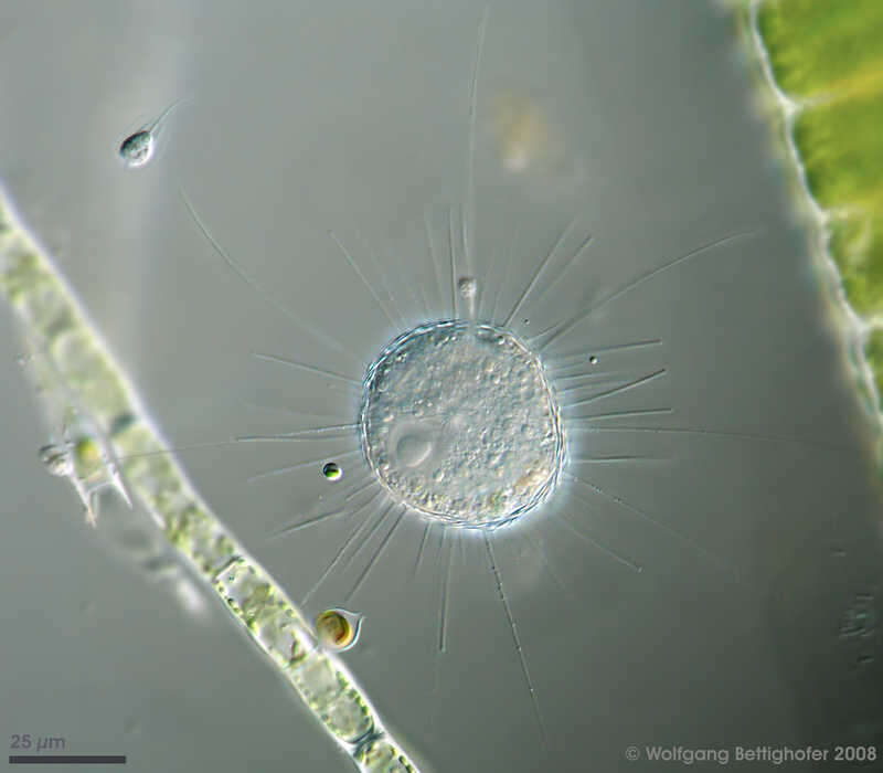
Acanthocystis penardi Acanthocystis penardi shows numerous siliceous spines and scales inserting and lying respectively upon the surface of the cell forming the periplast. The central body from which all the axopodia originate is clearly visible. Scale bar indicates 25 µm. Sample from sphagnum pond Dosenmoor near Neumuenster (Schleswig-Holstein, Germany). Images were taken using Zeiss Universal with Olympus C7070 CCD camera.Image under Creative Commons License V 3.0 (CC BY-NC-SA). Place name: Bog Dosenmoor near Neumuenster (Schleswig-Holstein, Germany) Latitude: 54.136219 Longitude: 10.026433 Acanthocystis penardi trägt zahlreiche kieselsäurehaltigen Stacheln und Schuppen, die auf der Oberfläche der Zelle ansetzen bzw. liegen und den Periplast formen. Der Zentralkörper, von dem alle Axopodien ausgehen, ist deutlich sichtbar. Der Messbalken markiert eine Länge von 25 µm. Probe aus dem Dosenmoor in der Nähe von Neumünster. Mikrotechnik: Zeiss Universal, Kamera: Olympus C7070. Creative Commons License V 3.0 (CC BY-NC-SA). For permission to use of (high-resolution) images please contact postmaster@protisten.de.
-
Acanthocystis penardi Acanthocystis penardi shows numerous siliceous spines and scales inserting and lying respectively upon the surface of the cell forming the periplast. The nucleus and several narrow axopodia are clearly visible, also a free swimming Salpingoeca species and a Chrysophyta called Chrysopyxis inaequalis fixed on a Tribonema filament with a mucilaginous string. Scale bar indicates 25 µm. Sample from sphagnum pond Dosenmoor near Neumuenster (Schleswig-Holstein, Germany). Images were taken using Zeiss Universal with Olympus C7070 CCD camera.Image under Creative Commons License V 3.0 (CC BY-NC-SA). Place name: Bog Dosenmoor near Neumuenster (Schleswig-Holstein, Germany) Latitude: 54.136219 Longitude: 10.026433 Acanthocystis penardi trägt zahlreiche kieselsäurehaltigen Stacheln und Schuppen, die auf der Oberfläche der Zelle ansetzen bzw. liegen und den Periplast formen. Der Kern und mehrere dünne Axopodien sind deutlich sichtbar, des Weiteren eine frei schwimmende Salpingoeca-Art und eine Chrysophycee namens Chrysopyxis inaequalis, die sich mit einem Gallertfaden auf einem Algenfaden (Tribonema) befestigt hat. Der Messbalken markiert eine Länge von 25 µm. Probe aus dem Dosenmoor in der Nähe von Neumünster. Mikrotechnik: Zeiss Universal, Kamera: Olympus C7070. Creative Commons License V 3.0 (CC BY-NC-SA). For permission to use of (high-resolution) images please contact postmaster@protisten.de.