-
This image was taken using differential interference contrast optics. This work was supported by the Australian Biological Resources Study.
-
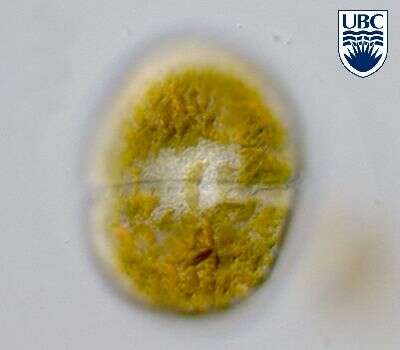
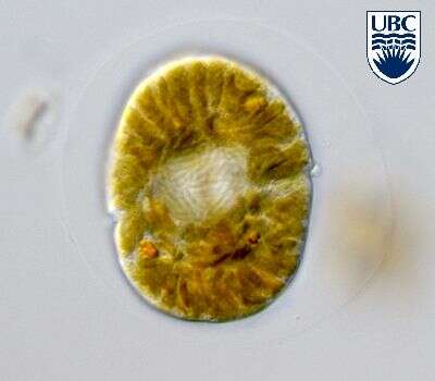
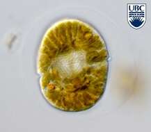
Durinskia_baltica. Collected by ATOL team at Oyster Pond near to Woods Hole, Massachusetts, during the Protistology Workshop at MBL. October-November 2005. Isolation and art by Adrian Reyes-Prieto.
-
Gymnodinium (Dinoflagellata) is the only genus of naked dinoflagellates found in Lake Kinneret. This genus never forms blooms but occurs quite often in small densities. Like other dinoflagellates, Gymnodinium has 2 unequal flagella, the longitudinal "whiplash" flagellum is seen in this picture, the second, transverse flagellum is hidden in the transverse groove, or cingulum.
-
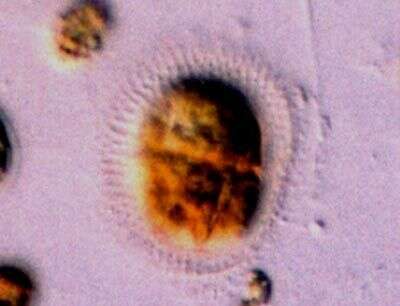
Spiniferodinium (spin-if-err-o-din-ee-um) galeiforme Horiguchi & Chihara 1987. The image shows a temporary cyst (non-motile cell stage). The cyst has many spines protruding on all sides. The cell contains yellow-brown plastids. The nucleus is in the anterior end of the cell.
-
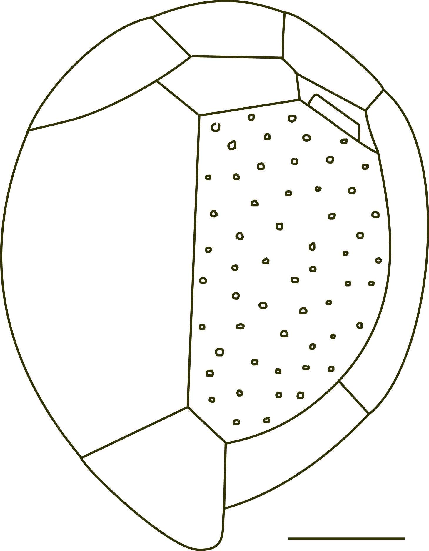
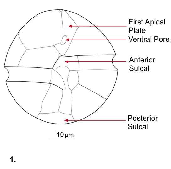
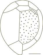
Fig 2: Coolia monotis Schematic diagram (epithecal view) redrawn from Tomas et al. 1997
-
This image was taken using differential interference contrast optics. This work was supported by the Australian Biological Resources Study.
-
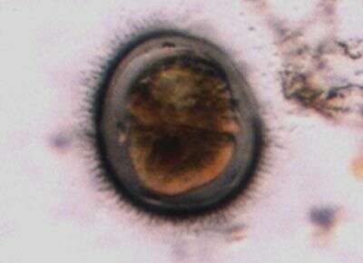
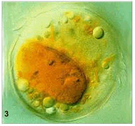
Sinophysis (sine-o-fi/fu-sis) grandis Hoppenrath 2000. The image shows a rectangular cell in left lateral view. The cell is laterally compressed. The epicone is much smaller than the hypocone. The cell contains no plastids, but different coloured food particles are visible. The cell is thecate and has cingular lists.
-
Gymnodinium spec. in dorsal view. Note the helical transverse flagellum running in the cingulum. The nucleus is lying in the cell centre.
-
Spiniferodinium (spin-if-err-o-din-ee-um) galeiforme Horiguchi & Chihara 1987. The image shows a temporary cyst (non-motile cell stage). The cyst has many spines protruding on all sides. The cell contains yellow-brown plastids. The nucleus is in the anterior end of the cell.
-
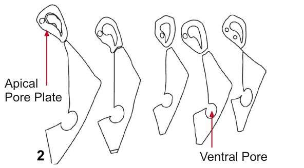
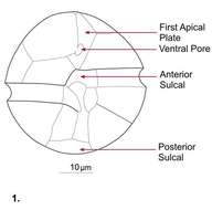
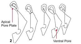
Fig 1: Alexandrium ostenfeldii Schematic drawing of a cell showing plate patterns on ventral side of cell
-
This image was taken using differential interference contrast optics. This work was supported by the Australian Biological Resources Study.
-
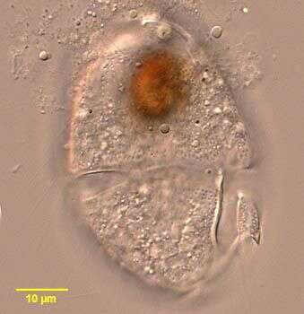
Sinophysis (sine-o-fi/fu-sis) grandis Hoppenrath 2000. The image shows a cell in right lateral view. The cell is laterally compressed. The epicone is much smaller than the hypocone. The cell contains no plastids. The cell is thecate and has cingular lists.
-
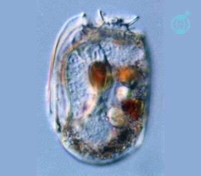
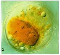
Gymnodinium spec. in mid cell focus showing the central nucleus with nicely visible condensed, rod-shaped chromosomes. The golden-brown chloroplasts are elongated radiating to the cell periphery. Note also the very faint, colorless round cyst around the cell. It is a vegetative division cyst.
-

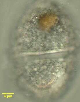
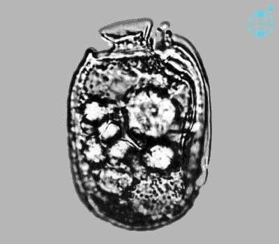
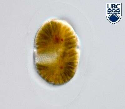
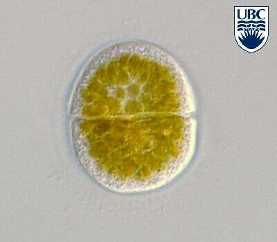
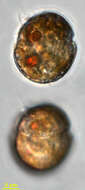
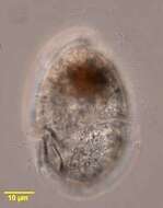
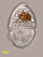
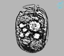
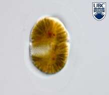
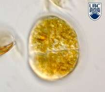
Spiniferodinium galeiforme motile cells are oval to round, slightly dorso-ventrally flattened. Length 23 34 microns, width 17 27 microns. Non-motile cell 25 - 35 microns in length, 20 - 30 microns in width. On motile cell, cingulum begins approximately 0.5 of the cell length from the apex, not displaced. Apical groove extending vertically towards the apex, until approximately 2 microns before the apex, where it turns to the left. The left side of the epicone overhangs slightly at the junction of the cingulum and sulcus. Sulcus initially narrow, then opening towards the antapex. Longitudinal flagellum arising in a pocket just below the junction of the cingulum and sulcus. Nucleus round, 10-12 microns diameter, in the epicone. Chloroplasts small, round to ellipsoidal, 2 - 3 microns diameter. Usually many chloroplasts present. Many colourless starch globules present. Non-motile cell appearing similar to motile cell, round, covered by a transparent helmet with small spines protruding.
-
Fig 2: Alexandrium ostenfeldii Schematic drawing of a cell showing the morphological variation in APC and 1'
-
This image was taken using differential interference contrast optics with a closed condenser iris. This work was supported by the Australian Biological Resources Study.
-
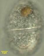
Sinophysis (sine-o-fi/fu-sis) stenosoma Hoppenrath 2000. The image shows a cell in right lateral view. The cell is laterally compressed. The epicone is much smaller than the hypocone. The cell contains no plastids. The cell is thecate and has cingular lists.
-
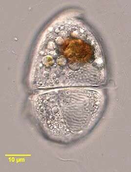
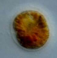
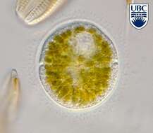
Gymnodinium spec.
-
Spiniferodinium galeiforme Horiguchi et Chihara 1987
-
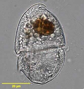
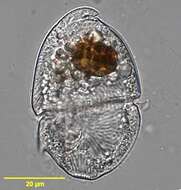
Fig 3: Alexandrium ostenfeldii whole cell with food vacuole
-
This image was taken using differential interference contrast optics. This work was supported by the Australian Biological Resources Study.
-
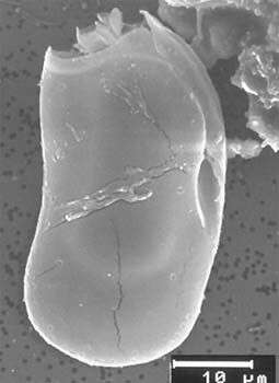
Sinophysis stenosoma, scanning electron microscope image. This image was taken by Mona Hoppenrath of a sample from Town Beach, Broome. This work was supported by the Australian Biological Resources Study.
-
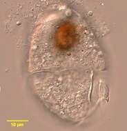
Gymnodinium spec. in ventral view. The sulcus is extending on the episome and the start of the acrobase (apical groove) is visible at the upper cell end.
-
Spiniferodinium galeiforme Horiguchi et Chihara 1987