-
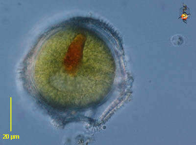
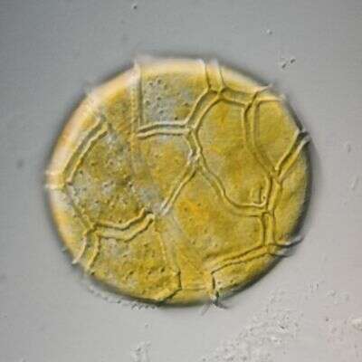
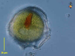
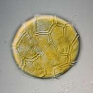
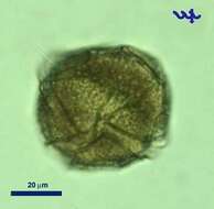
Peridinium (perry-din-ee-um) a dinoflagellate. This is one of the armoured dinoflagellate in which there are substantial skeletal elements in the cortical region of the cell. The groove which contains the circumferential flagellum has strongly developed margins. With chloroplasts. Encysted form - red inclusion resembles an eyespot but in this case we are advised this is more likely residues of food. Differential interference contrast.
-
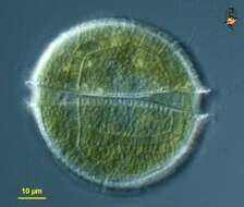
Peridinium (perry-din-ee-um) a dinoflagellate. This is one of the armoured dinoflagellate in which there are substantial skeletal elements in the cortical region of the cell. The groove which contains the circumferential flagellum has strongly developed margins. With chloroplasts. Differential interference contrast.
-
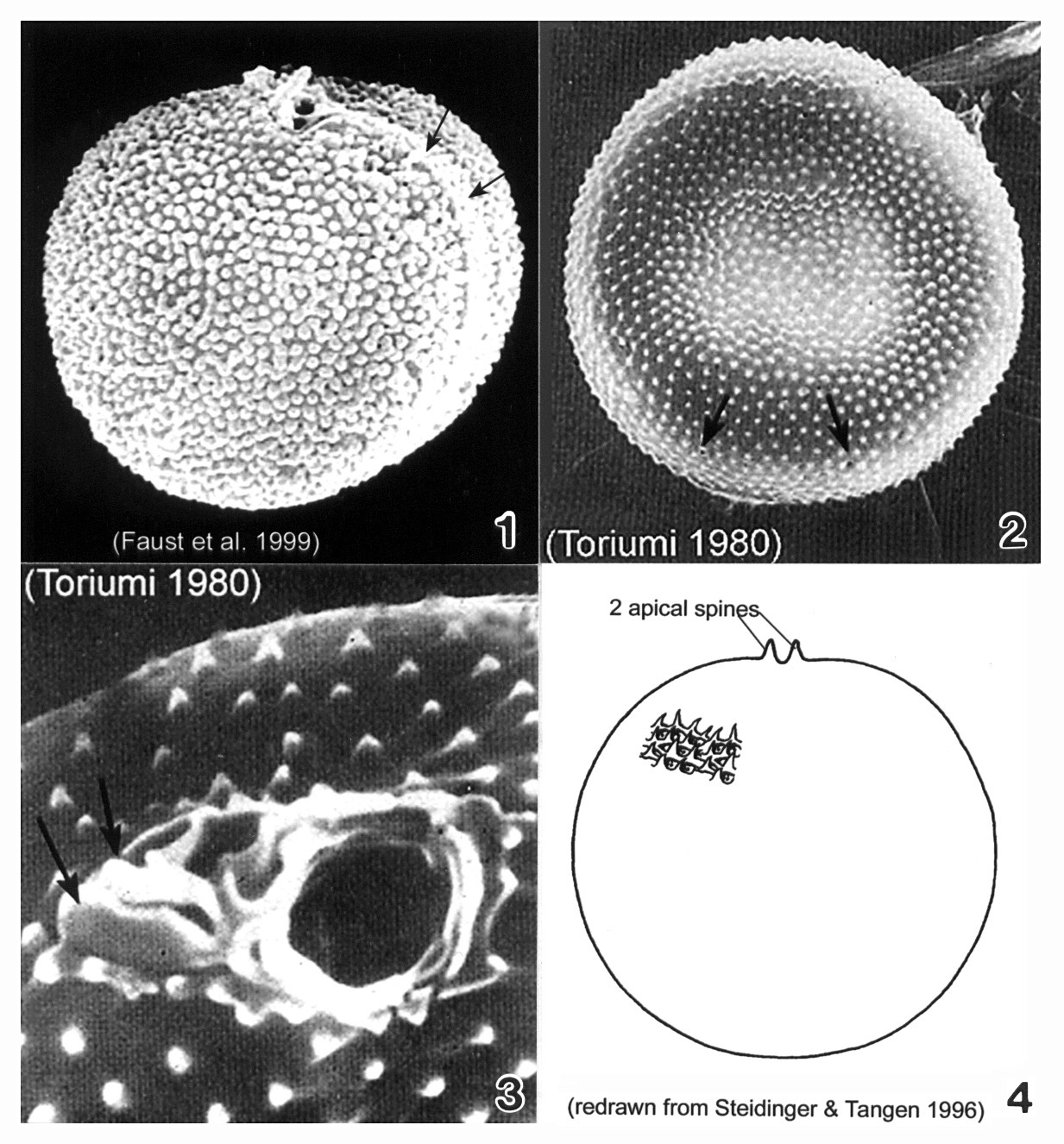
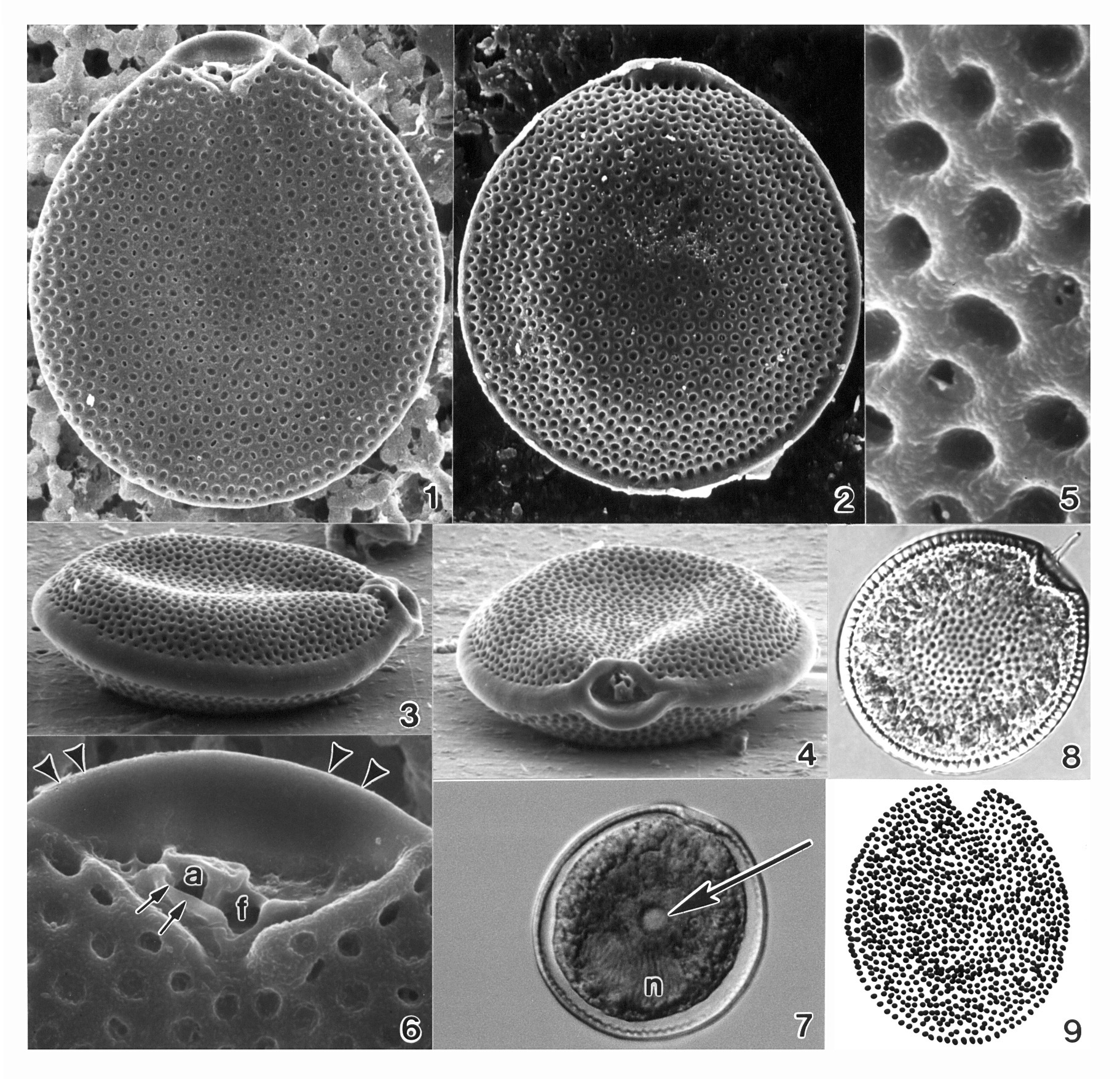
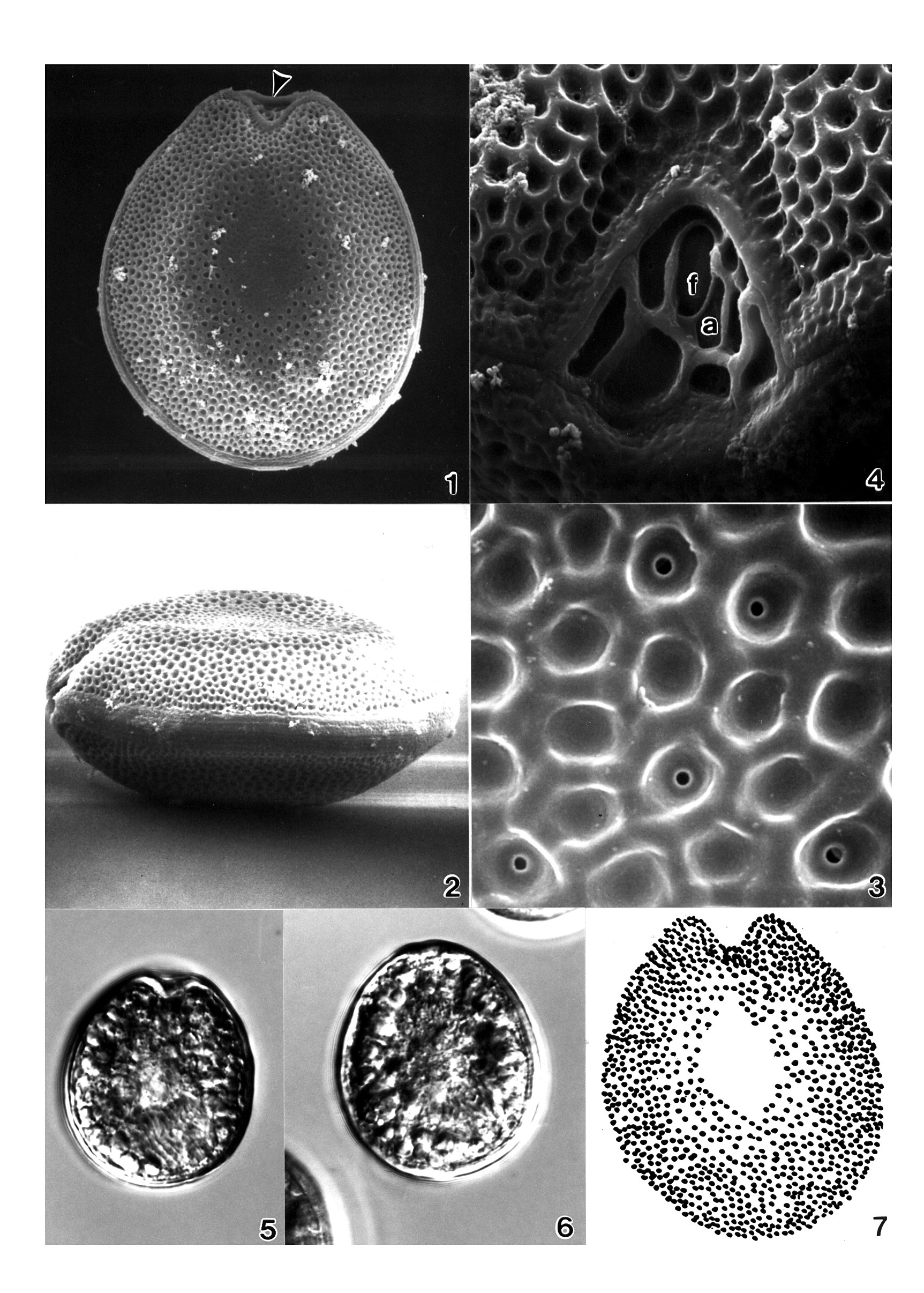
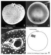
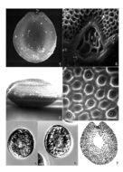
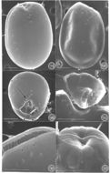
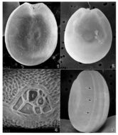
Plate 38. Prorocentrum balticum. Figs. 1-3. SEM. Fig. 1. Valve view: cell round to spherical, covered with many tiny spines. Apical spine apparent. Intercalary band broad, transversely striated (arrows). Fig. 2. Surface with scattered rimmed pores (arrows). Fig. 3. Periflagellar region: two different sized pores and two small apical projections (arrows). Fig. 4. Line drawing.
-
-
Ventral view of a dinoflagellate of the genus Peridinium (Ehrenberg,1832). Collected from a freshwater pond near Boise,Idaho. DIC
-

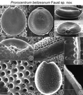
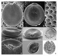
Figs. 1-10. Prorocentrum belizeanum sp. nov. Fig.1. Valve surface is areolated. Fig.2. Cells are round to oval in valve view. Fig.3. In side view, cells are ellipsoid, the apical area exhibits a rounded lip, and both left and right valves are excavated. Fig.4. Periflagellar area is a wide, V-shaped depression located in the right valve. It has a flagellar and auxiliary pore, equal in size. Fig.5. Auxiliary pore (A) is surrounded by a curved apical collar (arrowheads) and is adjacent to the flagellar pore (F). The left valve margin exhibits a wide, rounded ridge (arrow). The two flagella are not shown. Fig.6. Intercalary band is horizontally striated. Fig.7. Areolae are round to ovoid with a smooth margin. Some areolae have trichocyst pores (arrowheads). Fig.8 . The center of the inside valve surface is smooth. The arrangement of round trichocyst pores is shown. Fig. 9. Inside valve surface shown at higher magnification. Trichocyst pores (arrowheads) are arranged in an array along the intercalary band. Fig.10. Inside of the intercalary band is constructed of evenly spaced rectangular sections separated by shallow grooves. Scale bars =10 µm.
EMu: Holotype SEM negative # 104052; SEM stub #104; Field # n.a; Accession # 407164; Catalog # 34; Figure # 1.
-
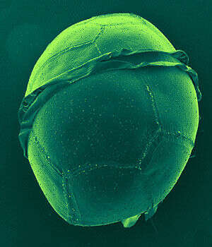
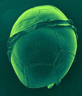
Dorsal view of the cell. The lists are the ridges on wirther side of the girdle.
-
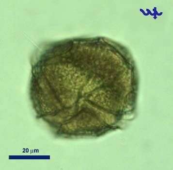
Peridinium penardii has skeletal elements (cellulose plates) in the cortical region of the cell. Species has chloroplasts. Collection from littoral region (stand of Phragmites) of a rain storage reservoir in Kiel (Schleswig-Holstein, Germany). This image was taken using Zeiss Universal with Olympus C7070 CCD camera.
-
Plate 39. Prorocentrum belizeanum. Figs. 1-6. SEM. Fig. 1. Right valve: cell round to oval; surface heavily areolated. Fig. 2. Left valve: anterior margin with flared curved apical collar. Marginal areolae visible. Fig. 3. Lateral view: valve center concave; intercalary band smooth and wide. Fig. 4. Apical view: apical area with rounded lip; both valves excavated. Fig. 5. Areolae round to ovoid with smooth margins; some with pores. Fig. 6. Periflagellar area: auxiliary pore (a) surrounded by curved periflagellar collar (arrows); adjacent to flagellar pore (f). Left valve with flared apical collar (arrowheads). Fig. 7. Left valve: central pyrenoid (arrow) and posterior nucleus (n). Fig. 8. LM: right valve; flagella present. Fig. 9. Line drawing: areolae arrangement (after Faust 1993a).
-
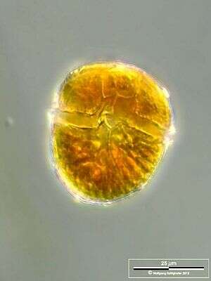
Scale bar indicates 25 µm. Sample from a wetland at the Pillersee (Tyrol, Austria). The image was built up using several photomicrographic frames with manual stacking technique. Images were taken using Zeiss Universal with Olympus C7070 CCD camera.Image under Creative Commons License V 3.0 (CC BY-NC-SA).
-
Cyst. Scale bar indicates 25 µm. Sample from a wetland at the Pillersee (Tyrol, Austria). The image was built up using several photomicrographic frames with manual stacking technique. Images were taken using Zeiss Universal with Olympus C7070 CCD camera.Image under Creative Commons License V 3.0 (CC BY-NC-SA).
-

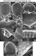
Figs. 17-27. Prorocentrum caribbaeum sp. nov. FIG.17. Cell shape is oval with a rounded anterior and a pointed posterior end. FIG.18. Valve surface is smooth with minute depressions. Radially arranged trichocyst pores are present on each valve. The two flagella are not shown. FIG.19. Cells are ovate to convex in side view. The periflagellar area is located in the right valve and is highly ornate. FIG.20. In valve view, the periflagellar area has a rectangular orientation and is composed of a curved apical collar (on the left) and a smaller protuberant apical plate (on the right). FIG.21. Curved apical collar (arrow) is the largest platelet situated adjacent to the auxiliary pore (A). The apical plate (arrowhead) is located next to the flagellar pore (F) and is separated by a rectangular platelet from the auxiliary pore. FIG.22. The trichocyst pores (arrow) are round with smooth edges and are similar in size. They are situated in furrowed depressions. Small, round pores (arrowhead) are also present, unevenly distributed on the valve surface. FIG.23. The posterior end is pointed and laced with trichocyst pores and small, round pores unique to this species. FIG.24.Ejected trichocysts are on valve surface. FIG.25. The intercalary band is transversally striated and sinous. FIG.6.Inner valve surface is smooth. The location of round trichocyst pores is illustrated, and a distinct striated intercalary band is present (arrowheads). FIG. 7. The inner face of the intercalary band is highly ornate and lacelike. Scale bars = 8 µm.
EMu: Holotype SEM negative # 103001; SEM stub #103; Field # 358-90 Accession # 407164; Catalog # 49; Figure # 17.
-
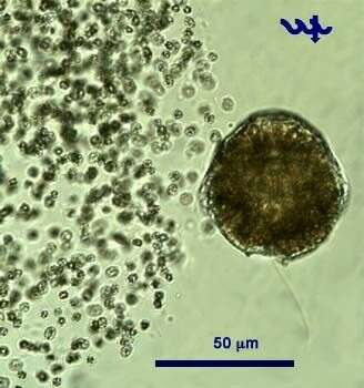
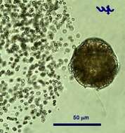
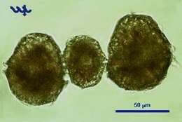
Peridinium gatunense is a large (ca. 50 um diameter) dinoflagellate, the most common bloom-forming species in Lake Kinneret, and the most studied species from this lake. Its winter-spring blooms give the water coffee-brown color. The blooms are patchy, the brown color patches can be observed from the distance. These blooms, reported to occur each spring since the 1950s, were characteristic of Lake Kinneret till the mid 1990s. In recent years Peridinium gatunense failed to bloom in some of the years, whereas in others its bloom was even more intense than recorded previously.
-
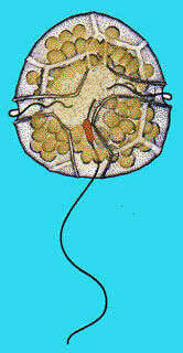
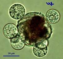
Peridinium gatunense, a large dinoflagellate (ca. 50 um diameter) is the main bloom-forming species in the plankton of Lake Kinneret. At the height of the bloom it forms patches in which cell densities are particularly high and the water gets coffee color. Peridinium has 2 unequal flagella, the longitudinal "whiplash" flagellum is seen in this picture, the second, transverse flagellum is hidden in the transverse groove or cingulum. A Microcystis colony is seen next to the Peridinium, note the large difference in cell size. This specimen was sampled in the littoral in June 2006.
-
Plate 40. Prorocentrum concavum. Figs. 1-4. SEM. Fig. 1. Right valve. Cell ovate and heavily areolate. Valve center devoid of areolae. Left valve with anterior apical ridge (arrowhead). Fig. 2. Lateral view. Valve center concave and flattened. Intercalary band granulated and horizontally striated. Fig. 3. Valve areolae round to oval with smooth edges; some with small central pores. Fig. 4. Periflagellar area a V-shaped depression. Two pores: small auxiliary pore (a); large flagellar pore (f). Figs. 5-6. LM (M.A. Faust). Fig. 5. Right valve. Fig. 6. Left valve. Fig. 7. Line drawing: areolae arrangement. (Figs. 1-4,7 after Faust 1990b)
-
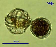
A protoplast of Peridinium is leaving its thecae. Usually this happens during adverse conditions, e.g. lack of nutrients or when being subjected to very strong light - or as part of cell division. In this case, there is no cell division.
-
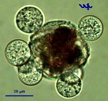
This cell of Peridinium gatunense is infected by several individuals of Phlyctochytrium sp, a chytrid fungus. Note that the Peridinium protoplasm is separated from the thecae and condensed, an indication that the cell is dead or dying. The fungal sporangia are at a developed stage with zoospores about to emerge out of in order to infect new Peridinium cells.
-

FIGS. 11-16. Prorocentrum elegans sp. nov. FIG 11. Cells are oval in valve view; the cell surface is smooth with few valve pores. The two flagella are not present. FIG.12. Left valve margin at the anterior end is flat or inclined. FIG.13. Cells are ovate in side view. The periflagellar area is large relative to the cell size. It has a flagellar (F) and auxiliary (A) pore and an angled flagellar plate (arrowhead adjacent to the auxiliary pore). Large pores (arrow) and small pores (arrowheads) are present on the valves. The small pores are better illustrated at higher magnification in Figure 15. FIG.14. Periflagellar area is detached from the right valve. It has a smooth inner surface, and discrete platelets are unequal in size. FIG.15. The intercalary band is transversely striated, and the inner surface appears ribbed. Small pores (arrowheads) are situated in an array along the intercalary band. FIG.16. The two apical pores are separated by a ridge in this naked cell. Scale bars = 5 µm.
Note:Holotype SEM negative # 86075A; SEM stub #86; Field # n.a; Accession # 407164; Catalog # 44; Figure # 11.
-
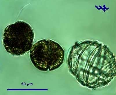
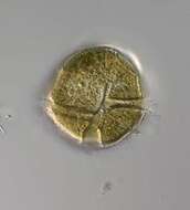
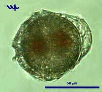
This cell of Peridinium gatunense is about to form a cyst in a process called encystation. Note the two red bodies within the protoplast, and the nearly perfect circular shape of the cyst emerging out of the thecae.
-
The two small daughter-cells have just emerged from the mother thecae. Note the considerably larger size of the empty, colorless thecae of the mother cell, note the flagellum of the cell on the left, note that both young cells are naked protoplasts, within a few hours they will be each covered by new thecae.
-
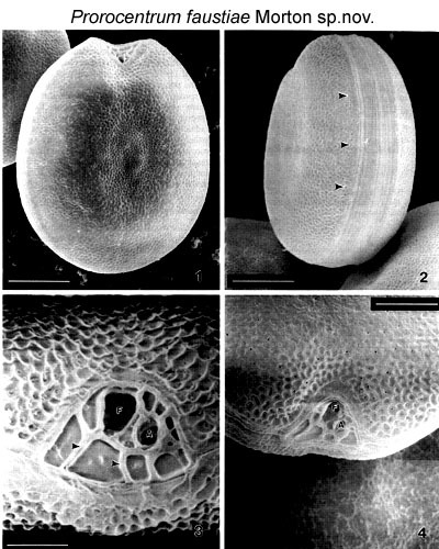
Figs. 1-4. Fig.1.Valve surface of Prorocentrum faustiae. Scale bar represents 5µm. Fig. 2. Intercalary band of Prorocentrum faustiae. Scale bar represents 5 um. Arrow heads point to the evenly spaced marginal pores. Fig. 3. Periflagellar area of Prorocentrum faustiae displays a large flagellar pore (F) and smaller auxiliary pore (A). Scale bar represents 1 µm. Arrow heads point to sutures which separate each periflagellar plate. Fig. 4. Periflagellar area in valve view. Scale bar represents 3 µm.
Note: Isolated from Heron Island, Australia (23.25° S, 151.55° E).
-
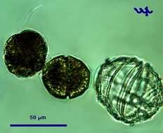
Since the 2 daughter cells of Peridinium gatunense are half the size of their mother cell, actively growing populations show a large variety of cell sizes, as seen in this photograph.
-
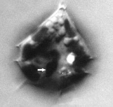
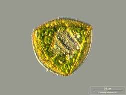
Peridinium quinqucorne cells are diamond shaped to ovoid, with a pointed apex, and a number of projections from the hypocone, 20 - 25 microns long, 15 - 18 microns wide. Thecal plates present: an apical pore, a canal plate, 3 apical plates, 1 anterior intercalary plate, 7 precingular plates, 5 cinguluar plates, sulcal plates not known, 5 postcingular plates, 2 posterior plates. Small apical pore present, in a short apical horn. Cingulum 4 - 5 microns wide, number of plates not recorded. The large anterior sulcal plate forming a list over the left sulcal plate. Four spines (4-5 microns long) project from the antapical plates. Thecal plates smooth, with scattered pores. Intercalary bands may be wide on some cells. Nucleus round, in the centre of the cell, approximately 7 microns diameter. Reddish stigma, present near the beginning of the sulcus. Yellow- brown plastids, 2 - 3 microns diameter, scattered throughout the cell.
-
Plate 41. Prorocentrum faustiae. Figs. 1-4. SEM. Fig. 1. Right valve. Cells broadly ovate to rotundate with slightly concave center. Valve surface rugose. Periflagellar area situated apically. Fig. 2. Left valve: apical region slightly excavated. Fig. 3. Intercalary band wide and transversely striated. Small marginal pores evenly spaced along cell perifery (arrows). Fig. 4. Periflagellar area: apical view. Broad V-shaped depression; larger flagellar pore (f) adjacent to smaller auxiliary pore (a). (All figures donated by S.L. Morton)