-
IZ_1453621_FrameGrab_03.jpg; EX1702_IMG_20170221T022553Z_ROVHD.jpg
-
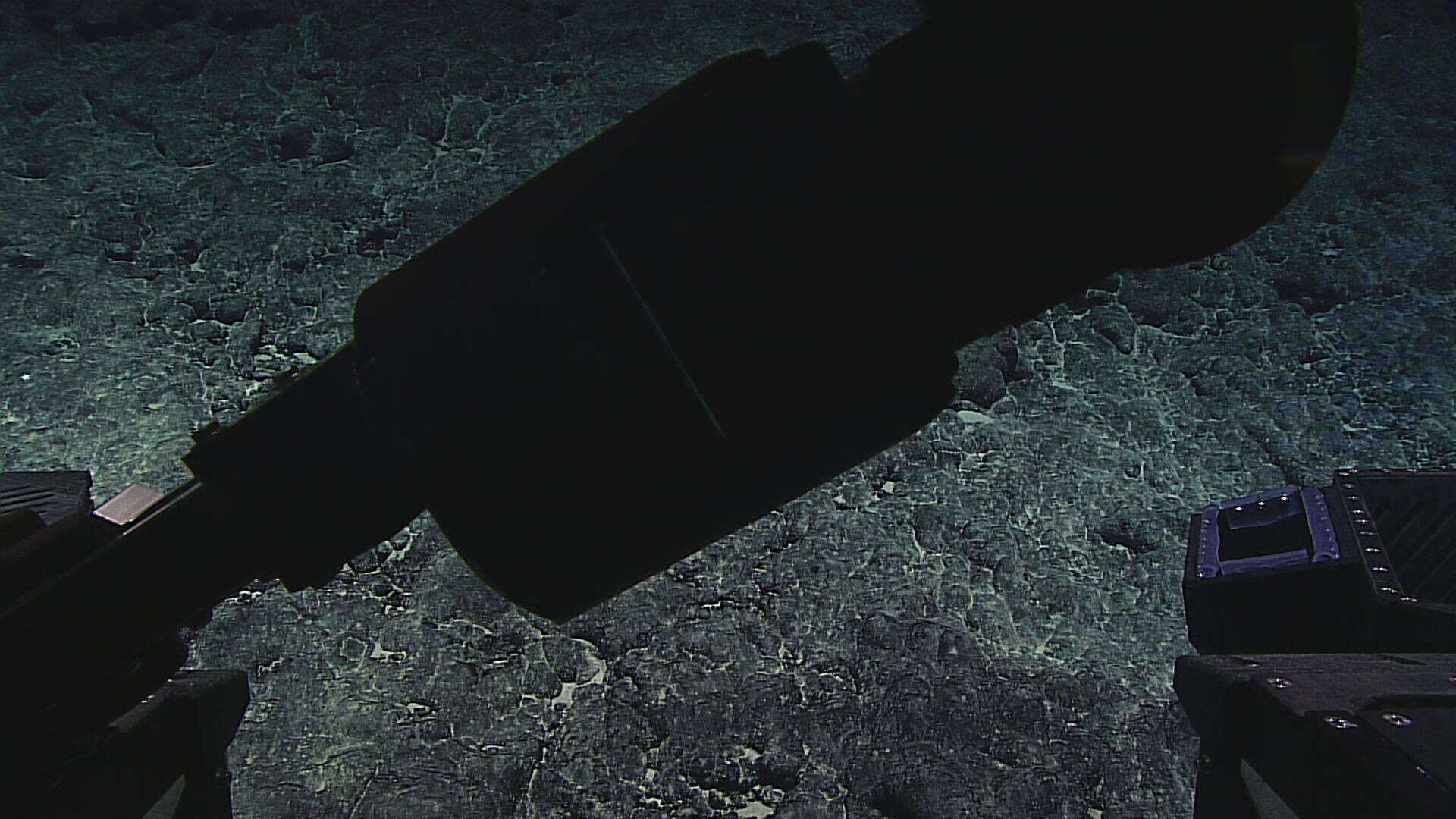
IZ_1453621_CPHD.jpg; EX1702_IMG_20170221T022322Z_CPHD.jpg
-
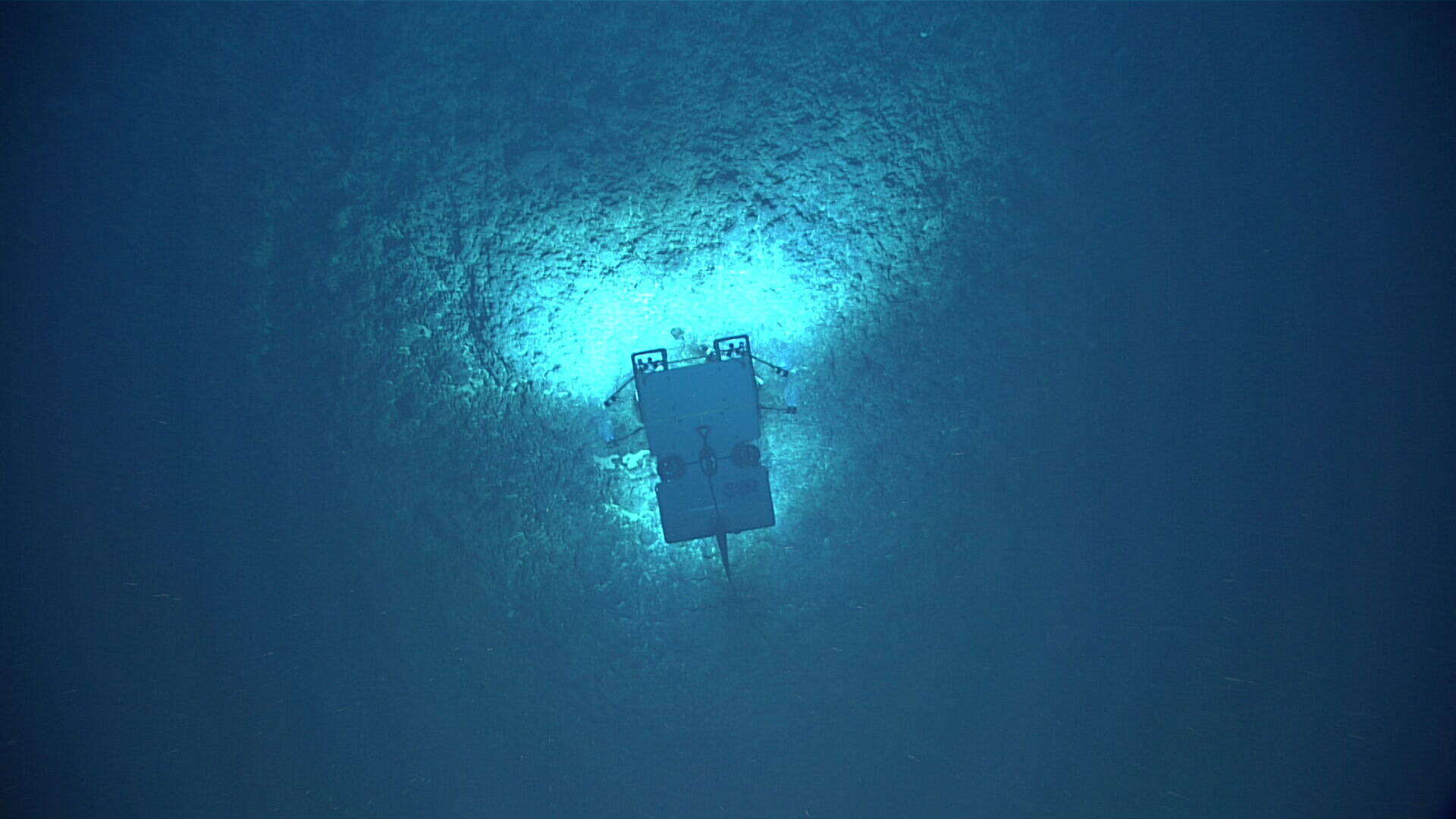
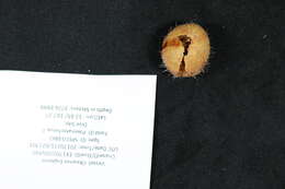
IZ_1453621_LabImage_01.jpg; EX1702_IMG_20170221T022703Z_SMPSTL_D2_DIVE05_SPEC03BIO_L01.JPG
-
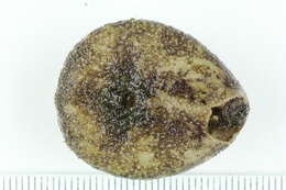
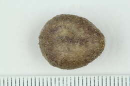
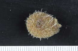
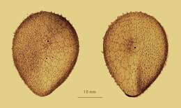
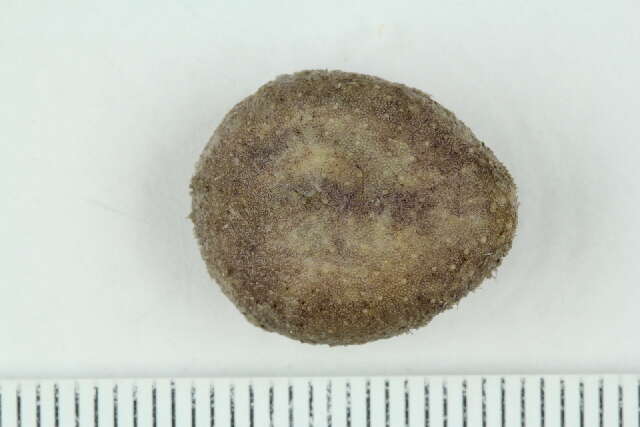
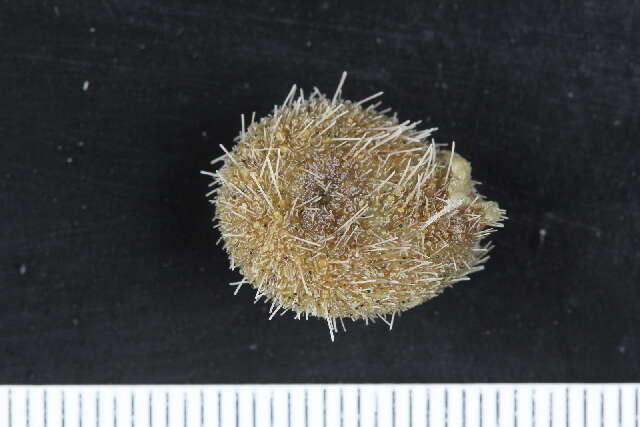
Ventral view, dry specimen
-
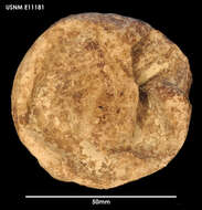
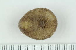
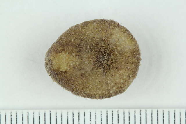
Dorsal view, dry specimen
-
-
-
University of Bergen. Natural History Collections. University of Bergen. Year: 2021. Contact: Katrine.Kongshavn@uib.no.
Barcode of Life Data Systems
Ventral. Catalog no.: 138773. Specimen ID: 13315479. Taxon rep.: Urechinus. Image quality: 1. Aspect ratio: 1.499.
-
University of Bergen. Natural History Collections. University of Bergen. Year: 2021. Contact: Katrine.Kongshavn@uib.no.
Barcode of Life Data Systems
Dorsal. Catalog no.: 138794. Specimen ID: 13315559. Image quality: 1. Aspect ratio: 1.499.
-
University of Bergen. Natural History Collections. University of Bergen. Year: 2021. Contact: Katrine.Kongshavn@uib.no.
Barcode of Life Data Systems
Ventral. Catalog no.: 138794. Specimen ID: 13315559. Image quality: 1. Aspect ratio: 1.499.
-
University of Bergen. Natural History Collections. University of Bergen. Year: 2020. Contact: Katrine.Kongshavn@uib.no.
Barcode of Life Data Systems
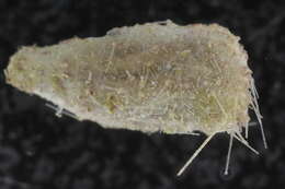
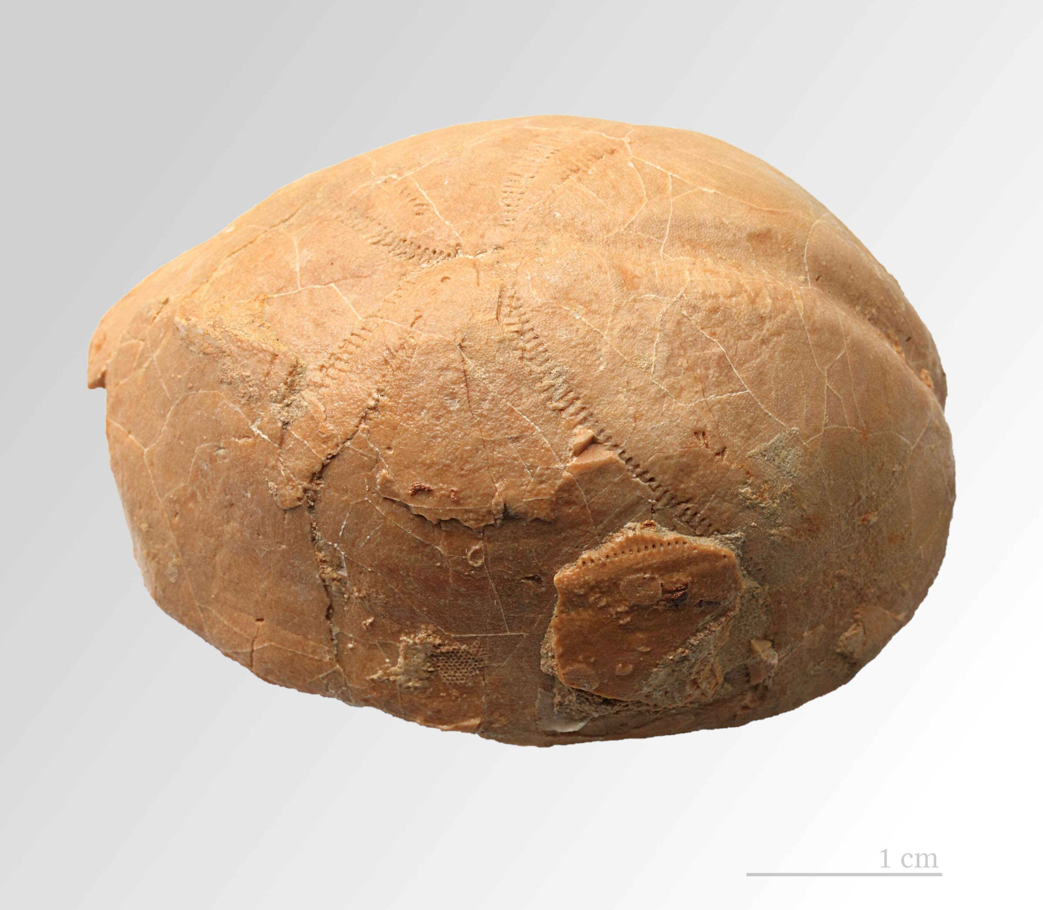
Lateral. Catalog no.: 136448. Specimen ID: 11943994. Taxon rep.: Pourtalesiidae. Image quality: 1. Aspect ratio: 1.499.
-
University of Bergen. Natural History Collections. University of Bergen. Year: 2021. Contact: Katrine.Kongshavn@uib.no.
Barcode of Life Data Systems
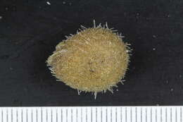
Dorsal. Catalog no.: 138756. Specimen ID: 13315579. Image quality: 1. Aspect ratio: 1.499.
-
University of Bergen. Natural History Collections. University of Bergen. Year: 2021. Contact: Katrine.Kongshavn@uib.no.
Barcode of Life Data Systems
Ventral. Catalog no.: 138756. Specimen ID: 13315579. Image quality: 1. Aspect ratio: 1.499.
-
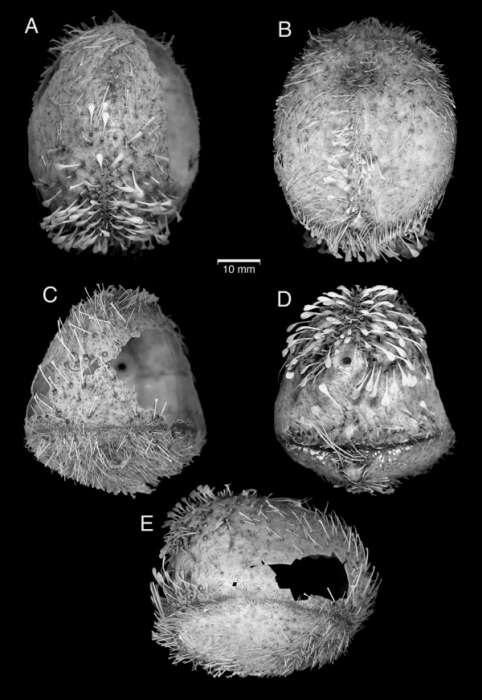
Specimen from R/V ‘Akademik Kurchatov’ cruise 37, station 3787. A, aboral surface. B, oral surface. C, anterior surface. D, posterior surface. E, right side. from Saucède, T., Mironov, A. N., Mooi, R. & David, B. 2009. The morphology, ontogeny, and inferred behaviour of the deep-sea echinoid Calymne relicta (Holasteroida). Zoological Journal of the Linnean Society 155, 630-648.
-
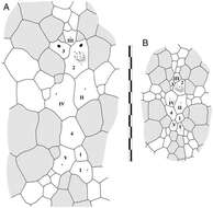
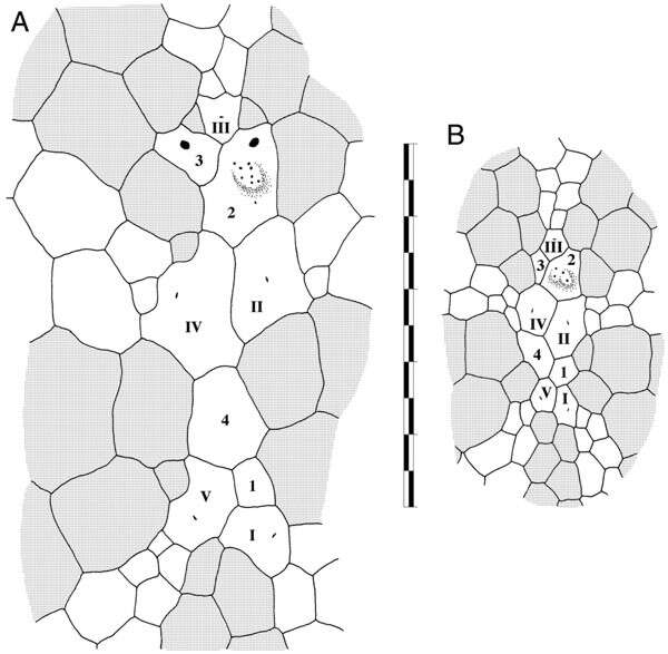
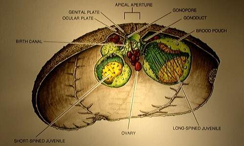
Apical system plate architecture of Calymne relicta (MCZ 8571) from R/V ‘Atlantis’ cruise 24, station 122, anterior towards top of page, interambulacral plates shaded, apical plates labelled according to Lovén’s system. A, adult specimen approximately 30 mm long. B, juvenile specimen (lacking gonopores) 11.3 mm in test length. from Saucède, T., Mironov, A. N., Mooi, R. & David, B. 2009. The morphology, ontogeny, and inferred behaviour of the deep-sea echinoid Calymne relicta (Holasteroida). Zoological Journal of the Linnean Society 155, 630-648.
-
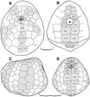
Plate architecture of Calymne relicta. For each, the fasciole is indicated by bands of small, open circles, the interambulacra are shaded, mouth and anal openings are black, some plate sutures are omitted because they were not visible on the specimen. The plates of the posterior interambulacrum are numbered according to Lovén’s rule. Scale bars are 5 mm long. A, oral surface of adult specimen from R/V ‘Akademik Kurchatov’ cruise 37, station 3787, anterior towards top of page. B, posterior surface of specimen in (A). C, left side of specimen from MCZ 8571, R/V ‘Atlantis’ cruise 24, station 122, position of mouth indicated by dashed line. D, posterior surface of specimen in (C), viewed slightly more from the oral surface than view shown in (B). from Saucède, T., Mironov, A. N., Mooi, R. & David, B. 2009. The morphology, ontogeny, and inferred behaviour of the deep-sea echinoid Calymne relicta (Holasteroida). Zoological Journal of the Linnean Society 155, 630-648.
-
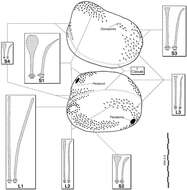
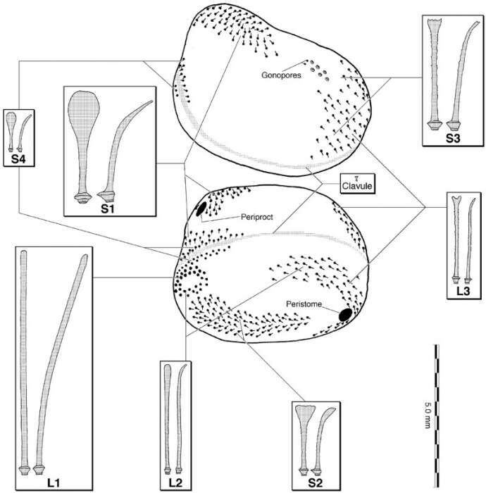
Location of spine types on Calymne relicta plotted on specimen from R/V ‘Akademik Kurchatov’ cruise 37, station 3787. For each view of entire specimen, the orientation of the power stroke as suggested by tubercle asymmetry is indicated by the direction of the small lines leading away from the dot. The upper drawing of the entire specimen is an oblique view from the anterior-dorsal, anterior to the right. The lower drawing of the entire specimen is an oblique view from the posterior-ventral, anterior to the right. Shaded bands indicate the position of the fasciole. For each spine drawing (except for the clavule), the image on the left is a ‘frontal’ view, the one on the right is a ‘side’ view. Scale bar at lower right is for the spines, which are all drawn to scale. from Saucède, T., Mironov, A. N., Mooi, R. & David, B. 2009. The morphology, ontogeny, and inferred behaviour of the deep-sea echinoid Calymne relicta (Holasteroida). Zoological Journal of the Linnean Society 155, 630-648.
-
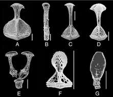
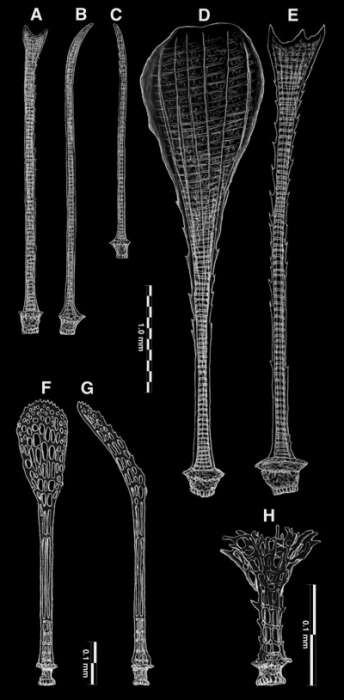
Drawings of spines of Calymne relicta, depicted as viewed with transmitted light microscopy. Scale bar at uppermost refers to (A)–(E), at lower left to (F) and (G), at lower right to (H). A, ‘frontal’ view of type L3. B, ‘side’ view of type L3. C, ‘side’ view of type L4. D, ‘frontal’ view of type S1. E, ‘frontal’ view of type S3. F, ‘frontal’ view of miliary. G, ‘side’ view of miliary. H, clavule from fasciole. from Saucède, T., Mironov, A. N., Mooi, R. & David, B. 2009. The morphology, ontogeny, and inferred behaviour of the deep-sea echinoid Calymne relicta (Holasteroida). Zoological Journal of the Linnean Society 155, 630-648.
-
Pedicellariae of Calymne relicta. A, scanning electron micrograph (SEM) of interior view of type 1 rostrate valve. B, SEM of interior view of type 1 rostrate stem. C, SEM of interior view of type 2 rostrate valve. D, SEM of interior view of type 3 rostrate valve. E, drawing of two of the three valves of an ophicephalous pedicellaria showing position in life relative to the stem. F, SEM of interior view of triphyllous valve. G, drawing of sphaeridium. Scale bars 0.1 mm. from Saucède, T., Mironov, A. N., Mooi, R. & David, B. 2009. The morphology, ontogeny, and inferred behaviour of the deep-sea echinoid Calymne relicta (Holasteroida). Zoological Journal of the Linnean Society 155, 630-648.
-
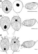
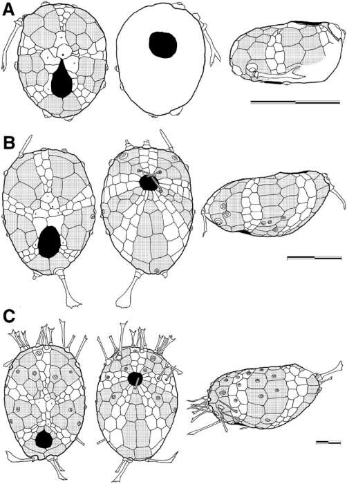
Ontogeny of Calymne relicta from MCZ 8571, R/V ‘Atlantis’ cruise 24, station 122. For each set of drawings, from left to right: apical view, oral view, and left side. The periproct and peristome are in black, and the interambulacral plates are shaded. In some specimens, the sutures are omitted because they were not discernible, and only primary tubercles are shown (some still with spines attached). All scale bars are 1 mm long. A, just-postlarval imago. B, very small juvenile. C, larger juvenile with periproct separated from apical system. from Saucède, T., Mironov, A. N., Mooi, R. & David, B. 2009. The morphology, ontogeny, and inferred behaviour of the deep-sea echinoid Calymne relicta (Holasteroida). Zoological Journal of the Linnean Society 155, 630-648.
-
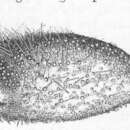
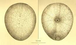
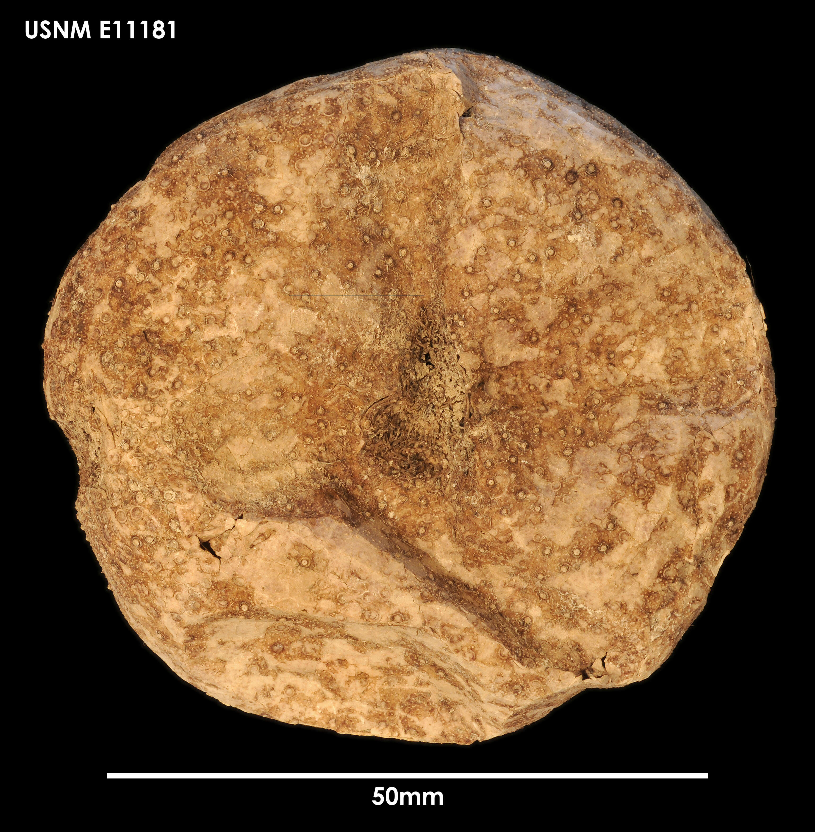
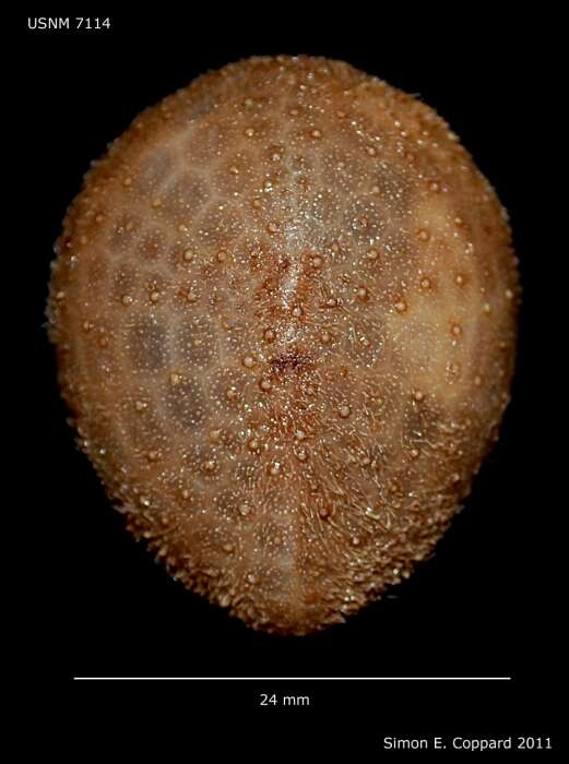
Urechinus naresianus (USNM 7114 (http://echinoderms.lifedesks.org/file-colorboxed/679), aboral view), collected of Georges Bank (Centroid Latitude: 41.7167, Centroid Longitude: -65.3639), Massachusetts, USA, by the R.V. Albatross, from a depth of 2394 m.
-
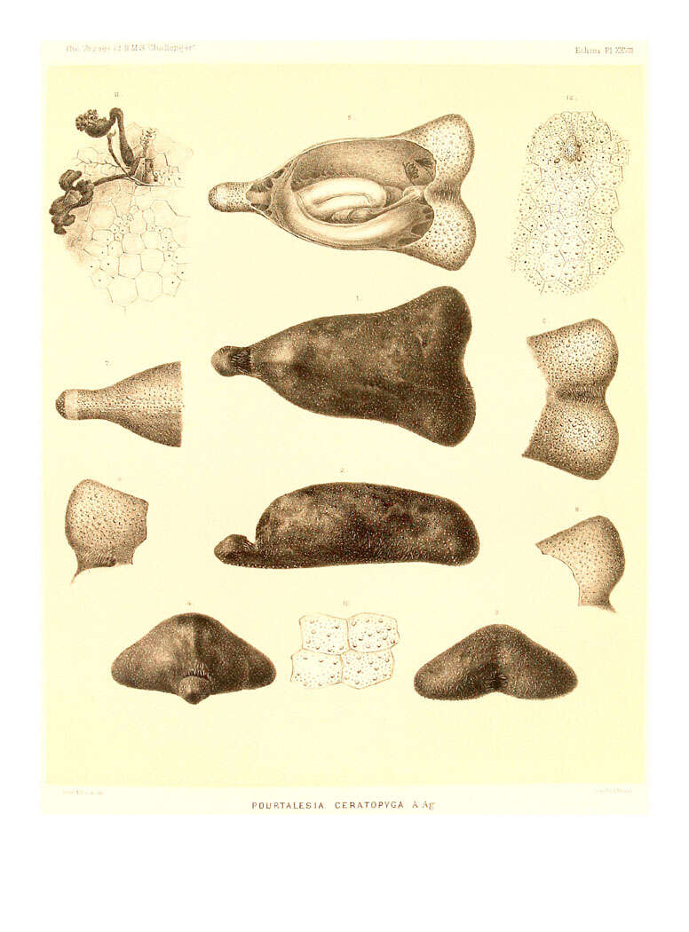
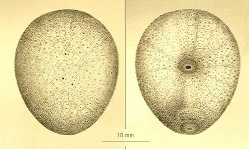
Urechinus naresianus / left: apical view, right: oral view, (after A. Agassiz, 1881).
-
Antrechinus drygalskii / left: apical view, right: oral view, (after Mortensen, 1909).
-
Antrechinus mortenseni (after Mooi & David, 1993).


