-
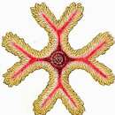
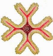
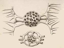
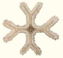
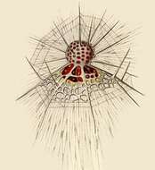
Dicranastrum furcatum.
-
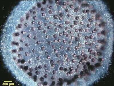
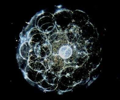
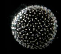
A colonial spumellarian radiolarian (Collosphaera sp.) composed of numerous central capsules (purple porous spheres) connected to one another by cytoplasmic strands and enclosed in a clear gelatinous sheath secreted by the radiolarian cytoplasm.
-
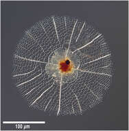
A light microscopic view of a living nassellarian radiolarian (Eucyrtidium acuminata) showing the reddish pigmented cytoplasm within the siliceous, conical shell.
-
-
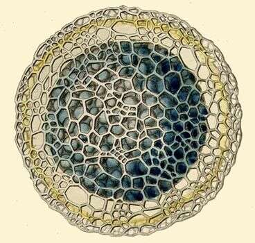
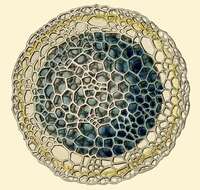
Section of cell with central capsule, associated black pigment, central nucleus, and calymma. Inset is an endoplasmic vacuole.
-
-
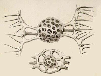
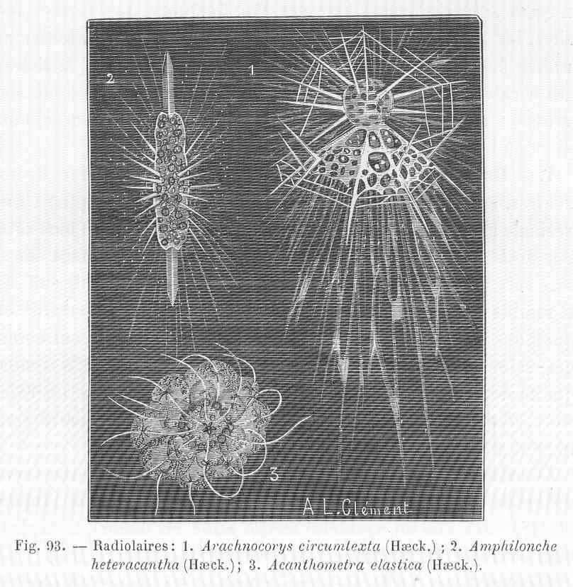
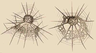
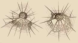
Radiolaires. 1, Arachnocorys circumtexta (Haeck.); 2, Amphilonche heteracantha (Haeck.)
-
Lipmanella dictyoceras (Haeckel) Kling, 1973. From the Bay of Villefranche on Jan 27, 2014. Z-stack of images made using a 10x objective and DIC optics.
-
Stylodictya multispina (probably) found in sample taken on March 10 2014 from the Bay of Villefranche. Z-stack of images made using a 20x objective and DIC optics.
-
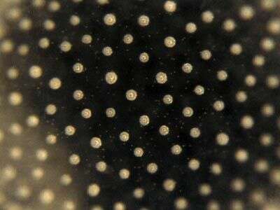
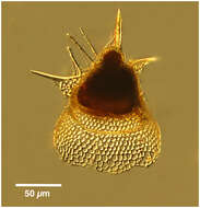
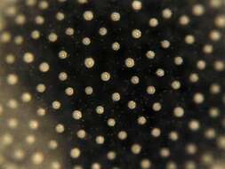
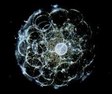
Radiolarian from the South Pacific Ocean
-
-
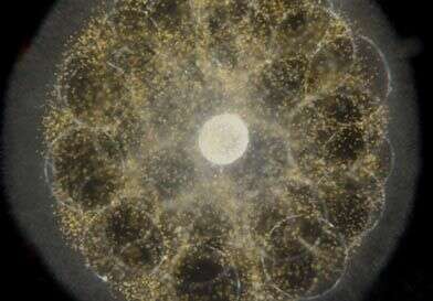
Sphaerozoum (sphere-owe-zoo-um), detail of the surface of a colony, in which many individual organisms can be seen. In the centre of each of the bright regions is the capsule. This is an example of one of the four types of large amoebae which is common in the marine water column. Dark ground image by Dave Caron.
-
Acrosphaera (ack-row-sphere-a) spinosa, spherical colonial radiolarian. This is an example of one of the four types of large amoebae which commonly occur in the marine water colum. Dark ground image by N. R. Swanberg.
-
-
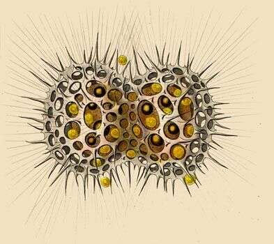
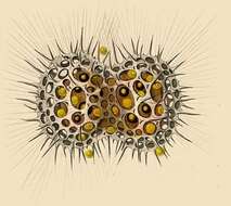
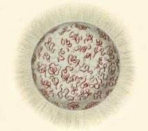
The central capsules are worm-like. With oil droplets, nuclei, small pigment spots and yellow symbiotic algae.
-
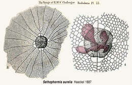
From the species description in the Challenger Reports
-
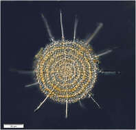
A living specimen of a spherical spumellarian radiolarian (Styptosphaera sp.) showing the spherical central capsule enclosed by a spongiose siliceous shell. Numerous cytoplasmic strands (axopodia) radiate outward from the central capsule and bear patches of golden-pigmented algal symbionts.
-
-
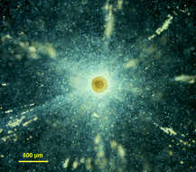
Thalassophysa (thal-ass-owe-fie-sa) is a large radiolarian protist has a cytoplasm that is full of bubbles. The bright area in the centre of the cell is the capsule and within this lie most of the cytoplasmic organelles. The yellow colour comes from symbiotic algae living in association with this protozoa. The algae are dinoflagellates from the genus Scrippsiella - the same genus is also found in symbiotic association with the by-the-wind sailor (Velella). The light region towards the outside is the region of the axopodia. This is an example of one of the four types of large amoebae which is common in the marine water column. Dark ground illumination, image by Dave Caron.
-
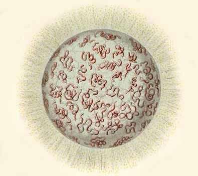
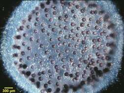
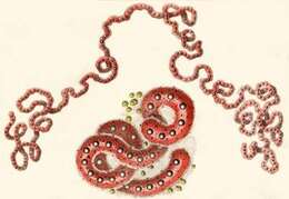
Living coenobium, with serpentine central capsules. Numerous yellow algal cells are scattered among the radial pseudopodia.
-
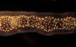
Collozoum - a colonial radiolarian, the bright spots being the central capsules of the organisms which make up the colony. Up to 5000 capsules may be present in a colony. These colonies are like long sausages up to several centimetres in length. There is a sheath of organic material, and this may be referred to as gelatin. The colony has a segmented appearance because the components of the colony lie in a matrix around large vacuoles or alveoli. This is an example of one of the four types of large amoebae which is common in the marine water column. Image by Dave Caron.
-
-
Thalassophysa (thal-ass-owe-fie-sa), a spumellarian radiolarian in which the cytoplasm is very clearly differentiated into the condensed inner region contained within the capsule, and the outer frothy later. Numerous symbiotic dinoflagellates (Scrippsiella - which also occurs in the cnidarian Velella) can be seen as orange spots in the cell. This is an example of one of the four types of large amoebae which is common in the marine water column. Dark ground image by N. R. Swanberg.
-
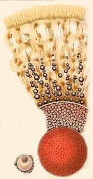
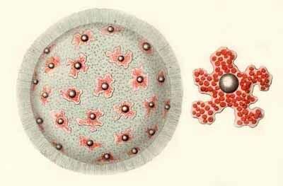
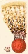
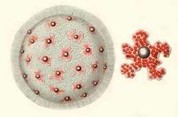
Large colonial coenobium or jelly colony, and a single isolated amoeboid central capsule with oil droplet.