-
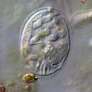
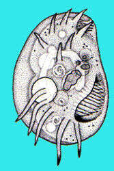
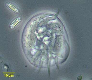
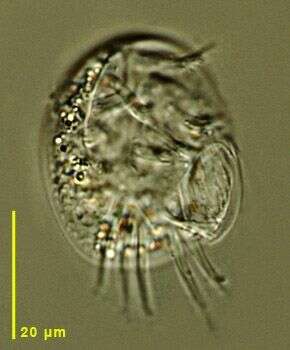
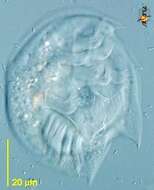
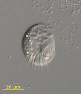
Portrait (ventral view) of the marine hypotrich ciliate, Aspidisca leptaspis (Fresenius, 1865). The cell outline is broadly oval and strongly dorsoventrally flattened. The pellicle is rigid and transparent. There are 2 left-anterolateral and several smaller posterior spines. The posterior spines vary from very small to quite prominent. In this individual they are not discernable. There are seven large and one small frontoventral (FV) cirri (including one buccal cirrus). There are five transverse cirri. The left-most transverse cirrus may be subdivided into several small bundles. There is a small left anterolateral âadoralâ zone of membranelles (AZM1), which is actually separate from the peristome. The more prominent adoral zone of membranelles (AZM2) borders the large obliquely situated left posterior peristome. There are longitudinal rows short dorsal cilia on subtle dorsal ridges. The macronucleus is horseshoe-shaped. Collected from a commercial saltwater aquarium in Boise, Idaho February 2003. DIC optics.
-
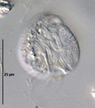
Aspidisca (as-pid-isk-a) is a hypotrich ciliate, and identifiable as a hypotrich because it uses clumps of cilia (cirri) on the ventral surface to walk over the substrate. Hypotrichs are part of the polyhymenophora, and usually feed using an extensive adoral zone of membranelles which extends from the front of the cell to a mouth in the posterior ventral part of the cell. However, in Aspidisca, the AZM has been greatly reduced and forms a kind of scrubbing brush on the ventral surface . Differential interference contrast.
-
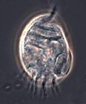
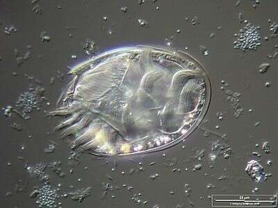
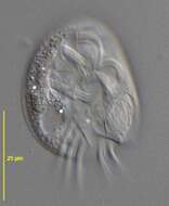
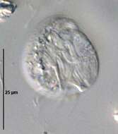
Ventral view of Aspidisca lynceus (MUELLER,1773) EHRENBERG, 1830.Collected from a freshwater irrigation canal in Boise,Idaho December 2007.DIC
-
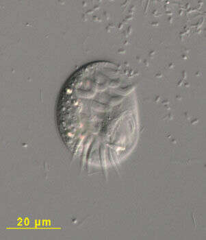
Aspidisca (as-pid-isk-a) is a hypotrich ciliate, and identifiable as a hypotrich because it uses clumps of cilia (cirri) on the ventral surface to walk over the substrate. Hypotrichs are part of the polyhymenophora, and usually feed using an extensive adoral zone of membranelles which extends from the front of the cell to a mouth in the posterior ventral part of the cell. However, in Aspidisca, the AZM has been greatly reduced and forms a kind of scrubbing brush on the ventral surface - and visible at 4 o clock on the right margin of the cell. Phase contrast
-
-
-
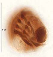
Lateral view of the hypotrich ciliate, Aspidisca turrita (Ehrenberg, 1831) Claparède & Lachmann, 1858. The thorn-like dorsal process is clearly seen here (to viewer's left). The first ventral cirrus is also visible projecting anteriorly. Collected from a freshwater pond near Boise, Idaho. June 2005.DIC
-
Aspidisca, small atypical hypotrich, seen here from ventral surface. With a few ventral cirri and adoral zone of membranelles, on the right, reduced to a small brush. Differential interference contrast.
-
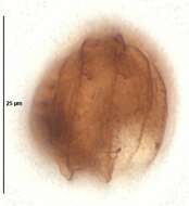
Ventral view of the hypotrich ciliate, Aspidisca turrita (Ehrenberg, 1831) Claparède & Lachmann, 1858. The cytostome with adoral zone of membranelles is senn at viewer's lower right.The ventral ciiri are seen anteriorly and the transverse cirri posteriorly.Collected from a freshwater pond near Boise, Idaho. June 2005.DIC
-
Aspidisca, a common, small hypotrich ciliate genus with many species. The cell body is rigid, colorless and dorsoventrally flattened sometimes with peripheral or dorsal spinous projections. The dorsum may be longitudinally ribbed (e.g. A. cicada). Marginal and caudal cirri are absent and ventral cirri prominent (frontoventral and transverse groups). The oral aperture is faintly visible on the organism's left posterior margin in this image (we are looking at it from the ventral side). The adoral zone of membranelles is divided into a small part at the anterior left side and a larger part around the peristome, neither is well seen in this image. Macronucleus is bipartite in some species but more usually "C" or horseshoe shaped. Usually small but one species, A. magna may exceed 150 microns in length. Probably polyphagous but mainly feeds on bacteria. From standing freshwater near Boise, Idaho. Phase contrast.
-
Originally described by Ehrenberg under the name Euplotes turritus.
-
Aspidisca is a small, atypical hypotrich ciliate with cilia clustered together to form little 'legs'. A small group of cilia, used for feeding, is near the back (lower right of the image) of the cell.
-
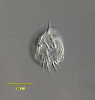
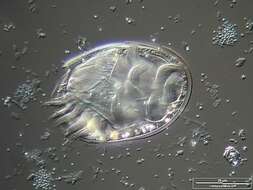
This image of Aspidisca was taken from an anaerobic marine sediment sample incubated under laboratory conditions.
-
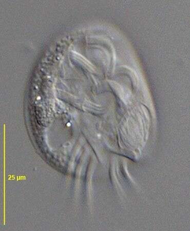
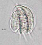
Differential Interference Contrast image showing the cirri and the mouthparts.
-
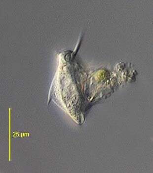
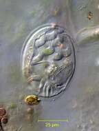
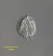
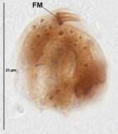
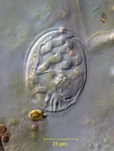
Scale bar indicates 25 µm. Collected from Bodden, the brackish waters lying between the isles of Hiddensee and Ruegen (German Baltic Sea). The image was built up using several photomicrographic frames with manual stacking technique. This image was taken using Zeiss Universal with Olympus C7070 CCD camera.
-
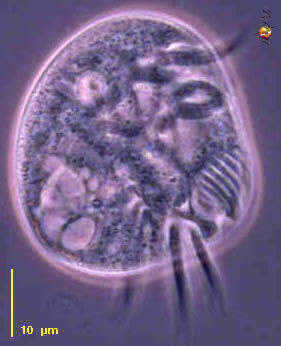
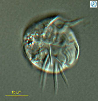
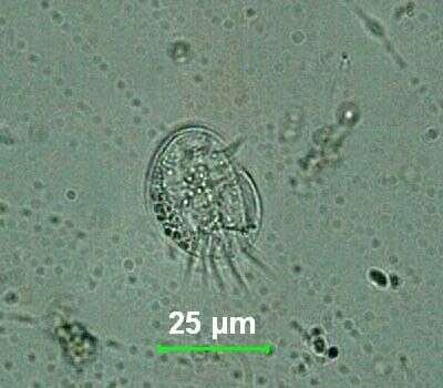
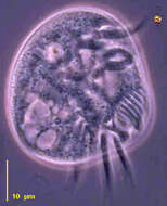
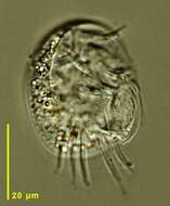
A hypotrich ciliate isolated after Uhlig extraction of sandy sediments from Little Sippiwissett salt marsh. Micrograph taken by Jeffrey Cole.
-
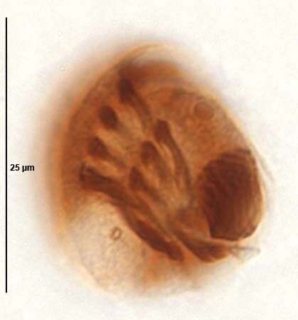
Ventral view of Aspidisca cicada (MUELLER,1786) CLAPARÃDE&LACHMANN,1858.DIC.
-
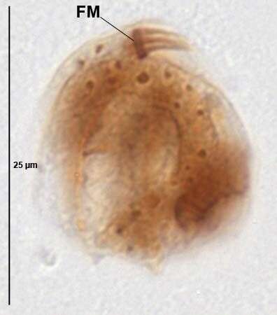
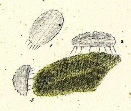
Ventral infraciliature of Aspidisca cicada (MUELLER,1786) CLAPARÃDE&LACHMANN,1858. Protargol protocol A. (see Foissner, W. Europ. J. Protistol., 27:313-330;1991).Brightfield.
-
Dorsum of Aspidisca cicada (MUELLER,1786) CLAPARÃDE&LACHMANN,1858. Protargol protocol A. (see Foissner, W. Europ. J. Protistol., 27:313-330;1991).Brightfield.
-
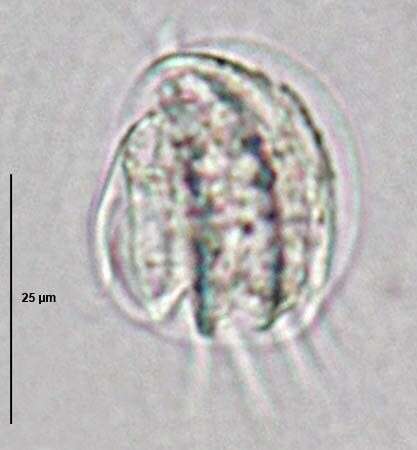
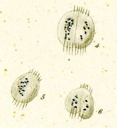
Frontal membranelle of Aspidisca cicada (MUELLER,1786) CLAPARÃDE&LACHMANN,1858. Protargol protocol A. (see Foissner, W. Europ. J. Protistol., 27:313-330;1991).Brightfield.
-
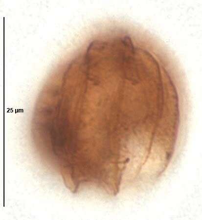
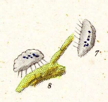
Dorsal view of Aspidisca cicada (MUELLER,1786) CLAPARÃDE&LACHMANN,1858.Brightfield,closed condenser.
-
Originally described by Ehrenberg under the name Oxytricha cicada.
-
Originally described by Ehrenberg under the name Oxytricha cicada.
-
Originally described by Ehrenberg under the name Oxytricha cicada.