Comprehensive Description
provided by Smithsonian Contributions to Zoology
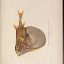
Triodon macropterus Lesson
MUSCLES OF THE CHEEK
ADDUCTOR MANDIBULAE (Figures 138, 139, 140: A 1, A 2α, A 2β, A 3).—A 1 is a single, undivided muscle which originates beneath the orbit as a flat, aponeurotic sheath attached primarily to the anterodorsal face of the hyomandibular. It inserts on the posteromedial face of the ventral half of the maxilla. A 2 is separated into two parts by the passage of ramus mandibularis V. A 2α originates exclusively from the anterodorsolateral face of the hyomandibular—a few fibers may attach to the ventral face of the aponeurosis of A 1 posteriorly. The extreme posterior fibers are covered laterally by the dilatator operculi. A 2β, which lies beneath A 2α, originates almost entirely from the dorsal and lateral faces of the preopercle. The posterodorsal fibers overlie the posteroventral region of A 2α. The two subdivisions insert together on the medial face of the dentary above the Meckelian fossa. A 3 is present at a thin sheet of fibers originating from the lateral metapterygoid and symplectic. It inserts medial to A 2 on the dentary by a somewhat sheetlike aponeurosis. It is partially confluent with A 2β posteriorly. A ω is well developed, and stretches along the medial surface of the quadrate and anterior metapterygoid. It inserts by a flat aponeurosis on the dentary, lying medial to the insertion of A 3.
LEVATOR ARCUS PALATINI (Figures 138, 140: L.A.P.).—From a straplike origin on the ventral sphenotic at the rear of the orbit, the muscle passes downward to insert on the posterodorsolateral face of the hyomandibular. This point lies medial to the dorsolateral flange of that bone, which has developed in association with the origin of A 2α. The fibers of the muscle fan out before their insertion, this being particularly marked anteriorly.
DILATATOR OPERCULI (Figure 138: D.O.).—Origin is from the ventrolateral sphenotic and adjoining pterotic. The muscle narrows to a tendon which inserts on the enlarged anterodorsal process of the opercle.
LEVATOR OPERCULI (Figure 138: L.O.).—This is a broad, well-developed muscle, originating in the groove between the pterotic and the large supracleithrum. It inserts broadly on the dorsomedial face of the opercle. A small superficial aponeurosis is present on the midlateral face of the muscle.
ADDUCTOR ARCUS PALATINI (Figure 140: A.A.P.).—The muscle is confined to the floor and rear of the orbit. Origin is from the lateral surface of the parasphenoid. Anteriorly it inserts on the dorsomedial face of the mesopterygoid. There is a gap where the fibers end in the tough connective tissue lining the buccal cavity. Posteriorly, the fibers insert on the posterodorsal faces of the metapterygoid and hyomandibular.
ADDUCTOR OPERCULI (Figure 142: AD.OP.).—The muscle originates from the prootic beneath the hyomandibular fossa ventral to, but dorsally continuous with, the origin of the adductor hyomandibulae. The fibers pass out laterally to insert on the dorsomedial face of the opercle, just posterior to its articulation with the hyomandibular.
ADDUCTOR HYOMANDIBULAE (Figure 142: A.H.).—The muscle originates from the ventrolateral prootic just above the adductor operculi, and inserts as a more or less flattened sheet on the posterodorsomedial face of the hyomandibular.
MUSCLES OF THE HYOID REGION
VALVULUS.—The muscle is present as a thin, flattened sheet of fibers, arising from the dorsolateral face of the protractor hyoidei at about the middle of its length and grading into the tissues of the buccal valve anterodorsally.
PROTRACTOR HYOIDEI (Figure 138: PR.HY.).—The fibers arise from the posteroventromedial dentary and insert on the posteroventral anterohyal and the adjoining posterohyal. It completely covers the first two branchiostegal rays, and is a long, cylindrical muscle.
HYOHYOIDEUS INFERIORIS.—The fibers of this muscle are partially fused with those of the protractor hyoidei. It arises from the ventral face of the anterohyal, sharing, to some degree, aponeuroses with the medial region of the protractor hyoidei. The fibers pass anteromedially and fuse with their antimeres. The muscle lies lateral to the origin of the infracarinalis anterior, and there is some intermingling of fibers with those of the protractor hyoidei.
HYOHYOIDEI ABDUCTORES (Figure 138: H.AB.).—The muscle is made up of three slips. That to the first ray originates from the medial face of the ventrohyal and inserts broadly over the ventromedial face of the ray. The slip to the second ray arises from the expanded dorsolateral surface of the first ray, while that to the third ray originates from the bases of the first and second rays.
HYOHYOIDEI ADDUCTORES (Figure 138: H.AD.).—This is a well-developed sheet of muscle fibers. It is firmly attached to the dorsomedial face of opercle dorsally, and fades out in the wall of the opercular chamber in the region of the dorsohyal anteroventrally.
STERNOHYOIDEUS.—The muscle is small, its origin being confined to the anterodorsal tip of the ventral part of the cleithrum, including a small fossa in that bone. Anteriorly, the fibers sweep up to insert on the ventral face of the ventrohyal.
VENTRAL BRANCHIAL MUSCLES
PHARYNGOCLAVICULARIS EXTERNUS (Figure 141: PHC.E.).—This is a straplike muscle originating from the lateral face of the cleithrum and inserting on the anteroventral face of ceratobranchial 5. The fibers pass posterodorsolaterally from their origin.
PHARYNGOCLAVICULARIS INTERNUS (Figures 141, 142: PHC.I.).—Orgin is from the anterodorsomedial face of the cleithrum, the fibers passing anteriorly to insert on the posterior face of ceratobranchial 5. It is a flattened muscle.
OBLIQUI VENTRALES II, III (Figure 141: OBL.V.).—The anterior muscle connects the ventral faces of the ceratobranchial and hypobranchial of the second arch. Obliquus III joins ceratobranchial 3 to the ventral process of hypobranchial 3 and the arch-shaped ligament.
TRANSVERSI VENTRALES IV, V (Figure 141: TR.v.).—The anterior muscle connects the anteromedial faces of the fourth ceratobranchials across the ventral midline. Transversus V is well developed, and joins the anteromedial faces of the fifth ceratobranchials.
RECTI VENTRALES I, IV (Figure 141: RECT.V.).—Rectus I is a fairly long, straplike muscle which connects the dorsal face of the dorsohyal and the ventrolateral face of the first ceratobranchial and hypobranchial. Rectus IV arises from the anteroventral face of ceratobranchial 4 and passes anteromedially to attach to the posterior surface of the arch-shaped ligament between the ventral processes of the third hypobranchials.
RECTUS COMMUNIS (Figure 141: R.COMM.).—The muscle originates anteriorly from the posterior face of the urohyal. The fibers pass posteriorly, grading into an aponeurosis at about the level of the third basibranchial which inserts on the ventrolateral face of ceratobranchial 5.
DORSAL BRANCHIAL MUSCLES
LEVATORES EXTERNI I–IV (Figure 142: L.EXT.).—The four muscles originate from the prootic just anteroventral to the hyomandibular fossa. They insert on the dorsal faces of epibranchials 1–4. The first two muscles are not very well developed, the fourth levator being the largest.
LEVATORES INTERNI II, III (Figure 142: L.INT.).—The muscles originate from the prootic beneath the hyomandibular fossa, levator II having a more medial origin than levator III. The former inserts partially on the medial face of infrapharyngobranchial 2 but mainly on the anteromedial face of infrapharyngobranchial 3. Levator III inserts on the dorsal face of infrapharyngobranchial 3.
OBLIQUUS DORSALIS III (Figure 142: OBL.D.).—Two heads of this muscle arise from the anterolateral face of epibranchial 3. The fibers coalesce and pass medially to attach to the posterodorsal face of infrapharyngobranchial 3.
OBLIQUUS POSTERIOR (Figure 142: OBL.P.).—Fibers pass from the dorsomedial face of ceratobranchial 5 to the posterior face of epibranchial 4.
TRANSVERSI DORSALES II, III (Figure 142: TR.D.).—The anterior muscle is well developed and partially subdivided. The dorsal layer arises from infrapharyngobranchial 2 and the surrounding connective tissue. The fibers pass posteriorly and then curve toward the midline just behind the basal process of the parasphenoid, and join their antimeres. Beneath this, the ventral layer passes outward from the midline to attach to the anterolateral face of epibranchial 4, the two layers separating completely. Transversus III arises from the dorsal midline and passes laterally to insert on the dorsal face of the medial process of epibranchial 3.
RETRACTOR DORSALIS (Figure 142: D.RETR.).—The muscle originates from the lateral face of the first vertebra and passes anteriorly to insert on the ventromedial faces of infrapharyngobranchials 3 and 4.
ADDUCTORES IV, V (Figure 142: AD.).—Adductor IV is well developed, and connects the medial faces of epibranchial 4 and ceratobranchial 4. Adductoi V is less well developed, and passes from the anterodorsal tip of ceratobranchial 5 to the posterolateral face of epibranchial 4.
SPHINCTER OESOPHAGI (Figures 141, 142: S.O.).—The anteroventral fibers of this muscle attach to the ventromedial face of ceratobranchial 5. There are two fairly distinct layers dorsally. The ventral one arises from infrapharyngobranchial 2, the fibers passing posteriorly and then medially before fading out in the connective tissue of the esophagus. This is covered posterodorsally by the main mass of the muscle, which does not extend anteriorly beyond the posteromedial face of epibranchial 4.
MUSCLES OF THE PECTORAL REGION
ABDUCTOR SUPERFICIALIS (Figure 138: ABD.S.).—The muscle arises from the posteroventral surface of the elongated cleithrum and inserts on the anterolateral bases of the principal fin rays.
ABDUCTOR PROFUNDUS (Figure 138: ABD.P.).—Origin is from the lateral face of the cleithrum. The fibers insert on the posterolateral bases of the principal rays and on that of the lateral half of the vestigial fin ray.
ARRECTOR VENTRALIS (Figure 138: ARR.V.).—The muscle lies above the abductor superficialis and originates from the lateral face of the cleithrum beneath the flange. It inserts on the anterior face of the medial half of the vestigial fin ray. There may be a few fibers attaching to the lateral half of the ray posteriorly.
ADDUCTOR SUPERFICIALIS (Figure 143: ADD.S.).—The fibers originate from the anteromedial face of the cleithrum and insert on the dorsomedial faces of the principal rays distal to their bases. The more ventrolateral fibers serve the more dorsal rays.
ADDUCTOR PROFUNDUS (Figure 143: ADD.P.).—The muscle is well developed, and originates from the medial face of the cleithrum and coracoid. It inserts on the ventromedial bases of the principal fin rays, the tendons to the ventral two rays being covered medially by the main mass of the muscle.
ARRECTOR DORSALIS (Figure 143: ARR.D.).—This is a fairly small muscle, originating from the medial face of the cleithrum and inserting on the base of the medial half of the vestigial fin ray. It is partially covered medially by the adductor profundus.
CORACORADIALIS (Figure 143: COR.R.).—A somewhat flattened muscle, arising from the dorsal face of the posterior process of the coracoid and inserting on the ventromedial face of the fourth radial.
PROTRACTOR PECTORALIS (Figures 138, 142: P.P.).—The muscle originates from the ventrolateral tip of the pterotic and fans out posteroventrally to attach to the dorsal region of the cleithrum.
LEVATOR PECTORALIS (Figure 138: TR.).—Origin is from the posterior face of the pterotic. The fibers pass mainly posteriorly, but also somewhat laterally, to insert on the extreme posterodorsal tip of the cleithrum. The muscle is almost entirely covered by the supracleithrum, and is closely associated with the fibers of the epaxialis dorsally.
MUSCLES OF THE DORSAL FIN
INCLINATORES DORSALES (Figure 144: INC.).—Each fin ray possesses an inclinator which arises in the fascia between the skin and the epaxialis and inserts on the lateral base of the fin ray a little dorsal to the insertions of the erector and depressor muscles. The inclinators decrease in size posteriorly.
ERECTORES DORSALES (Figure 144: EREC.).—There are two small, anterior erectors, which originate from the lateral faces of the first two pterygiophores and insert on the bases of the spines of the spiny dorsal fin. Each fin ray has an erector inserting on its anterolateral base. These muscles arise from the lateral faces of the pterygiophores and the neural spines and centra of the eighth to fourteenth vertebrae.
DEPRESSORES DORSALES (Figure 144: DEPR.).—The first dorsal spine only has a depressor muscle, which originates from the lateral base of the first pterygiophore and inserts on the posteroventral base of the spine. Each ray in the soft dorsal fin has a depressor inserting on its posterolateral base. Origin is as for the erectors, the depressor to each ray lying just behind the erector.
SUPRACARINALIS ANTERIOR.—This is a well-developed muscle arising from the posterior and lateral faces of the supraoccipital. The fibers pass posteriorly to attach to the anterior face of the first pterygiophore. There is some intermingling of fibers from the left and right sides in the midline.
SUPRACARINALIS POSTERIOR (Figures 144, 145: s. POST.).—The muscle stretches from the posterior face of the last pterygiophore of the dorsal fin and the neural spine of the fourteenth vertebra to the anterodorsal regions of the neural spines of the third and fourth last vertebrae.
MUSCLES OF THE ANAL FIN
INCLINATORES ANALES (Figure 144: INC.).—An inclinator arises from the fascia over the obliquus inferioris and inserts on the anterolateral base of each fin ray. The medial fibers of the more anterior inclinators attach to the myocommata of the obliquus superioris, making separation of the muscle difficult. In addition, there is a large, residual inclinator inserting on the anteroventrolateral base of the first pterygiophore. Its ventromedial region is more or less continuous with the ventral fibers of the obliquus inferioris.
ERECTORES ANALES (Figure 144: EREC.).—These muscles, which insert on the anterolateral bases of the anal fin rays, originate almost entirely from the lateral faces of the pterygiophores, with only a few fibers from the tips of the haemal spines of the associated vertebrae.
DEPRESSORES ANALES (Figure 144: DEPR.).—Origin is as for the erectors, the depressor of each fin ray inserting on the posterolateral base of the ray.
INFRACARINALIS ANTERIOR (Figure 138: INF.A.).—This is a well-developed muscle which originates from the anteroventral tip of the cleithrum and inserts in the anteroventral groove in the pelvis. Evidence of its originally paired nature is visible externally, but the deeper fibers of either side have fused in the midline.
INFRACARINALIS MEDIUS (Figure 138: INF.M.).—A fairly well-developed section, which attaches to the ventrolateral face of the pelvis just anterior to the middle of its length. It grades into the obliquus inferioris at about the level of the postcleithrum, the more posterolateral fibers intermingling somewhat with those of the residual inclinator analis.
INFRACARINALIS POSTERIOR (Figures 144, 145: INF.P.).—The muscle arises from the posterolateral faces of the last anal pterygiophore and the haemal spine of the fifteenth vertebra and attaches to the anteroventral face of the haemal spines of the third and fourth last vertebrae.
MUSCLES OF THE CAUDAL FIN
INTERRADIALIS (Figure 145a: INT.).—The muscle is developed between the middle twelve caudal fin rays. The fibers span a few rays between their sites of attachment. Those of the dorsal rays pass anteroventrally, while those of the ventral rays pass anterodorsally.
HYPOCHORDAL LONGITUDINALIS (Figure 145b: H.L.).—The muscle is well developed, and originates from the dorsolateral face of the ural centrum, and the lateral faces of the hypurals 1 and 2, parhypural, and hypurapophysis. The muscle inserts on the bases of rays D 3–5.
FLEXOR DORSALIS (Figure 145c: F.D.).—Origin is restricted to the lateral faces of the epural, the first uroneural and the dorsal crest of the ural centrum The tendons insert on the bases of rays D 2–6.
FLEXOR DORSALIS SUPERIOR (Figures 145b,c: F.D.s.).—The muscle originates from the lateral faces of the epural and the neural spines of the second and third last vertebrae. Insertion is on the lateral base of ray D 7.
FLEXOR VENTRALIS (Figure 145c: F.V.).—The muscle is fairly well developed, and lies beneath the flexor ventralis externus. Origin is from the lateral faces of the centra and haemal spines of the last three vertebrae (including the ural centrum), the parahypural, and the hypurapophysis. The muscle inserts on the bases of rays V 1–7.
FLEXOR VENTRALIS EXTERNUS (Figure 145c: F.V.E.).—The fibers arise from the lateral faces of the centra and haemal spines of the third and fourth last vertebrae and pass posteriorly to insert on the bases of rays V 1–2.
FLEXOR VENTRALIS INFERIOR (Figure 145b,c: F.V.I.).—Origin is primarily from the lateral face of the haemal spine of the penultimate vertebra, the muscle inserting on the lateral base of V 7.
TRANSVERSUS CAUDALIS (Figure 145c: TR.C.).—The muscle is poorly developed. It originates from the lateral face of the fourth hypural and inserts on the dorsal base of ray V 1.
LATERAL BODY MUSCLES
EPAXIALIS (Figures 138, 145a,b: EPAX.).—The subdivision of this muscle into dorsal and ventral moieties is incomplete, particularly medially. Anteriorly, the dorsal section attaches to the posterodorsal faces of the frontal, epiotic, pterotic, supraoccipital, and exoccipital. The myocommata become increasingly oblique posteriorly until they are almost horizontal (behind the dorsal fin). Anteromedially, some of the fibers arise from the neural spines and centra of the vertebrae and the pterygiophores. Posteriorly, the fibers grade into a flattened tendon which joins a similar tendon from the obliquus inferioris. Together, these two tendons form an aponeurotic sheath inserting on the bases of all the procurrent and principal caudal fin rays. The ventral section of the epaxialis is virtually continuous with the obliquus superioris. The fibers arise anteriorly from the posterior faces of the pterotic and exoccipital. In the posterior region the muscle inserts on the lateral bases of rays D 1–6, the more ventral tendons passing medial to the hypochordal longitudinalis.
OBLIQUUS SUPERIORIS (Figures 138, 145a,b: OBL s.).—This section arises anteriorly as a small slip of fibers from the posterodorsal tip of the cleithrum. It fans out posteroventrally, attaching to and between the pleural ribs, with the ventral fibers intermingling with the obliquus inferioris. Behind the anal fin, these two sections are relatively well separated. It inserts tendinously on the bases of rays V 1–6. A large bundle, which appears to be mostly continuous with the obliquus superioris, but which also receives fibers from the epaxialis, passes somewhat dorsally in the peduncular region to insert on the lateral crest of the ural centrum. It is slightly overlain posteriorly by the hypochordal longitudinalis. It is somewhat reminiscent of a proximalis muscle, but is not separated from the body muscles.
OBLIQUUS INFERIORIS (Figures 138, 144, 145a: OBL.I.).—The muscle has a number of subsections. The anteroventral section arises from the posterior and posteroventral faces of the coracoid and cleithrum, and attaches to the anteroventral face of the postcleithra. Two vague myocommata are present, and occasional fibers are to be found in the connective tissue between the ventral border of this section and the infracarinalis medius below it. A thin sheet of fibers passes obliquely upward from the posterodorsal faces of the postcleithra and fades into the fascia overlying the obliquus superioris. Posteroventrally, the fibers become more horizontally oriented, and intermingle with those of the obliquus superioris, infracarinalis medius, and residual inclinator analis. Myocommata, which are continuous with those of the obliquus superioris, are present in the fibers behind the postcleithra. From the ventromedial surface of the posterior myocommata of the eleventh myomere, and closely associated with the medial faces of the infracarinalis medius and the rest of the obliquus inferioris, there arises a short bundle of fibers which passes posterodorsomedially to insert tendinously on the anteroventral face of the haemal spine of the fourteenth vertebra. The fibers pass medial to the obliquus superioris but lateral to the erectores and depressores anales.
Myological Descriptions of Representative Tetraodontids
The members of this family (see Figure 8 for general outline) as recognized here contain ten genera (representatives of six genera were dissected for this study), which were divided into four families by Fraser-Brunner (1943). Although his families may well represent distinct phyletic lines, the status accorded them by Fraser-Brunner has not been followed by other workers (e.g., Tyler, 1970a).
The skin is smooth or covered with small prickles, and the fish are capable of considerable inflation when threatened. Some forms have an almost lunate caudal fin (e.g., Lagocephalus), but a rounded caudal is more common. Dorsal and anal fins are usually small, and normally beat synchronously from side to side. The pectoral fins are large, and often used in locomotion. The powerful beaklike jaws are well adapted for crushing the mollusks and brachyurans which form a major part of the puffer-fish diet. They are generally tropical to subtropical in distribution (with a few fluviatile forms), and are commonly found over sea-grass meadows, sandy bottoms, and the adjoining coral reefs.
The family is considered to be closely related to the Canthigasteridae (which latter has often been included in the Tetraodontidae), and also close to the Diodontidae.
v
Lagocephalus lunaris (Bloch and Schneider)
MUSCLES OF THE CHEEK
ABDUCTOR MANDIBULAE (Figures 139b, 146, 147, 148, 149: A 1α, A 1β, A 2α, A 2β, A 3).—A 1 consists of two well-developed sections. A 1β has a complicated fiber structure. Fibers originating from the lateral faces of the palatine, vomer, and ethmoid pass ventrolaterally. They form a bipinnate structure with the fibers arising from the anterior face of the prefrontal (which pass ventromedially). Fibers from the ventrolateral tip of the prefrontal course anteroventrally, the fibers from the ventral face of that bone at first pass ventrally, and then (as they become more posterior in origin) anteroventrally. The ventral surface is largely aponeurotic. Beneath this, another bundle of fibers is present, arising from the lateral parasphenoid. It is well separated from the above-mentioned aponeurosis posteriorly, but grades into it anteriorly. The muscle inserts on the dorsomedial faces of the premaxilla and maxilla. A 1 α is smaller. It originates from an aponeurosis on the floor of the orbit (attaching posteriorly to the hyomandibular medial to A 2 α) and from a small head on the posterolateral preopercle. Insertion is on the posteromedial face of the maxilla. A 2 α is very well developed and is expanded ventrally to originate from two heads. The anteroventral one arises from the quadrate, symplectic, lateral preopercle, and the anteroventral tip of the hyomandibular. The other head arises from the dorsolateral preopercle and posterolateral hyomandibular. There is a considerable variety of fiber direction. Anteriorly, a tough aponeurosis joins A 2β. The section lies lateral to ramus mandibularis V apart from a small fiber bundle from the dorsolateral quadrate. A 2β, which lies medial to the above nerve, has shifted dorsally in origin, and lies above A 3. It originates from the lateral parasphenoid and prootic. The fibers pass laterally and anteroventrally to grade into a tendinous aponeurosis which joins that of A 2 α. The two sections insert on the medial face of the dentary. A 3 is a distinct muscle arising from the lateral faces of the metapterygoid, quadrate, and ectopterygoid and inserting by a flat, tendinous aponeurosis on the dorsal surface of Meckel’s cartilage. A ω is a small muscle (Figures 147, 149). It arises from the anteromedial edge of the ectopterygoid and inserts on the ventral rim of the Meckelian fossa. It lies medial to the tendon of A 3.
LEVATOR ARCUS PALATINI (Figures 146, 147: L.A.P.).—The fibers of this muscle are continuous dorsolaterally with the dilatator operculi. Origin is from the ventrolateral tip of the frontal, and the fibers fan out to insert in the fossa on the posterodorsal face of the hyomandibular, just anterior to the articulation with the skull.
DILATATOR OPERCULI (Figure 146: D.O.).—This muscle is very well developed. The anterior fibers are almost parallel, but the muscle becomes bipinnate posteriorly. Origin is from the lateral faces of the frontal and sphenotic, and the dorsolateral hyomandibular about its articulation with the skull. Insertion is on the dorsolateral opercle, mainly around the strong, dorsoventrally oriented crista, but also on the anterolateral surface of the bone. It is overlain dorsally by an aponeurotic sheath from the epaxialis. Anterolaterally, a strap of muscle fibers attaches to the posteroventral face of the preopercle.
LEVATOR OPERCULI (Figure 146: L.O.).—This is a flattened muscle, which arises from the anteroventrolateral pterotic and the ventrolateral face of the posterior process of the frontal. It inserts on the posterodorsal face of the opercle and its crista. Some of the ventral fibers are closely associated with the dorsal fibers of the protractor pectoralis.
ADDUCTOR ARCUS PALATINI (Figures 147, 148: A.A.P.).—The muscle is extensively developed, particularly anteriorly. It originates from the anteroventral prootic, lateral parasphenoid, and posteroventrolateral vomer. The fibers beneath the vomer stretch across the midline. Insertion is on the medial face of the palatine, and the dorsomedial faces of the ectopterygoid, metapterygoid, and hyomandibular.
ADDUCTOR OPERCULI (Figures 147, 150: AD.OP.).—The muscle originates from the anteroventral face of the pterotic and passes outward as a thin sheet to insert on the dorsomedial face of the opercle just medial to the insertion of the levator operculi.
MUSCLES OF THE HYOID REGION
VALVULUS (Figure 149: VA.).—The muscle, which is not fully separated from the protractor hyoidei posteriorly, arises from the anteroventral surface of the interopercle. It passes anteriorly, and then curves upward to face out in the tissues of the mandibular buccal valve.
PROTRACTOR HYOIDEI (Figure 146: PR.HY.).—Origin is from the ventral margins of the dentary, angular, anterior preopercle, and interopercle. The fibers insert on the lateral faces of branchiostegal rays 2–4, and the anterohyal between these rays. The more posterior fibers tend to course more toward the midline from their sites of insertion.
HYOHYOIDEI ABDUCTORES (Figures 146, 150: H.AB.).—A bundle of fibers passes posteroventrally from the base of the second ray to the medial faces of the third and fourth rays. The section serving the second ray arises from the posterior face of the lateral process of the enlarged first ray. The muscle to the first ray is massively developed. The ray forms a flat, horizontal plate extending posteriorly from the anterohyal, which lies lateral to it. The muscle has a large number of fiber directions. The ventral surface of the ray gives rise to a large bundle of fibers that pass anteriorly and a little dorsally to originate from the ventral and lateral anterohyal. The more lateral fibers, including those from the lateral process of the ray, tend to sweep anterodorsally, and then medially to the midline, where they join their antimeres. A large volume of fibers inserts on the dorsomedial face of the ray. They originate from the fascia overlying the sternohyoideus, the lateral face of the first basibranchial and the medial anterohyal. Most of these fibers, however, arise from the midline, where they join with their antimeres. Although undoubtedly functioning as two muscles, there is no physical hiatus, and the fibers from the two bundles intermingle to a considerable extent. The posterodorsomedial edge of the ray gives rise to a small bridge of fibers across the midline, which lies beneath the ventral branchial musculature at about the level of the third basibranchial.
HYOHYOIDEI ADDUCTORES (Figures 146, 147, 150: H.AD.).—The muscle is somewhat subdivided. The main mass passes dorsoventrally in the lateral wall of the opercular cavity, with a dorsal bundle attaching to the dorsomedial face of the hyomandibular. Ventrally, the fibers (to which the medial faces of branchiostegal rays 2–6 attach) pass anteromedially to meet another sheet of fibers from the medial fioor of the opercular cavity, the latter passing anterolaterally. Posterolaterally, a bundle arises from the posteroventral face of the supracleithrum and a myocomma with the anterior part of the obliquus inferioris. These fibers pass anteroventrally and then posteroventrally in a semicircle to attach ventrally to the dorsolateral tip of the coracoid. From the anteroventral margin of this semicircle, a bundle passes anteriorly to attach along the ventrolateral surface of the cleithrum. Anterodorsally, fibers from this arch are continuous with the rest of the muscle. Posterodorsomedially, a bundle passes upward to grade into the posterior fibers of the levator externus IV.
STERNOHYOIDEUS (Figures 146, 150: STH.).—The muscle is somewhat, conical, and is poorly developed. It originates from the anteroventral tip of the cleithrum and inserts on the dorsomedial face of the anterior region of the anterohyal.
VENTRAL BRANCHIAL MUSCLES
PHARYNGOCLAVICULARIS EXTERNUS (Figures 150, 151: PHC.E.).—Origin is from the ventromedial face of the cleithrum, the fibers passing dorsally to insert on the anteroventral face of ceratobranchial 5.
PHARYNGOCLAVICULARIS INTERNUS (Figures 150, 151: PHC.I.).—The muscle arises from the medial face of the cleithrum, and consists of two sections. The anterior section passes anterodorsally, medial to the pharyngoclavicularis externus, to insert on the posteroventral face of basibranchial 3. The more medial fibers of this section join their antimeres in a midventral raphe, while some of the more dorsal ones arise from ceratobranchial 4 and pass to basibranchial 3. The more posterior section arises from the anteroventromedial cleithrum and inserts on the ventral face of ceratobranchial 5.
OBLIQUUS VENTRALIS III (Figure 151: OBL.V.).—The muscle originates from the anteroventral region of ceratobranchial 3 and the posteroventral tip of hypobranchial 3. The fibers pass anteromedially to join with the fibers of rectus IV in an insertion on the anteroventral process of hypobranchial 3.
TRANSVERSI VENTRALES IV, V (Figure 151: TR.V.).—These muscles connect the anteromedial faces of the ceratobranchials of the fourth and fifth arches across the ventral midline. Both muscles are small.
RECTI VENTRALES I–IV (Figures 150, 151: RECT.v.).—Rectus I is well developed, passing from the dorsolateral face of the anterohyal to the anterolateral surface of hypobranchial I. The lateral surface of the muscle gives rise to a bundle of fibers passing posterodorsally, which grade into a tendon attaching to the ventromedial face of the hyomandibular just lateral to its articulation with the skull. Recti II and III connect the hypobranchials of the first and second and second and third arches respectively. Rectus IV passes from the anteroventral face of ceratobranchial 4 to the posterior face of the ventral process of hypobranchial 3. The anterodorsolateral fibers are continuous with the anteromedial fibers of obliquus ventralis III.
RECTUS COMMUNIS (Figure 151: R.COMM.).—The muscle arises from the posterior face of the ventrohyal. It passes posteriorly as a tendon, and develops fibers at the level of hypobranchial 1, where it splits into left and right halves. The fibers then pass posteriorly, and grade into tendon at the level of ceratobranchial 4. The tendon inserts on the ventrolateral face of ceratobranchial 5.
DORSAL BRANCHIAL MUSCLES
LEVATORES EXTERNI I–IV (Figure 150: L.EXT.).—These muscles originate from the ventral faces of the prootic and pterotic beneath the hyomandibular fossa. Levator I inserts on the dorsolateral face of epibranchial 1, with a few posterior fibers passing to the anteroventral face of epibranchial 2. Levator II inserts on the dorsolateral faces of epibranchials 2 and 3, and apparently represents fusion with the third levator. Levator IV is well developed. It inserts along the dorsal length of epibranchial 4, and continues in the wall of the opercular cavity behind the bone, reaching the dorsomedial face of the cleithrum.
LEVATORES INTERNI II, III (Figure 152: L.INT.).—These muscles have altered somewhat from the usual condition. Levator II originates from the posteroventromedial face of the pterotic. It passes anteroventrally to insert partly on the dorsomedial face of infrapharyngobranchial 3, and partly on the dorsolateral face of infrapharyngobranchial 2. The medial face of the muscle shares a strong aponeurosis with the dorsolateral fibers of transversus dorsalis III. Behind this, the muscle passes medial to levator III to its origin. Levator III originates from the ventrolateral sphenotic beneath the hyomandibular fossa. It passes ventrally to insert on the anterodorsolateral face of infrapharyngobranchials 2 and 3.
OBLIQUUS DORSALIS III (Figure 152: OBL.D.).—A fairly well-developed muscle, which passes anteromedially from the dorsomedial faces of epibranchials 3 and 4 to the posterodorsal face of infrapharyngobranchial 2.
OBLIQUUS POSTERIOR (Figure 150: OBL.P.).—The muscle appears to be present, but is not clearly separated from the sphincter oesophagi.
TRANSVERSI DORSALES II, III (Figure 152: TR.D.).—In transversus II, the fibers arise from the ventrolateral face of the parasphenoid. The posterior fibers almost intermingle with the anterior fibers of the lateral bundle of the sphincter oesophagi. The main mass of the fibers passes laterally and then curves anteriorly. Dorsolaterally, a strong aponeurosis attaches to the medial face of levator internus II. The ventrolateral fibers attach to the dorsal faces of epibranchials 1 and 2 and infrapharyngobranchial 1. The more anterior fibers pass medially to a middorsal raphe, and are not attached to the skull. Posteriorly, the muscle gives rise to a bundle that passes laterally into the posterodorsal wall of the opercular cavity. Transversus III is a well-developed muscle, which passes across the dorsal midline between the third and fourth epibranchials of either side. It lies ventral to transversus II, which covers it both anteriorly and posteriorly.
RETRACTOR DORSALIS (Figure 152: D.RETR.).—Origin is from the lateral and ventrolateral faces of vertebrae 1 and 2. The fibers course anteroventrally, fuse briefly with their antimeres in the midline, and insert on the posteromedial faces of infrapharyngobranchials 2 and 3. It is overlain dorsally by a small bundle of the sphincter oesophagi.
ADDUCTORES I–V (Figures 150, 151, 152: AD.).—Adductors I–III all connect the anteromedial faces of ceratobranchials 1–3 to the anterolateral faces of epibranchials 1–3. Adductor IV connects the medial faces of ceratobranchial 4 and epibranchial 4. Adductor V passes from the lateral region of ceratobranchial 5 to the posterior face of epibranchial 4.
SPHINCTER OESOPHAGI (Figures 150, 151, 152: S.O.).—The muscle has a very complicated form. Ventrally, two longitudinal bundles arise from the posterior face of basibranchial 3. They fuse at the level of ceratobranchial 5, and form a longitudinal sheath on the floor of the esophagus. Beneath this, a thick, almost transverse bundle connects the medial faces of the fifth ceratobranchials. Ventral to the latter bundle is a thin sheet of fibers, also between the fifth ceratobranchials posteriorly, but becoming longitudinally oriented and fading out above transversus ventralis V. Dorsally, three bundles pass anteriorly beneath the infrapharyngobranchials, attaching to the ventromedial faces of infrapharyngobranchials 1–3. They fade out posteriorly in the tissue of the esophagus. The muscle passes across the midline below the retractor dorsalis posteriorly, except for a small bundle of fibers lying above and in front of the retractor.
MUSCLES OF THE PECTORAL REGION
ABDUCTOR SUPERFICIALIS (Figures 146, 150: ABD.S.).—The muscle originates from the posterior, medial, and lateral faces of the cleithrum. It passes posteriorly to insert on the lateral faces of the anterodorsal processes of the bases of the principal fin rays.
ABDUCTOR PROFUNDUS (Figure 150: ABD.P.).—The muscle originares from the posteroventral cleithrum and lateral faces of the coracoid and actinosts. It inserts on the ventrolateral bases of the principal rays and the lateral base of the lateral half of the vestigial ray.
ARRECTOR VENTRALIS (Figure 146: ARR.V.).—Origin is from the ventrolateral face of the dorsal region of the cleithrum. The muscle inserts on the anterolateral base ot the medial half of the vestigial fin ray.
ADDUCTOR SUPERFICIALIS (Figure 153: ADD.S.).—The muscle originates from the dorsomedial cleithrum. The fibers to the dorsalmost rays pass posteriorly, the slip to each succeeding ray passing increasingly ventromedially. Thus, the slip to the ventralmost ray passes ventrally, and lies medial to all the others. The ventral six rays have dorsal processes, to which the tendons attach, the more dorsal tendons inserting on the dorsomedial faces of the rays distal to their bases.
ADDUCTOR PROFUNDUS (Figure 153: ADD.P.).—The muscle originates from the medial faces of the coracoid and the radials, and inserts on the posteromedial flanges of all but the six ventral rays, where insertion is on the ventromedial shaft of the ray.
ARRECTOR DORSALIS (Figure 153: ARR.D.).—The muscle is small, arising from the medial face of the scapula, and inserting on the medial face of the medial half of the vestigial fin ray.
CORACORADIALIS (Figure 153: COR.R.).—The muscle is poorly developed. It passes from the dorsomedial face of the posterior coracoid process to the posteroventral face of the fourth radial.
PROTRACTOR PECTORALIS (Figure 146: P.P.).—From its origin on the ventrolateral pterotic, the muscle passes ventrally to the anteromedial face of the supracleithrum and the posterodorsal cleithrum.
LEVATOR PECTORALIS (Figure 146: TR.).—The fibers pass from the posteroventral face of the pterotic to the extreme dorsolateral tip of the cleithrum medial to the supracleithrum.
MUSCLES OF THE DORSAL FIN
INCLINATORES DORSALES (Figure 154: INC.).—The muscles are represented by a flat sheet of transverse fibers. They have lost their association with the fin rays, and now form the dorsal part of the transverse cutaneous system.
ERECTORES DORSALES (Figure 154: EREC.).—The erectors originate from the lateral faces of the pterygiophores and neural spines, and the dorsolateral faces of the sixth to thirteenth centra. They insert on the anterolateral bases of all the fin rays (including the small first ray).
DEPRESSORES DORSALES (Figure 154: DEPR.).—These muscles arise in the same area as the erectors, and insert on the posterolateral bases of all the fin rays.
SUPRACARINALIS ANTERIOR (Figures 146, 154: S.ANT.).—The muscle is well developed, and arises from the posterior faces of the supraoccipital and epiotic. It passes posteriorly to attach to the anterolateral face of the first dorsal pterygiophore.
SUPRACARINALIS POSTERIOR (Figures 154, 155: S.POST.).—The fibers arise from the posterodorsal face of the last dorsal fin pterygiophore, and attach to the dorsolateral faces of the neural spines of the next three vertebrae to end on the neural spine of the seventeenth vertebra.
MUSCLES OF THE ANAL FIN
INCLINATORES ANALES (Figures 146, 154: INC.).—The residual inclinator arises from the posteroventral tip of the postcleithrum and attaches to the lateroventral face of the first pterygiophore of the anal fin. The first inclinator of the fin is well developed, but all the others are poorly so, and do not insert on the fin rays, but on the cartilagenous distal pterygiophores. These muscles arise from the fascia over the obliquus inferioris.
ERECTORES ANALES (Figure 154: EREC.).—The muscles originate from the lateral faces of the pterygiophores and the haemal arches and spines of the ninth to thirteenth vertebrae, inserting on the anterolateral bases of all the fin rays.
DEPRESSORES ANALES (Figure 154: DEPR.).—The depressors arise just behind the erectors to any particular ray, but insert on the posterolateral bases of the rays.
INFRACARINALIS ANTERIOR.—The muscle is apparently represented by fibers arising from the ventral tip of the cleithrum and passing posteriorly in the subdermal tissue to intermingle with those of the infracarinalis medius, helping to form the longitudinal cutaneous system.
INFRACARINALIS MEDIUS (Figures 146, 154: INF.M.).—It seems likely, as discussed above, that this muscle forms part of the longitudinal cutaneous system. It arises from the first pterygiophore of the anal fin and passes anteriorly into the subdermal tissue.
INFRACARINALIS POSTERIOR (Figures 154, 155: INF.P.).—The muscle arises from the posteroventral face of the last anal pterygiophore and its posterior process. Fibers attach to the ventrolateral faces of the haemal spines of the fourteenth to sixteenth vertebrae, ending on the spine of the seventeenth vertebra.
MUSCLES OF THE CAUDAL FIN
INTERRADIALIS (Figure 155a: INT.).—The muscle passes toward the midline from the lateral faces of the caudal rays, a little distal to their bases. The fibers are superficially continuous.
HYPOCHORDAL LONGITUDINALIS (Figure 155: H.L.).—This muscle originates from the dorsal surface of the ridged parhypural and the lateral faces of the ventral fused hypurals, as well as from the ural centrum. It inserts on the ventrolateral faces of rays D 3 and 4.
FLEXOR DORSALIS (Figure 155b: F.D.).—Origin is from the dorsolateral faces of the third and fourth last vertebral centra and the anterolateral face of the epural. The tendons insert on the anterolateral bases of rays D 1–4.
FLEXOR DORSALIS SUPERIOR (Figure 155: F.D.S.).—A well-developed muscle, originating from the dorsolateral faces of the thirteenth to fifteenth vertebral centra. The fibers give rise to a tendon which joins its antimere before inserting on the anterodorsal base of D 5.
FLEXOR VENTRALIS (Figure 155b: F.v.).—The muscle originates from the ventrolateral face of the hypural plate, the ural centrum, and the centrum and haemal spine of the penultimate vertebra. It inserts on the bases of rays V 3 and 4.
FLEXOR VENTRALIS EXTERNUS (Figure 155b: F.V.E.).—The fibers arise from the ventrolateral faces of the centra and haemal spines of the second and third last vertebrae and insert on the bases of rays V 1 and 2.
FLEXOR VENTRALIS INFERIOR (Figure 155: F.v.I.).—Origin is from the ventrolateral faces of the centra of the thirteenth to fifteenth vertebrae. The fibers give rise to a tendon which meets its antimere before inserting on the base of ray V 5.
TRANSVERSUS CAUDALIS (Figure 155b: TR.C.).—The muscle arises from the ventral face of the epural and the lateral surface of the upper part of the hypural plate. It passes posteroventrally as a thin tendon to insert on the dorsolateral face of ray V 3.
LATERAL BODY MUSCLES
EPAXIALIS (Figures 146, 154, 155a: EPAX.).—Anteriorly, the muscle is attached to the posterior faces of the supraoccipital, exoccipital, pterotic, epiotic, frontal, and supracleithrum. Fibers also arise from the lateral faces of the neural spines and centra of the vertebrae. The dorsal moiety is well separated from the main mass at about the level of the dorsal fin. It passes posteriorly, becoming increasingly aponeurotic, and eventually forms a flat, sheetlike tendon which joins a similar tendon from the obliquus inferioris to insert on the lateral bases of all the caudal fin rays. The anterior region covers the anterior part of the obliquus superioris. Fiber direction superficially tends to be from anteroventral to posterodorsal. A few of the ventromedial fibers attach to the peritoneum overlying the swim bladder near the vertebral centra. Posteriorly, the fibers grade into a flat aponeurosis which inserts on the lateral bases of rays D 1–4.
OBLIQUUS SUPERIORIS (Figures 146, 154, 155a: OBL.S.).—The muscle arises from the posterodorsal tip of the cleithrum. It broadens out rapidly, becoming closely associated with the epaxialis dorsally and the vertebral centra and haemal spines medially. Posteriorly, it grades into an aponeurosis which inserts on the lateral bases of rays V 1–4.
OBLIQUUS INFERIORIS (Figures 146, 154, 155a; OBL.I.).—The anteroventral section consists of two well-separated parts. The dorsal one connects the anteroventral face of the postcleithrum to the posterior and ventral faces of the coracoid. Beneath this, the ventral part passes back from the posteroventral cleithrum and coracoid to attach to the medial face of the postcleithurm via the third myocomma, the more posterior fibers being continuous with the anterodorsal part. The anterodorsal portion arises from the fascia overlying the epaxialis, being partly overlain itself by the superficialis lateralis. The fibers attach to the posterodorsal face of the postcleithrum, the more medial ones being continuous with those of the anteroventral section. Posteriorly, the fibers become increasingly horizontally oriented, most of them attaching to the ventrolateral face of the first anal pterygiophore. Some of the posterior fibers join to a myocomma of the deeper section of the obliquus inferioris. This deeper part is continuous anteriorly with the fibers of the infracarinalis medius, and now forms the longitudinal cutaneous system. A small bundle of fibers passes posterodorsomedially from this part of the muscle. It soon grades into a tendon which courses lateral to the erector-depressor complex of the anal fin to attach to the anteroventral tip of the haemal spine of the twelfth centrum.
CUTANEOUS MUSCULATURE
This system, which lies immediately below the skin, is very complicated and difficult to follow. These difficulties arise largely because of the numerous elastic connective tissue fibers which intermingle with the muscle fibers. Owing to the difficulty in following the system, only cursory attention will be paid to it. The fibers pass in two main directions, transverse and longitudinal, of which the transverse is the more superficial. The fibers are concentrated in the regions where the spines or prickles are best developed—mainly along the stomach and back. The central bases of the spines are closely associated with the muscle fibers, but histological sections are necessary to determine whether the fibers actually insert on these bases. Transverse fibers are also present behind the dorsal and anal fins, across the midline of the skull, and beneath the eye. The ring of fibers encircling the mouth in the tissues of the lips may also be derived from this system. A well-developed bundle of fibers passes anteriorly from the dorsal region of the prefrontal to the lips, where it is joined by another bundle arising from the anteroventral corner of the eye.
The longitudinal system is best developed ventrally, between the anus and the lower jaw. This part of the system presumably arose from the fibers of the infracarinales anterior and medius after the loss of the pelvis, while the transverse system may have been initially derived from the inclinators of the dorsal and anal fins. There is an enigmatic bundle (present in this species only of those examined) which arises from the peritoneum overlying the swim bladder and passes anterolaterally between the posterodorsal tip of the cleithrum and the anterior margin of the obliquus inferioris. It grades into the connective tissue overlying the dilatator operculi.
- bibliographic citation
- Winterbottom, Richard. 1974. "The familial phylogeny of the Tetraodontiformes (Acanthopterygii: Pisces) as evidenced by their comparative myology." Smithsonian Contributions to Zoology. 1-201. https://doi.org/10.5479/si.00810282.155