-
Tomislav Karanovic, Mark J. Grygier, Wonchoel Lee
Zookeys
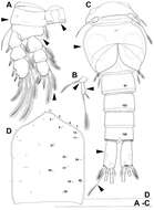
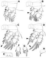
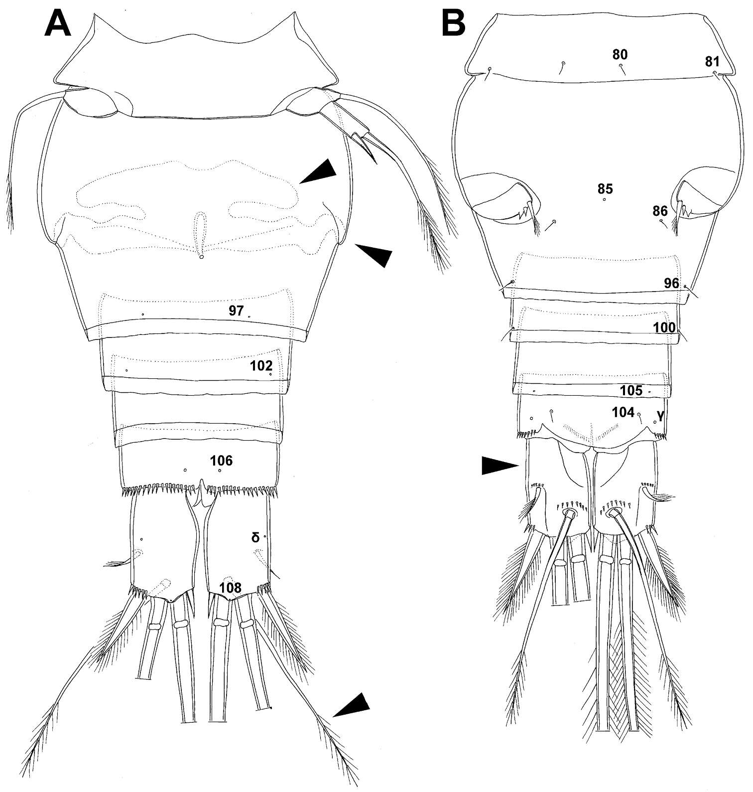
Figure 12.Diacyclops parasuoensis sp. n., A–B holotype female C–D allotype male. A fourth swimming leg, anterior view B fifth leg, anterior view C urosome, ventral view D cephalothorax, dorsal view. Arabic numerals indicating sensilla and pores presumably homologous to those in Diacyclops ishidai sp. n. Arrows pointing most prominent specific features. Scale bars 100 μm.
-
Tomislav Karanovic, Mark J. Grygier, Wonchoel Lee
Zookeys
Figure 13.Diacyclops parasuoensis sp. n., allotype male: A habitus, dorsal view B urosome, lateral view C antennula, flattened and slightly uncoiled, ventral view D sixth leg, ventro-lateral view. Arabic numerals indicating sensilla and pores presumably homologous to those in Diacyclops ishidai sp. n. Roman numerals indicating pores not present in Diacyclops ishidai sp. n. Arrows pointing most prominent specific features. Scale bars 100 μm.
-
Tomislav Karanovic, Mark J. Grygier, Wonchoel Lee
Zookeys
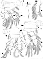
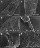
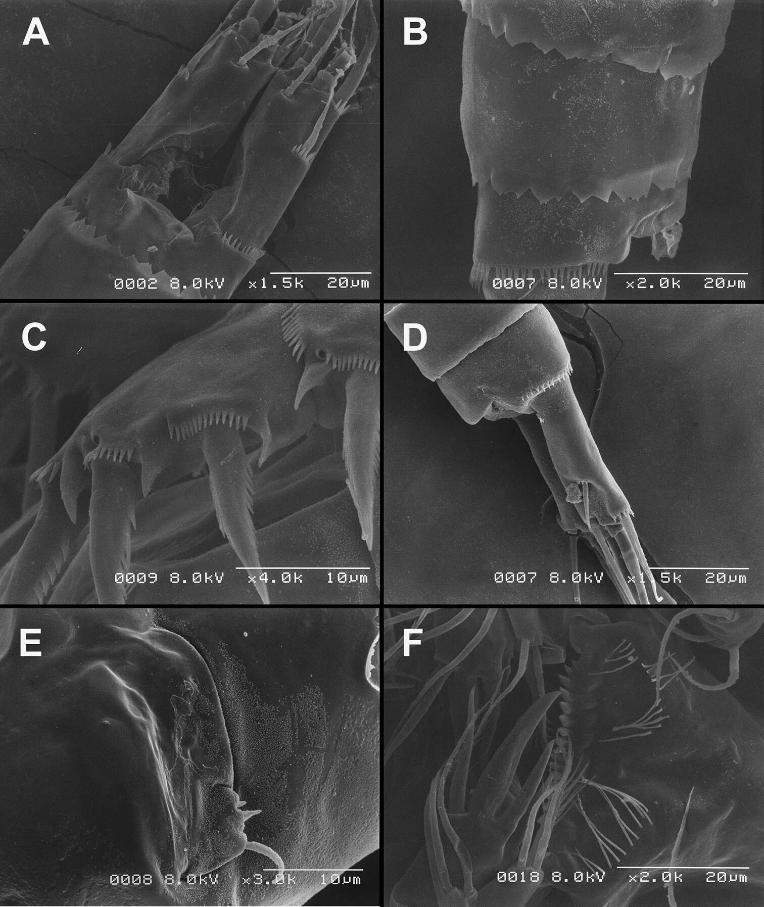
Figure 26.Scanning electron micrographs, A–C Diacyclops ishidai sp. n. D–E Diacyclops parasuoensis sp. n. F Diacyclops suoensis Ito, 1954: A anal somite and caudal rami, dorsal view, paratype female 1 B preanal and anal somites, lateral view, paratype female 2 C last two exopodal segments of second swimming legs, lateral view, paratype female 2 D anal somite and caudal rami, lateral view, paratype female E sixth leg, lateral view, paratype female F labrum and maxillulae, ventral view. Scale bars 20 μm (A, B, D, F) and 10 μm (C, E).
-
Tomislav Karanovic, Mark J. Grygier, Wonchoel Lee
Zookeys
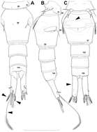
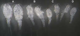
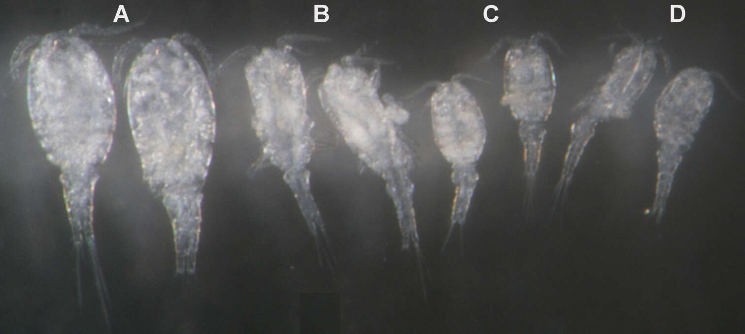
Figure 27.Light photograph of four sympatric Korean species of the Diacyclops/Acanthocyclops complex from Seomjin River: A Acanthocyclops sensitivus (Graeter & Chappuis, 1914), two adult females B Diacyclops languidoides s.l. (Lilljeborg, 1901), two adult females C Diacyclops parasuoensis sp. n., two adult females D Diacyclops hanguk sp. n., two adult females.
-
Tomislav Karanovic, Mark J. Grygier, Wonchoel Lee
Zookeys
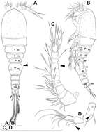
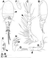
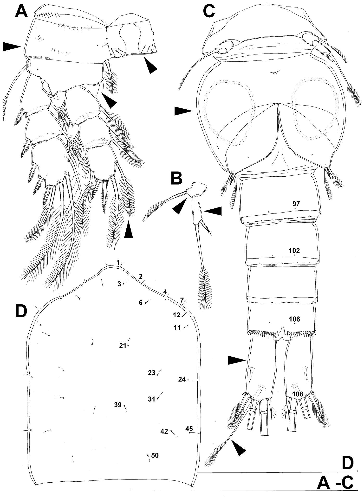
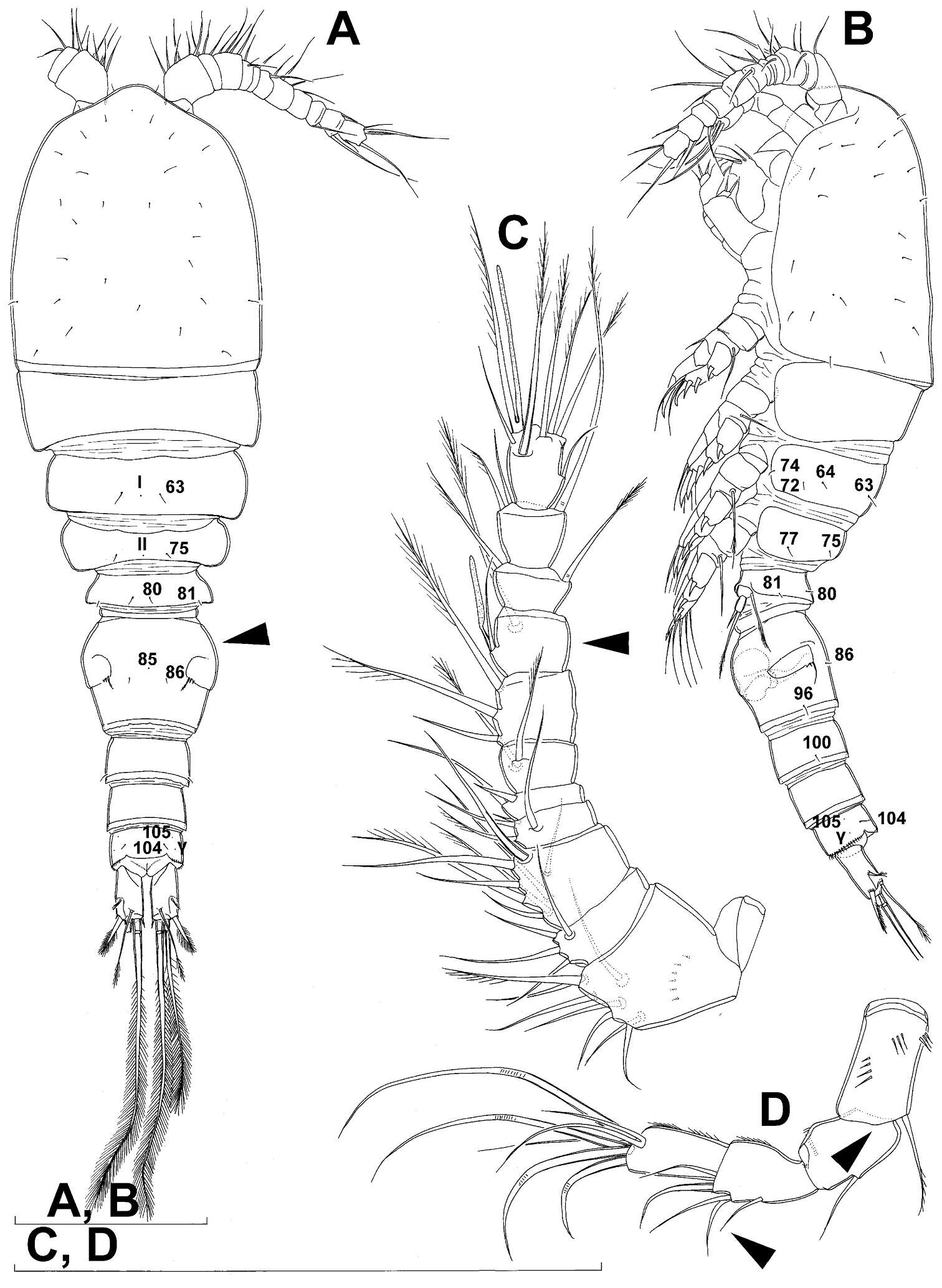
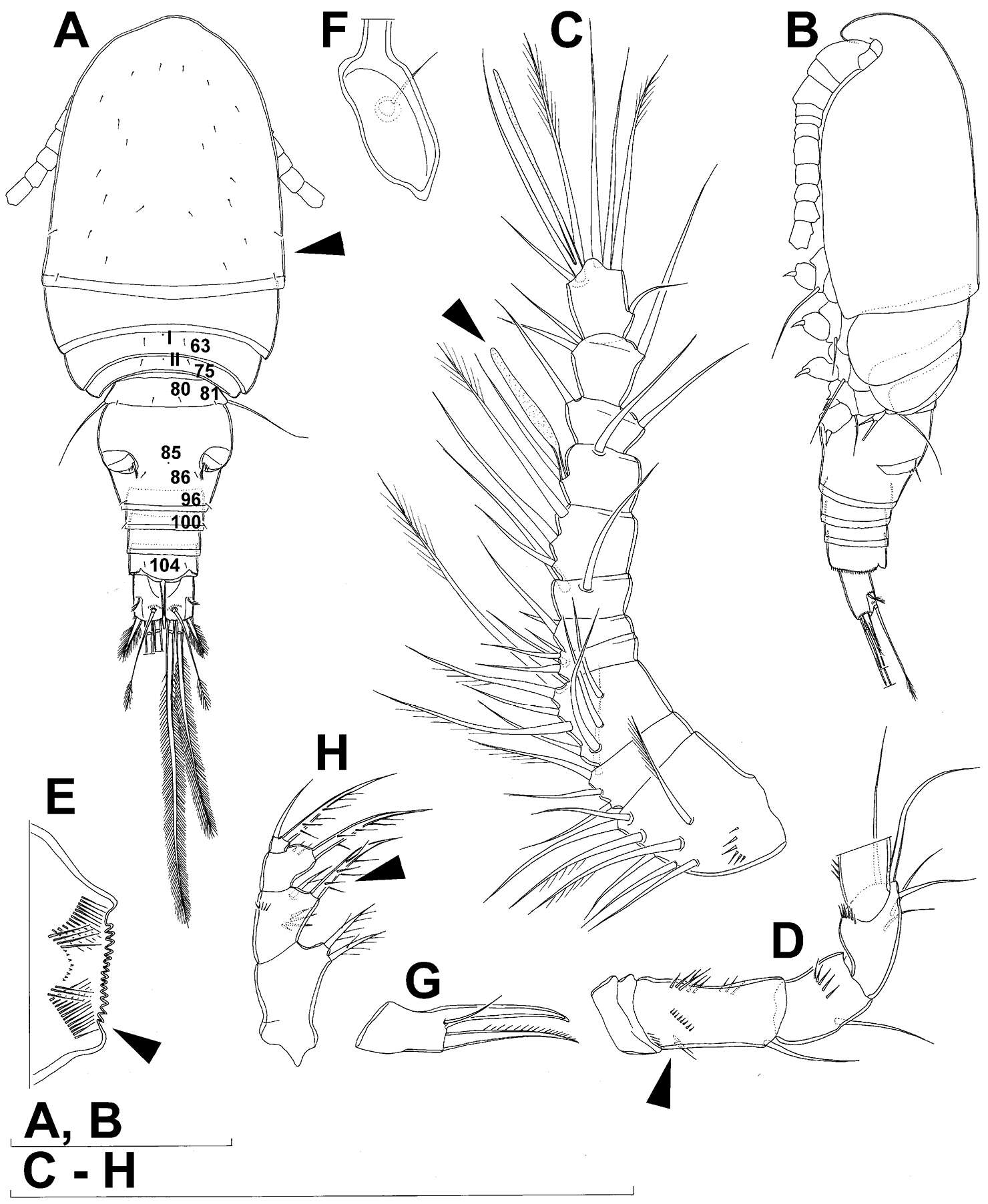
Figure 14.Diacyclops suoensis Ito, 1954, female: A habitus, dorsal view B antennula, ventral view C antenna, ventral view. Arrows pointing most prominent specific features. Scale bars 100 μm.
-
Tomislav Karanovic, Mark J. Grygier, Wonchoel Lee
Zookeys
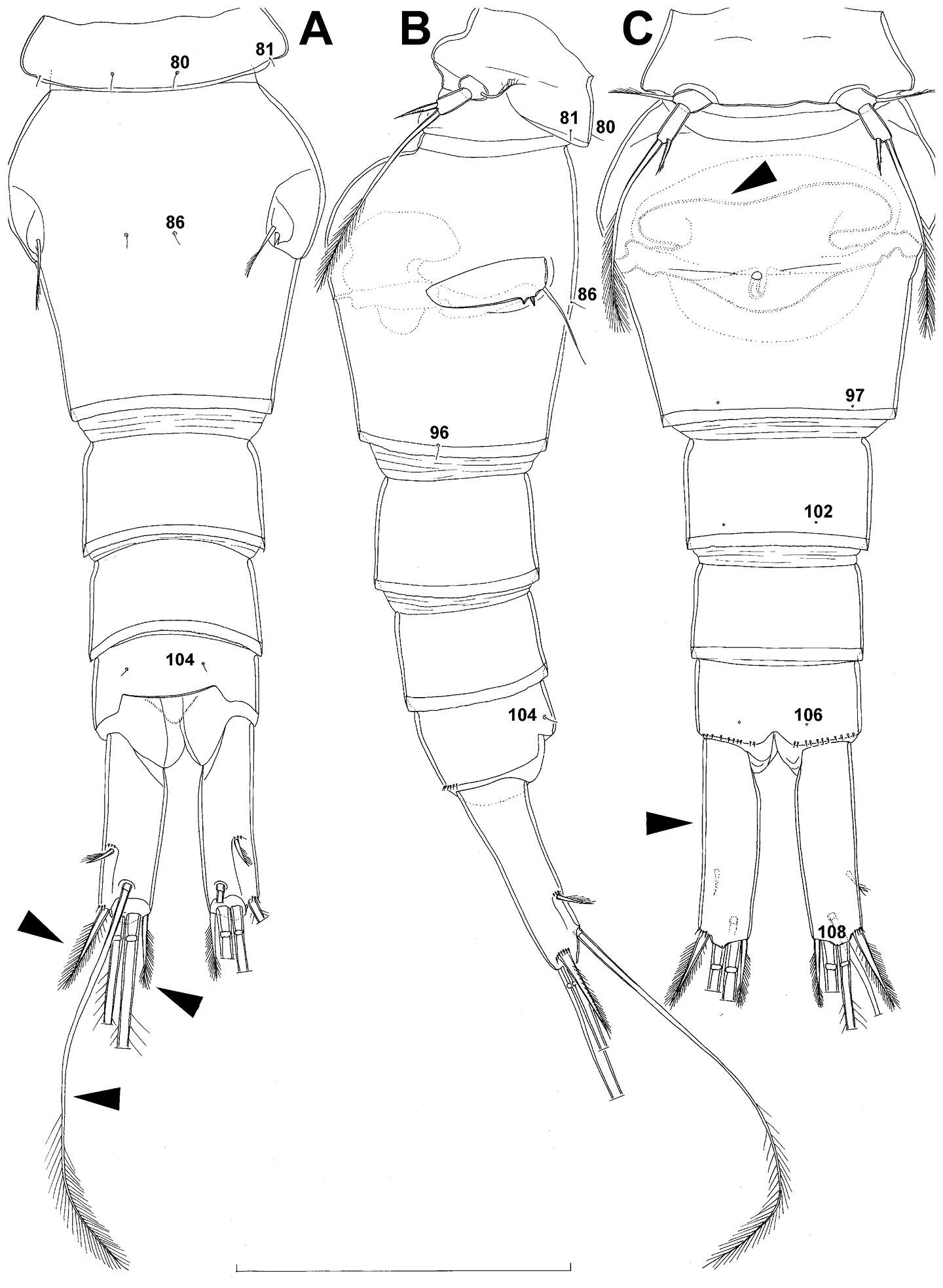
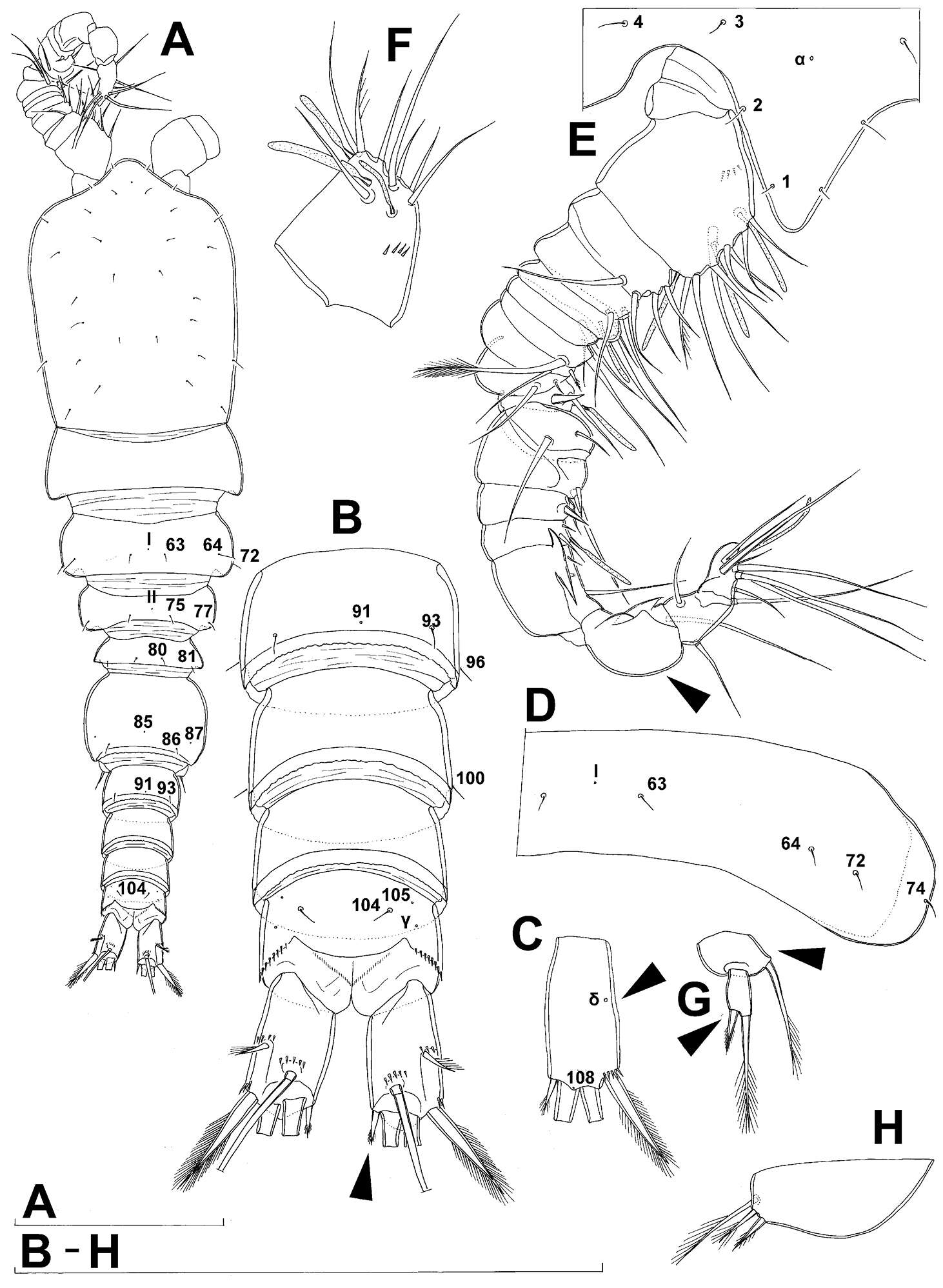
Figure 15.Diacyclops suoensis Ito, 1954, female: A urosome, dorsal view B urosome, lateral view C urosome, ventral view. Arabic numerals indicating sensilla and pores presumably homologous to those in Diacyclops ishidai sp. n. Arrows pointing most prominent specific features. Scale bar 100 μm.
-
Tomislav Karanovic, Mark J. Grygier, Wonchoel Lee
Zookeys
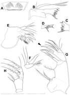
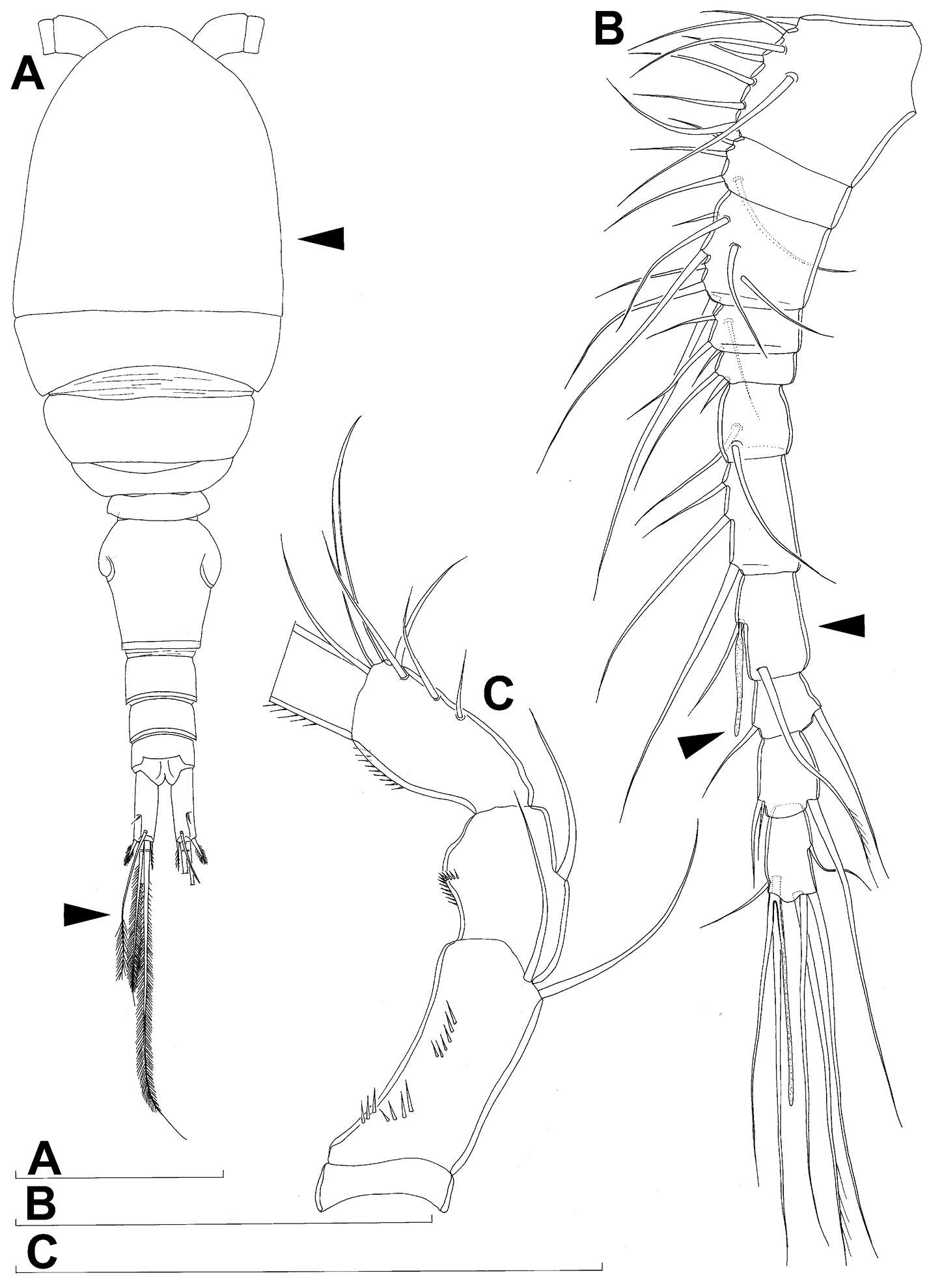
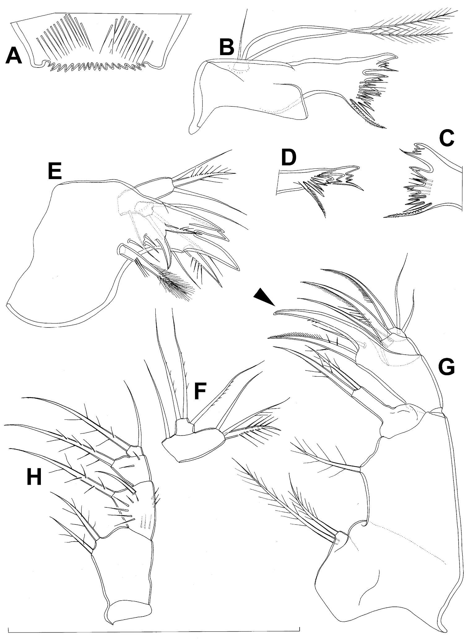
Figure 16.Diacyclops suoensis Ito, 1954, female: A labrum, anterior view B mandibula, anterior view C cutting edge of mandibula, posterior view D cutting edge of mandibula, dorsal view E maxillula, posterior view F maxillular palp, posterior view G maxilla, anterior view H maxilliped, anterior view. Arrows pointing most prominent specific features. Scale bar 100 μm.
-
Tomislav Karanovic, Mark J. Grygier, Wonchoel Lee
Zookeys
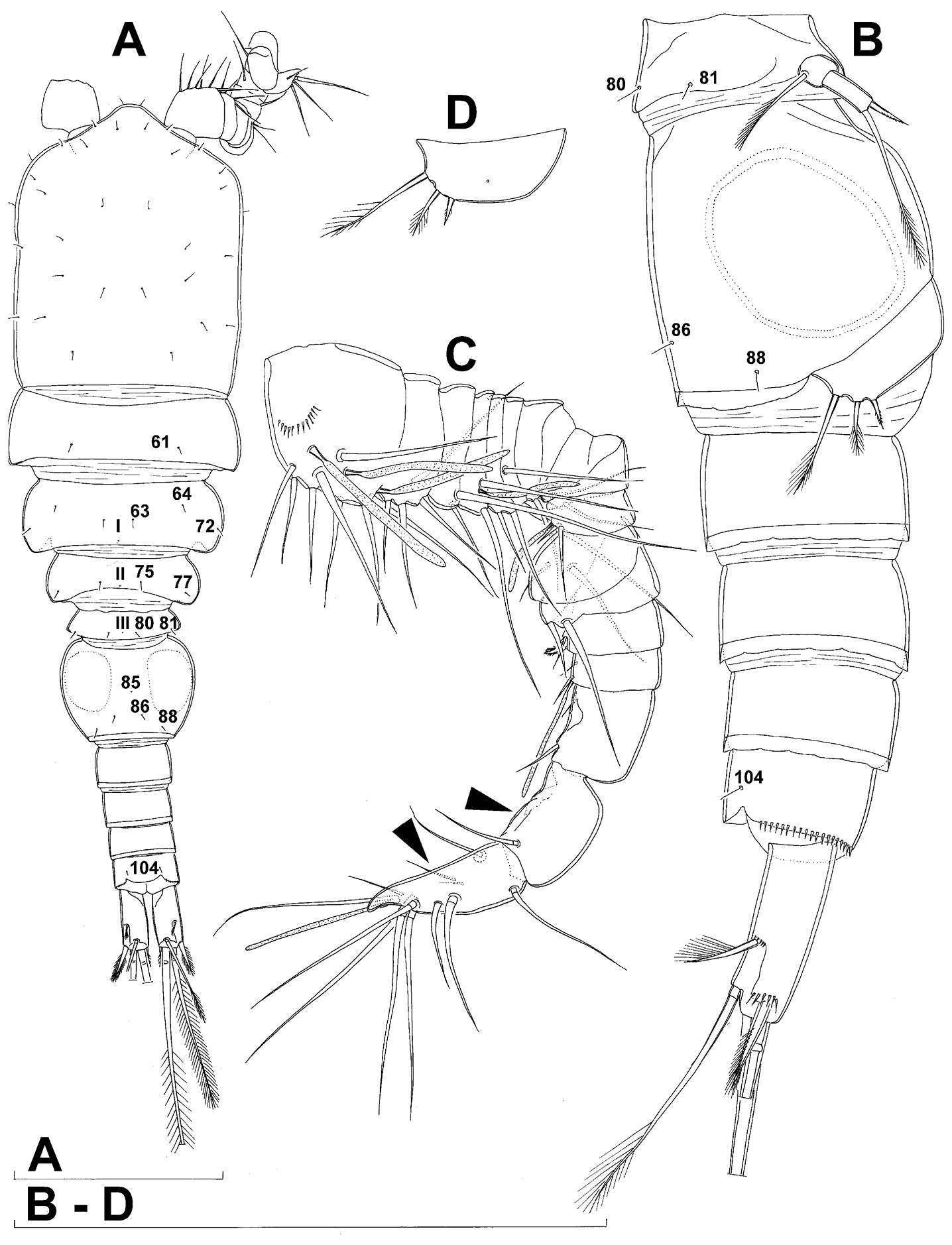
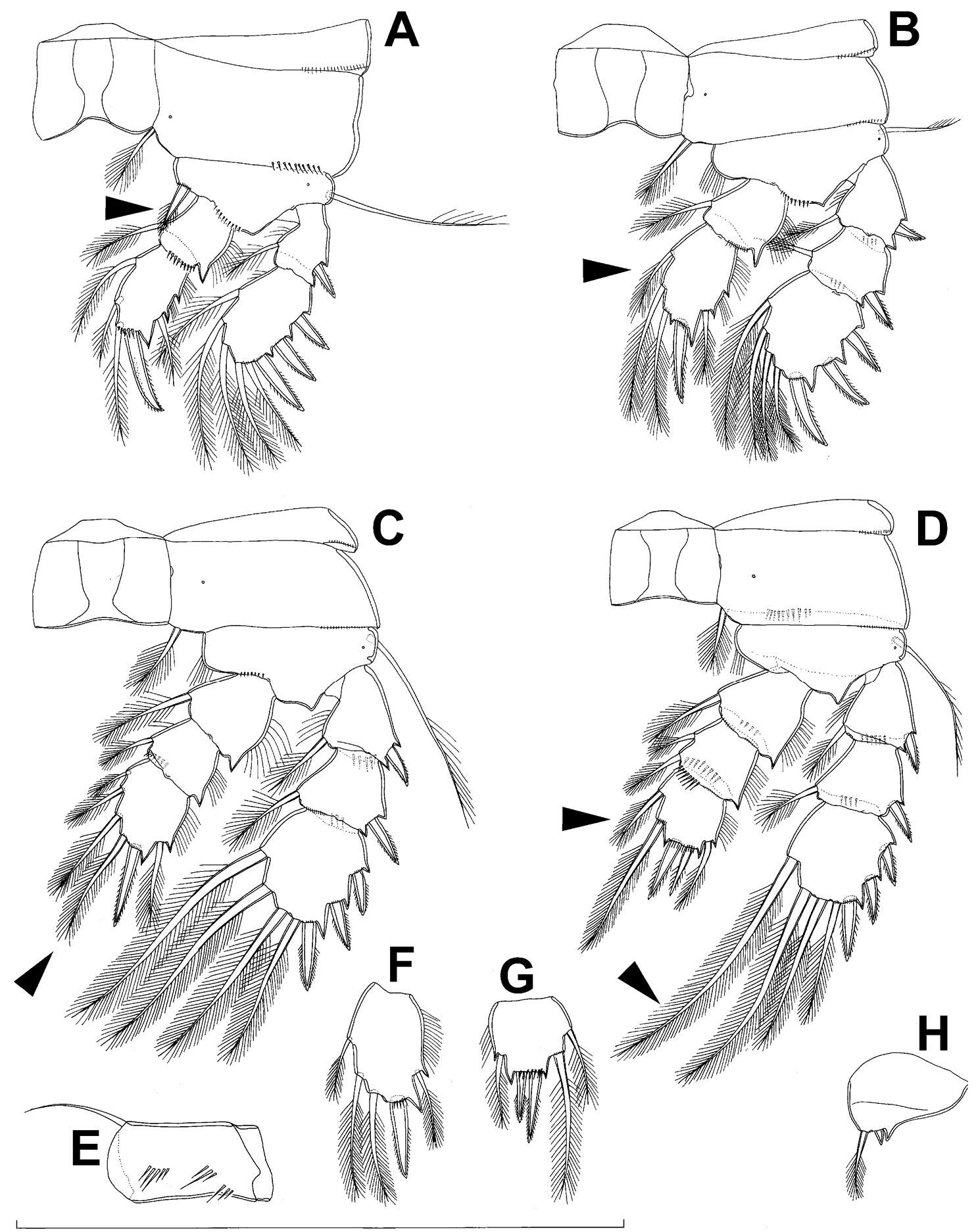
Figure 17.Diacyclops suoensis Ito, 1954, female: A first swimming leg, anterior view B endopod of second swimming leg, anterior view C third swimming leg, anterior view D fourth swimming leg, anterior view E fifth leg, anterior view. Arrows pointing most prominent specific features. Scale bar 100 μm.
-
Tomislav Karanovic, Mark J. Grygier, Wonchoel Lee
Zookeys
Figure 26.Scanning electron micrographs, A–C Diacyclops ishidai sp. n. D–E Diacyclops parasuoensis sp. n. F Diacyclops suoensis Ito, 1954: A anal somite and caudal rami, dorsal view, paratype female 1 B preanal and anal somites, lateral view, paratype female 2 C last two exopodal segments of second swimming legs, lateral view, paratype female 2 D anal somite and caudal rami, lateral view, paratype female E sixth leg, lateral view, paratype female F labrum and maxillulae, ventral view. Scale bars 20 μm (A, B, D, F) and 10 μm (C, E).
-
Tomislav Karanovic, Mark J. Grygier, Wonchoel Lee
Zookeys
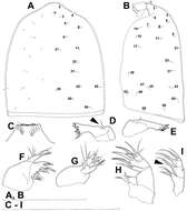
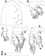
Figure 18.Diacyclops hanguk sp. n., holotype female: A habitus, dorsal view B habitus, lateral view C antennula, dorsal view D antenna, ventral view. Arabic numerals indicating sensilla and pores presumably homologous to those in Diacyclops ishidai sp. n. Roman numerals indicating pores homologous to those in Diacyclops parasuoensis sp. n. Arrows pointing most prominent specific features. Scale bars 100 μm.
-
Tomislav Karanovic, Mark J. Grygier, Wonchoel Lee
Zookeys
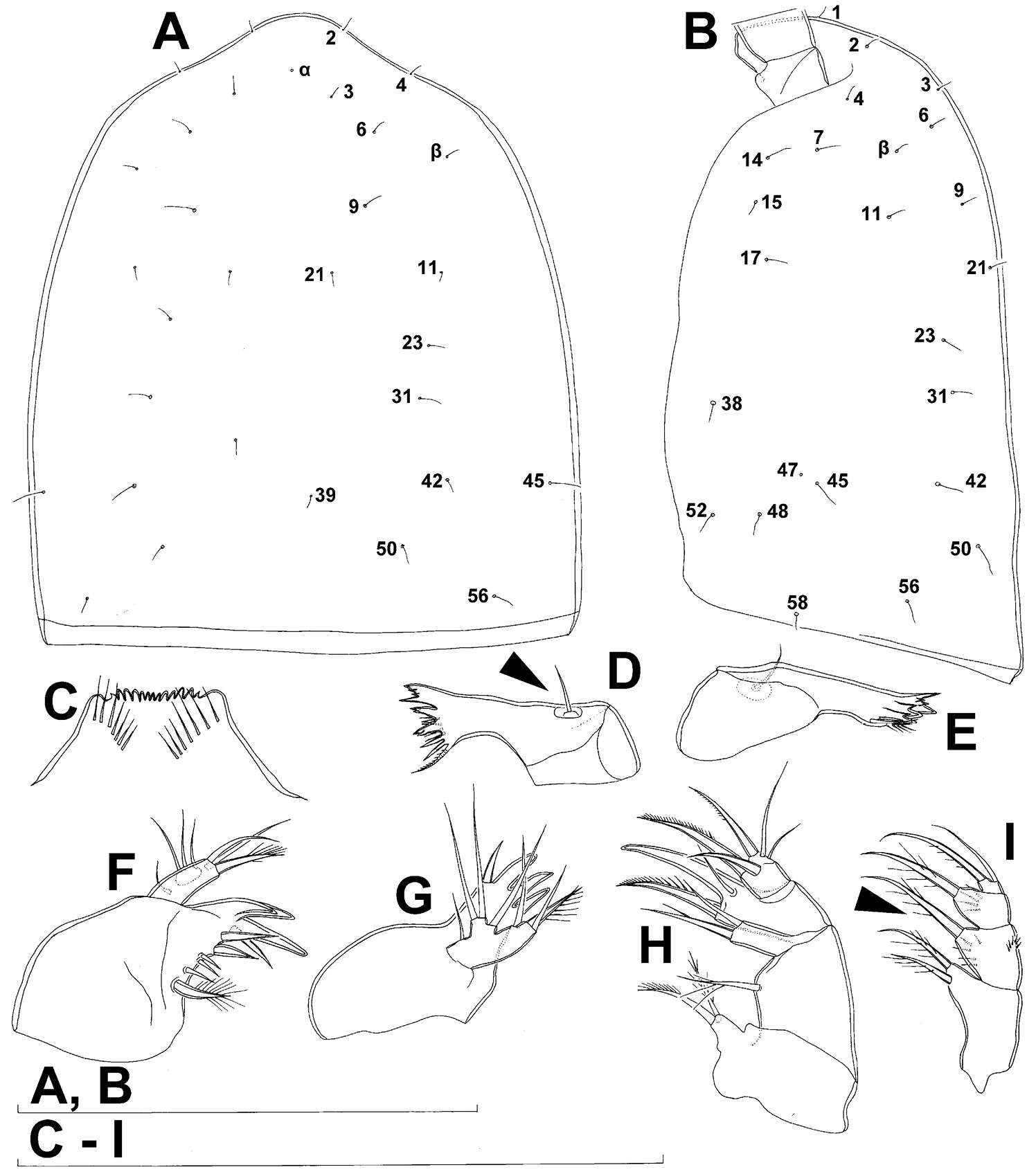
Figure 19.Diacyclops hanguk sp. n., holotype female: A cephalothorax, dorsal view B cephalothoracic shield, lateral view C labrum, anterior view D mandibula, posterior view E mandibula, antero-ventral view F maxillula, posterior view G maxillula, anterior view H maxilla, posterior view I maxilliped, posterior view. Arabic numerals indicating sensilla and pores presumably homologous to those in Diacyclops ishidai sp. n. Greek letters indicating unique pores and sensilla. Arrows pointing most prominent specific features. Scale bars 100 μm.
-
Tomislav Karanovic, Mark J. Grygier, Wonchoel Lee
Zookeys
Figure 20.Diacyclops hanguk sp. n., holotype female: A urosome, ventral view B usorome, lateral view C anal somite and caudal rami, dorsal view. Arabic numerals indicating sensilla and pores presumably homologous to those in Diacyclops ishidai sp. n. Greek letters indicating unique pores and sensilla. Arrows pointing most prominent specific features. Scale bar 100 μm.
-
Tomislav Karanovic, Mark J. Grygier, Wonchoel Lee
Zookeys
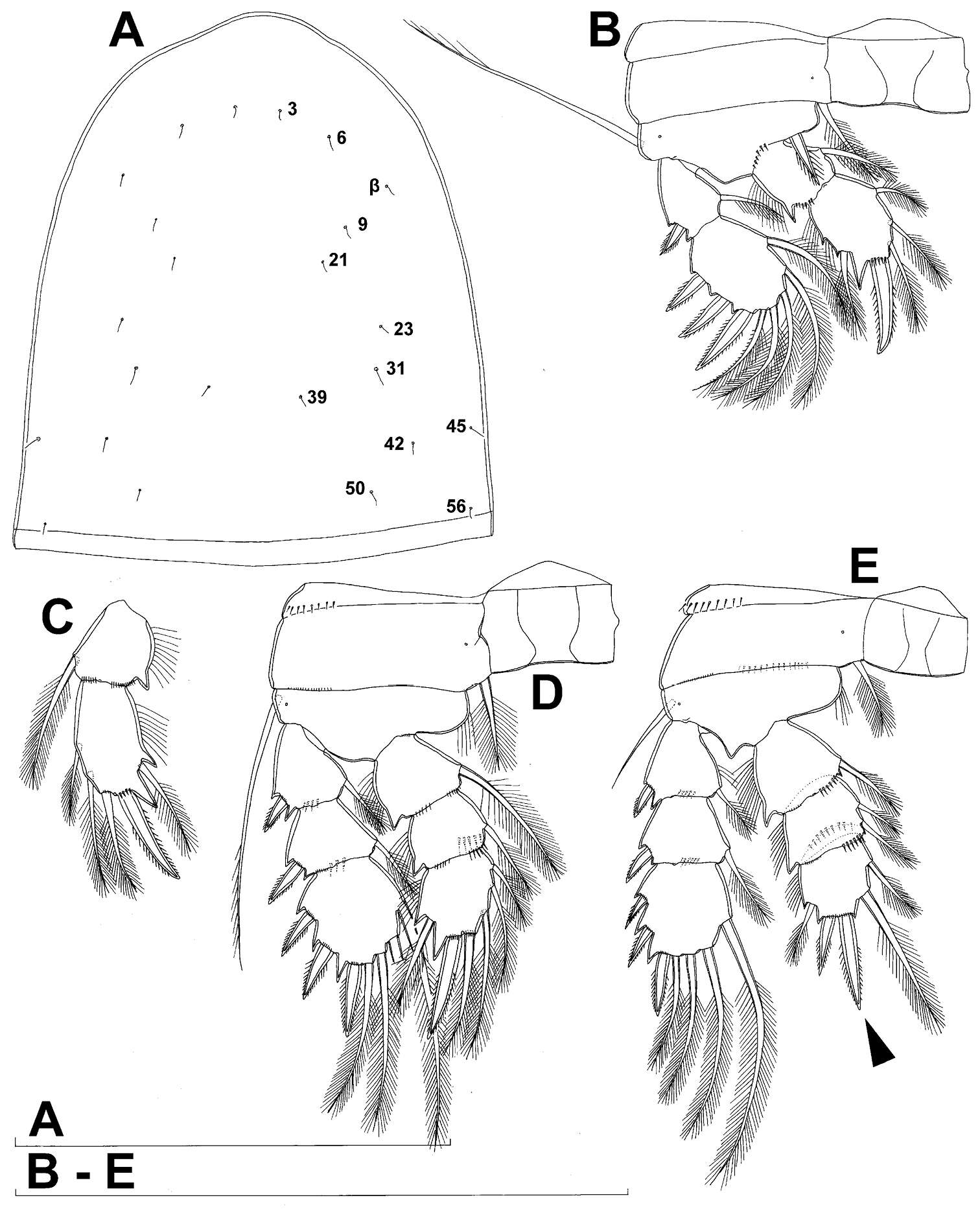
Figure 21.Diacyclops hanguk sp. n., A–D holotype female E–H paratype female A first swimming leg, anterior view B second swimming leg, anterior view C third swimming leg, anterior view D fourth swimming leg, anterior view E coxa and basis of antenna, ventral view F second endopodal segment of second swimming leg, anterior view G third endopodal segment of fourth swimming leg, anterior view H sixth leg, lateral view. Arrows pointing most prominent specific features. Scale bar 100 μm.
-
Tomislav Karanovic, Mark J. Grygier, Wonchoel Lee
Zookeys
Figure 22.Diacyclops hanguk sp. n., allotype male: A habitus, dorsal view B last four urosomites and caudal rami, dorsal view C left caudal ramus, ventral view D pleuron of second free prosomite (third pedigerous somite), flattened E rostrum and antennula, flattened, dorsal view F first antennular segment, ventral view G fifth leg, anterior view H sixth leg, ventro-lateral view. Arabic numerals indicating sensilla and pores presumably homologous to those in Diacyclops ishidai sp. n. Roman numerals indicating pores homologous to those in Diacyclops parasuoensis sp. n. Greek letters indicating unique pores and sensilla. Arrows pointing most prominent specific features. Scale bars 100 μm.
-
Tomislav Karanovic, Mark J. Grygier, Wonchoel Lee
Zookeys
Figure 27.Light photograph of four sympatric Korean species of the Diacyclops/Acanthocyclops complex from Seomjin River: A Acanthocyclops sensitivus (Graeter & Chappuis, 1914), two adult females B Diacyclops languidoides s.l. (Lilljeborg, 1901), two adult females C Diacyclops parasuoensis sp. n., two adult females D Diacyclops hanguk sp. n., two adult females.
-
Tomislav Karanovic, Mark J. Grygier, Wonchoel Lee
Zookeys
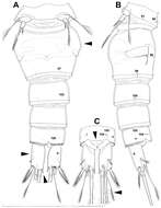
Figure 23.Diacyclops parahanguk sp. n., holotype female: A habitus, dorsal view B habitus, lateral view C antennula, ventral view D antenna, ventral view E labrum, anterior view F mandibula without cutting edge, dorsal view G basis of maxilla, posterior view H maxilliped, posterior vew. Arabic numerals indicating sensilla and pores presumably homologous to those in Diacyclops ishidai sp. n. Roman numerals indicating pores homologous to those in Diacyclops parasuoensis sp. n. Arrows pointing most prominent specific features. Scale bars 100 μm.
-
Tomislav Karanovic, Mark J. Grygier, Wonchoel Lee
Zookeys
Figure 24.Diacyclops parahanguk sp. n., holotype female: A urosome, ventral view B urosome, dorsal view. Arabic numerals indicating sensilla and pores presumably homologous to those in Diacyclops ishidai sp. n. Greek letters indicating pores and sensilla homologous to those in Diacyclops hanguk sp. n. Arrows pointing most prominent specific features. Scale bar 100 μm.
-
Tomislav Karanovic, Mark J. Grygier, Wonchoel Lee
Zookeys
Figure 25.Diacyclops parahanguk sp. n., holotype female: A cephalothorax, dorsal view B first swimming leg, anterior view C endopod of second swimming leg, anterior view D third swimming leg, anterior view E fourth swimming leg, anterior view. Arabic numerals indicating sensilla and pores presumably homologous to those in Diacyclops ishidai sp. n. Greek letters indicating pores and sensilla homologous to those in Diacyclops hanguk sp. n. Arrows pointing most prominent specific features. Scale bars 100 μm.