-
Susumu Ohtsuka, Michitaka Shimomura, Kota Kitazawa
Zookeys
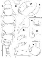
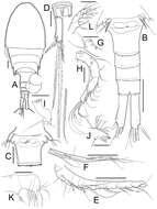
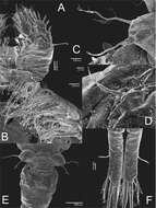
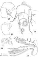
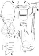
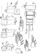
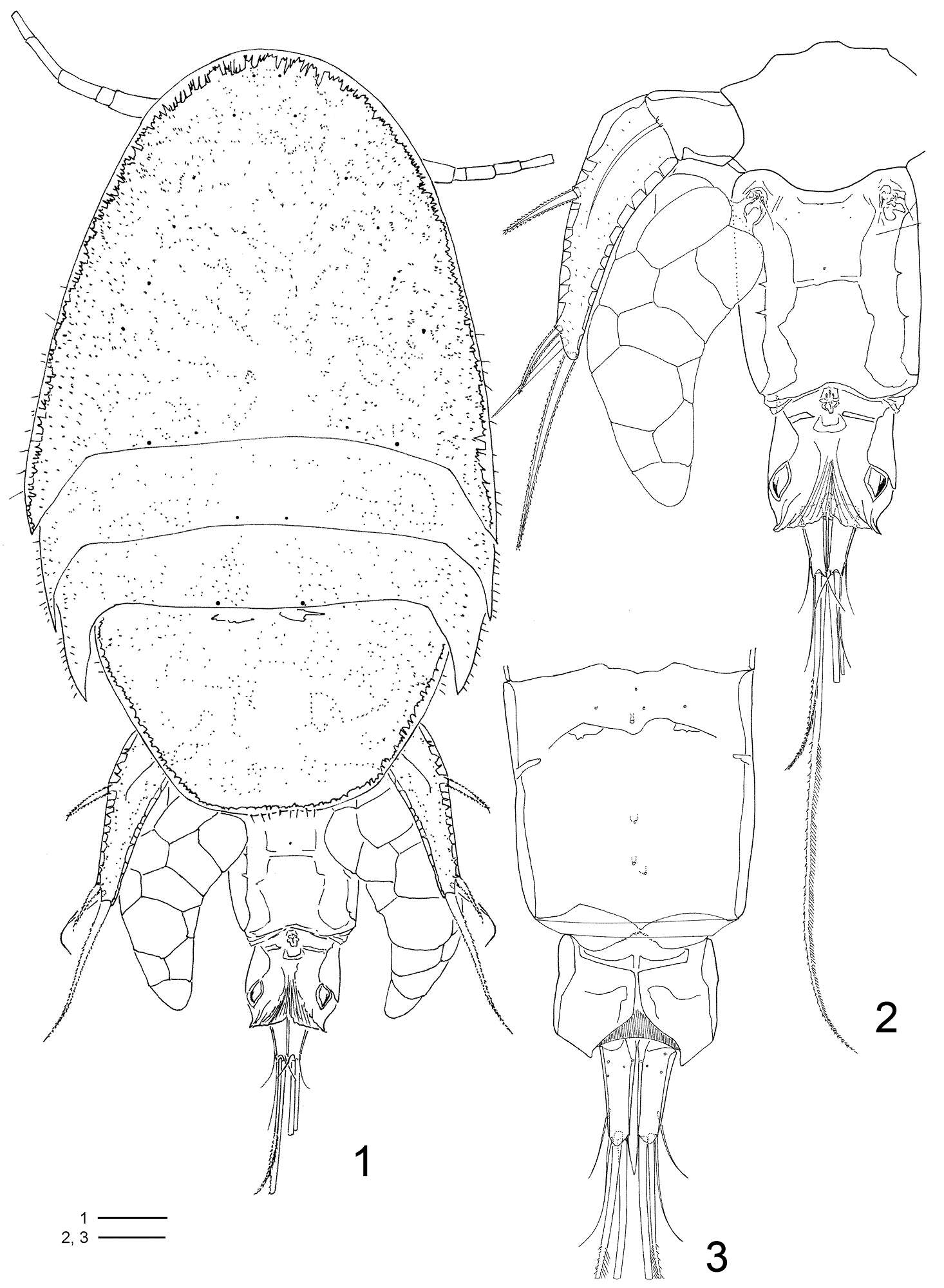
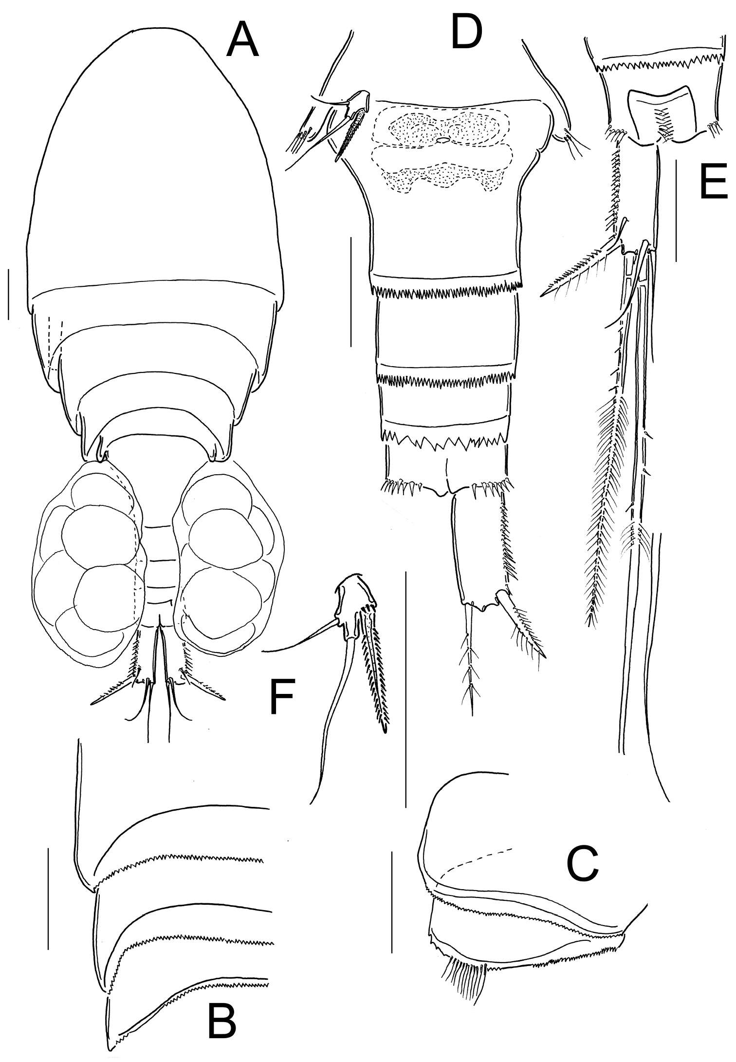
Figure 1. Enterognathus inabai sp. n. holotype female: A Habitus, dorsal view B Rostrum, dorsal view C Genital opening, right, dorsal view D Caudal ramus, left, dorsal view E Antennule F Antenna G Mandible H Labrum and paragnath, ventral view I Maxillule J Other maxillule K Maxilla. Scales in mm.
-
Daisuke Uyeno, Kazuya Nagasawa
Zookeys
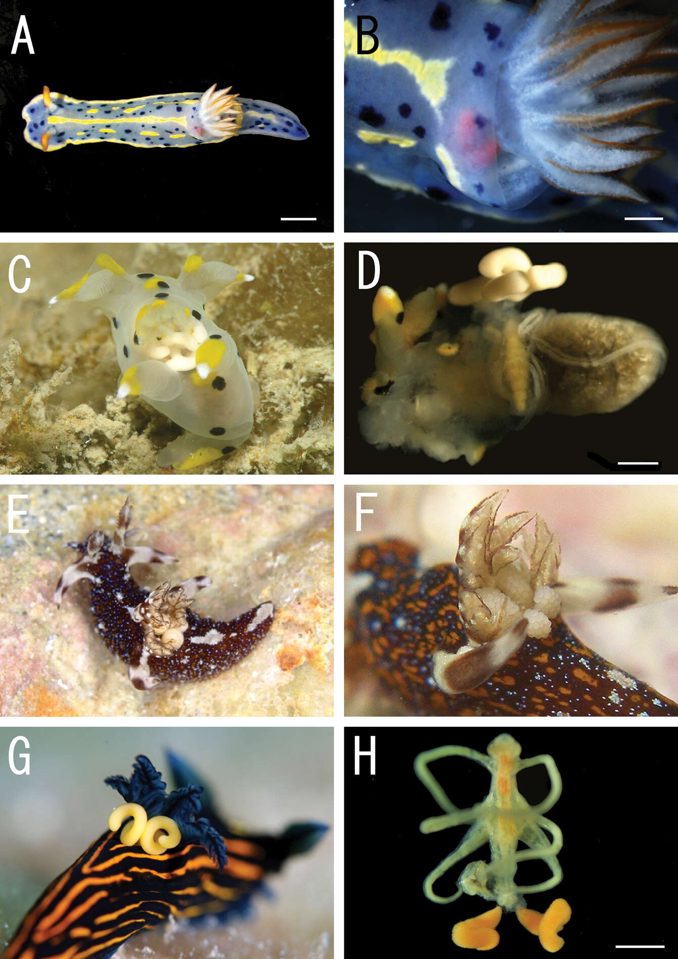
Figure 1.Live coloration of the host nudibranchs and the splanchnotrophids. A Hypselodoris festiva infected by an ovigerous specimenof Certosomicola japonica sp. n. B an egg sac of Ceratosomicola japonica sp. n. and the gill circle of Hypselodoris festiva with the mantle malformed into an elongate tube C Thecacera pennigera infected by an ovigerous specimen of Splanchnotrophus helianthus sp. n. D Trapania pennigera with the mantle removed to show a female specimen of Splanchnotrophus helianthus on the visceral sac E Trapania miltabrancha infected by an ovigerous specimen of Splanchnotrophus imagawai sp. n (photo by K. Imagawa) F gill circle of Trapania miltabrancha with egg sacs of Splanchnotrophus imagawai sp. n. (photo by K. Imagawa) G Roboastra luteolineata infected by an ovigerous specimen of Majimun shirakawai gen. et sp. n. (photo by N. Shirakawa) H female Majimun shirakawai gen. et sp. n. with dwarf male attached to the posterior part of the body. Scale bars = 5 mm in A; 1 mm in B, D, H.
-
Tomislav Karanovic, Mark J. Grygier, Wonchoel Lee
Zookeys
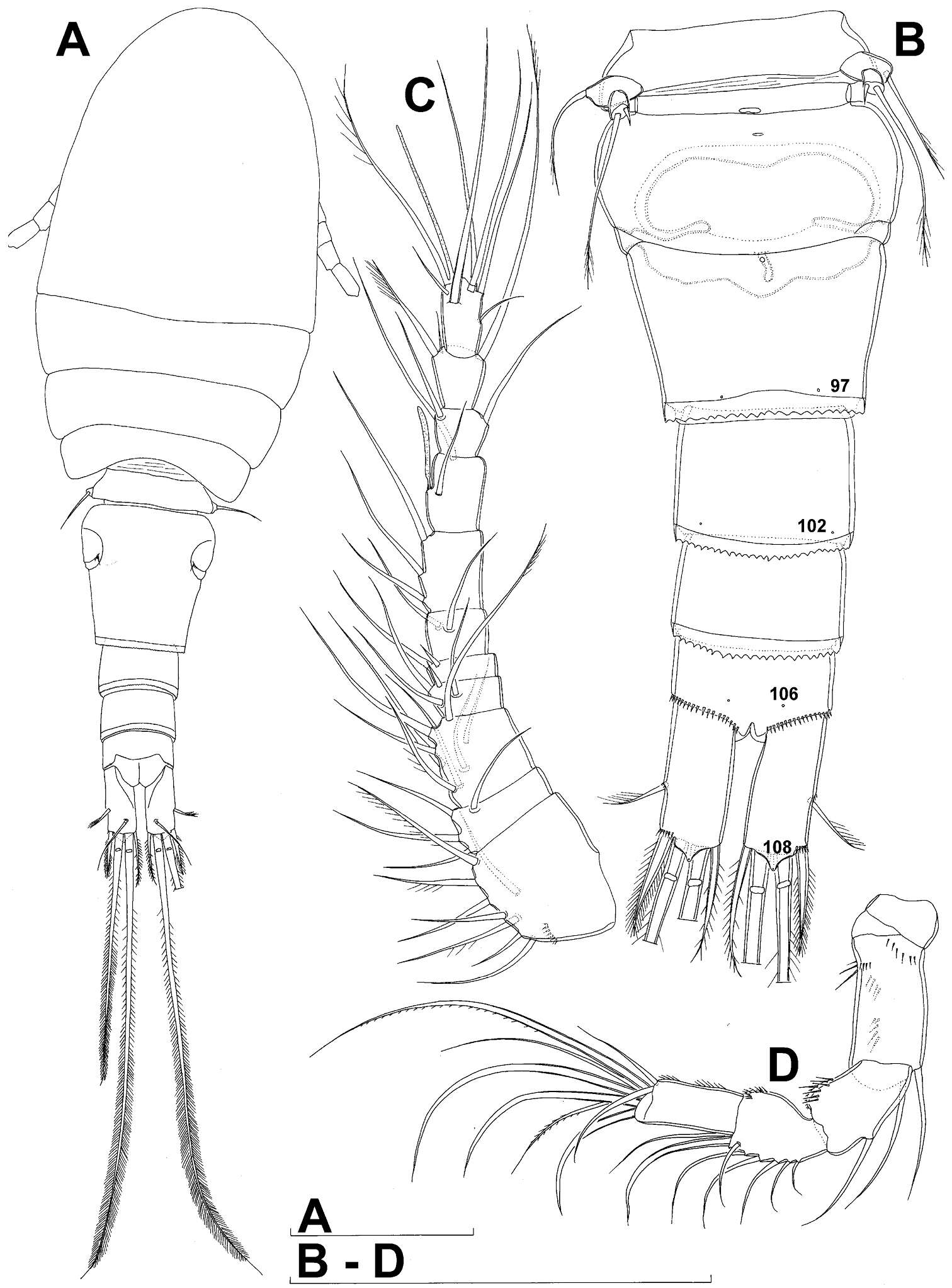
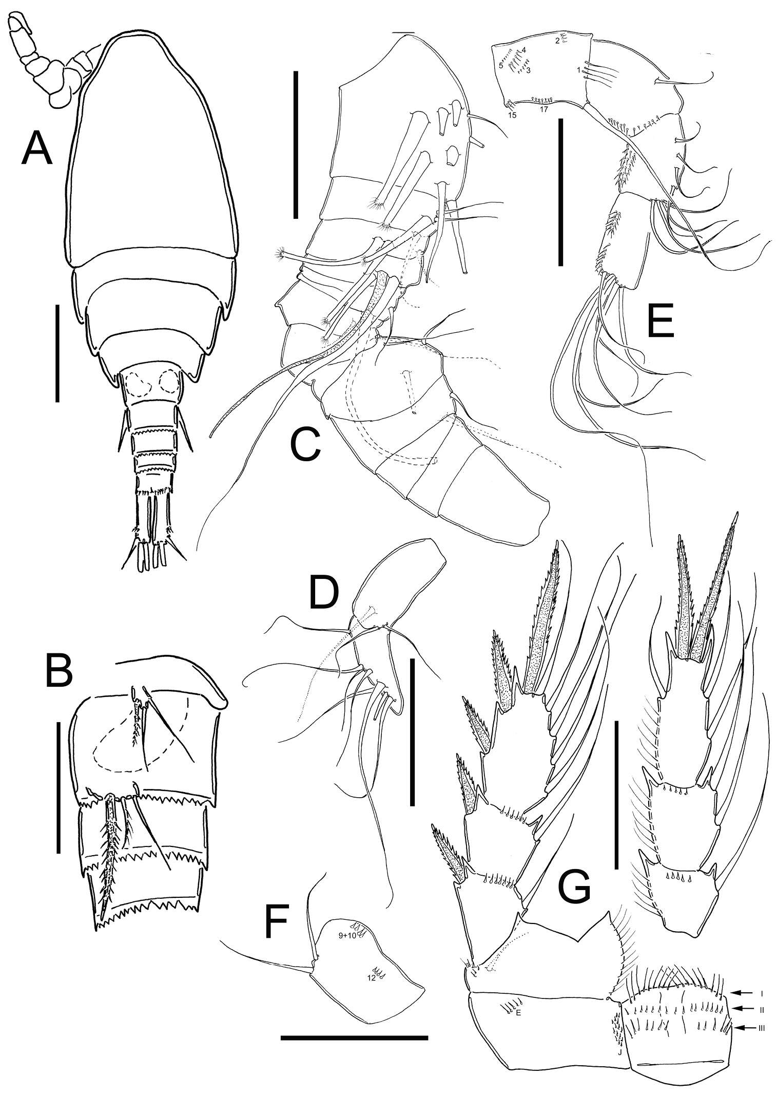
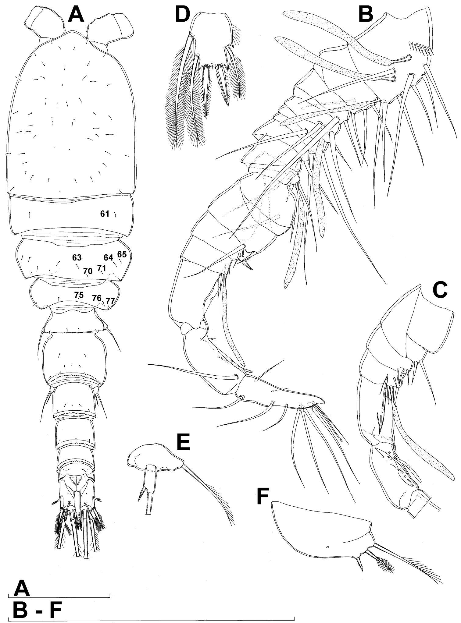
Figure 1.Diacyclops ishidai sp. n., holotype female: A habitus, dorsal view B urosome, ventral view C antennula, dorsal view D antenna, dorsal view. Arabic numerals numbering sensilla and pores consecutively from anterior to posterior end of body, and from dorsal to ventral side (excluding appendages). Scale bars 100 μm.
-
Nancy F. Mercado-Salas, Eduardo Suárez-Morales, Alejandro M. Maeda-Martínez, Marcelo Silva-Briano
Zookeys
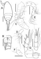
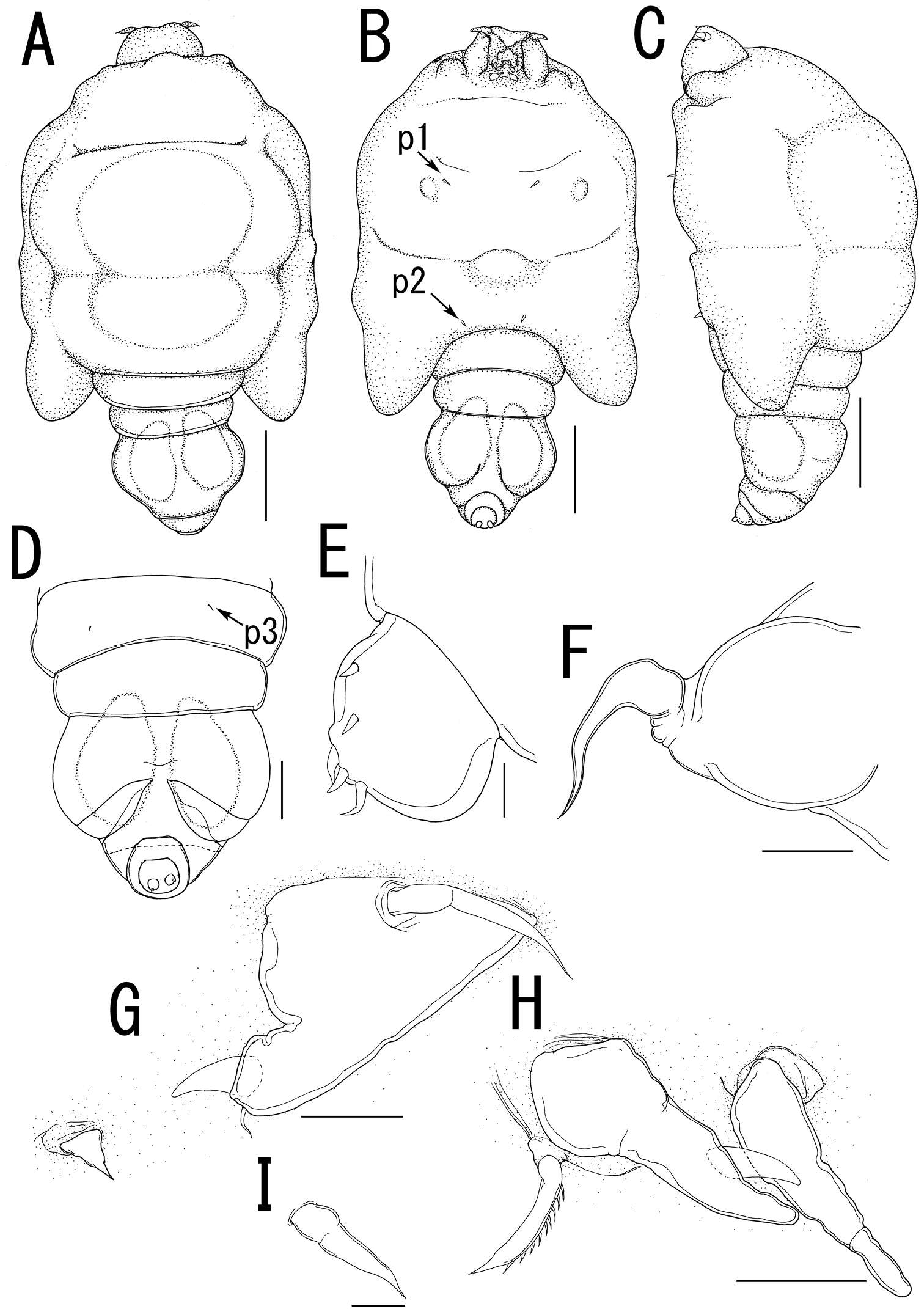
Figure 1.Metacyclops deserticus sp. n., female holotype from Coahuila, Mexico. A habitus, dorsal view B urosome, ventral view C antennule D antenna E mandible F maxillule G maxilla H maxilliped I anal operculum. Scales bars A–B= 100µm; C–I= 50 µm.
-
Terue C. Kihara, Carlos E. F. Rocha
Zookeys
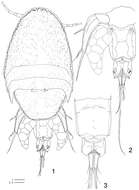
Figures 1–3.Clausidium rodriguesi sp. n. Female: 1 habitus, dorsal 2 urosome, dorsal 3 urosome lacking somite bearing P5, ventral. Scale bars: 1 = 100 μm; 2, 3 = 50 μm.
-
Martha Angélica Gutiérrez-Aguirre, Nancy Fabiola Mercado-Salas, Adrián Cervantes-Martínez
Zookeys
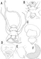
Figure 2.Eucyclops tziscao sp. n. A, C, D paratype B, E–L holotype from Laguna Tziscao, Chiapas. A Habitus, dorsal B Urosome C Genital double-somite, ventral D Anal somite and caudal ramus, dorsal E Antennule, segments 1–9 F Antennule, segments 10–12 G Antenna, caudal H Antenna, frontal I Mandible J Maxillule, caudal K Maxilla, frontal L Maxilliped, frontal. Scales bars: K = 20 µm; A, C, D, G, H, I, J, L = 50 µm; B, E, F = 100 µm.
-
Susumu Ohtsuka, Michitaka Shimomura, Kota Kitazawa
Zookeys
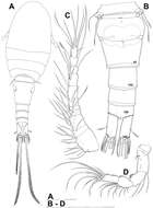
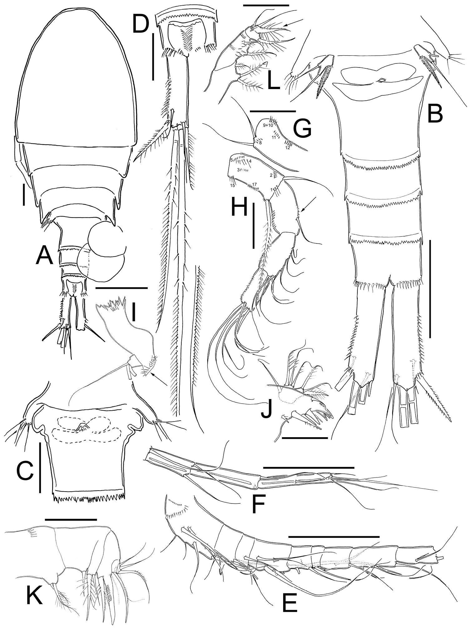
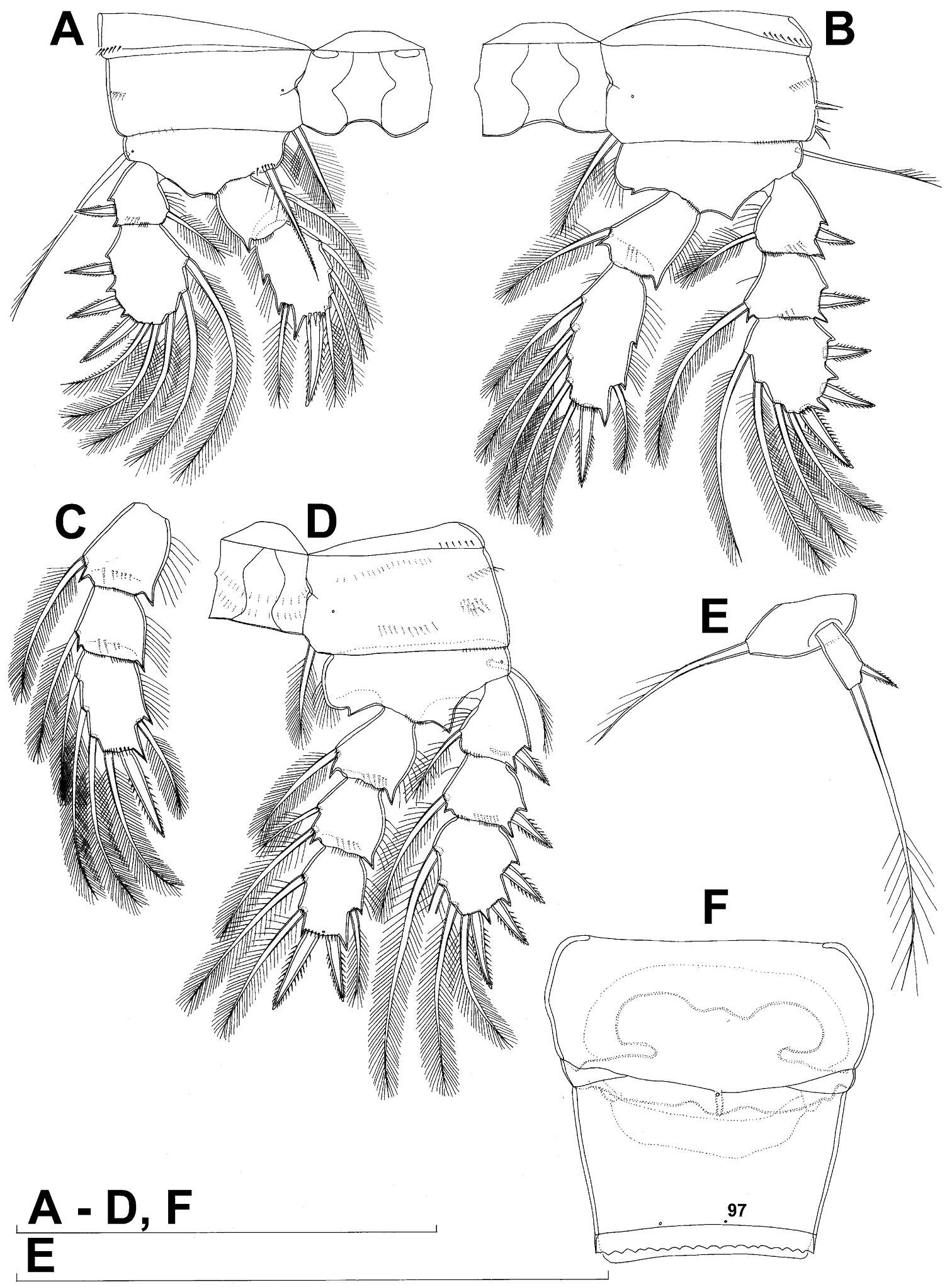
Figure 2. Enterognathus inabai sp. n. holotype female: A Leg 1, posterior view B Leg 1 excluding coxa (more or less flattened), posterior view C Leg 2, posterior view D Leg 2 excluding coxa, anterior view E Leg 3, posterior view F Leg 3 excluding coxa, anterior view G Leg 4, posterior view H Leg 4 excluding coxa and second endopodal segment, anterior view. Scales in mm.
-
Daisuke Uyeno, Kazuya Nagasawa
Zookeys
Figure 2.Ceratosomicola japonica sp. n., female, holotype NSMT–Cr 22240 (A–C), female, paratype NSMT–Cr 22243 (D–F). A habitus dorsal B habitus, ventral, p1 = leg 1, p2 = leg 2 C posterior portion of body, ventral D urosome, ventral E caudal ramus, ventral F egg sac. Scale bars = 1 mm in A, B; 300 μm in C; 100 μm in D; 10 μm in E; 500 μm in F.
-
Tomislav Karanovic, Mark J. Grygier, Wonchoel Lee
Zookeys
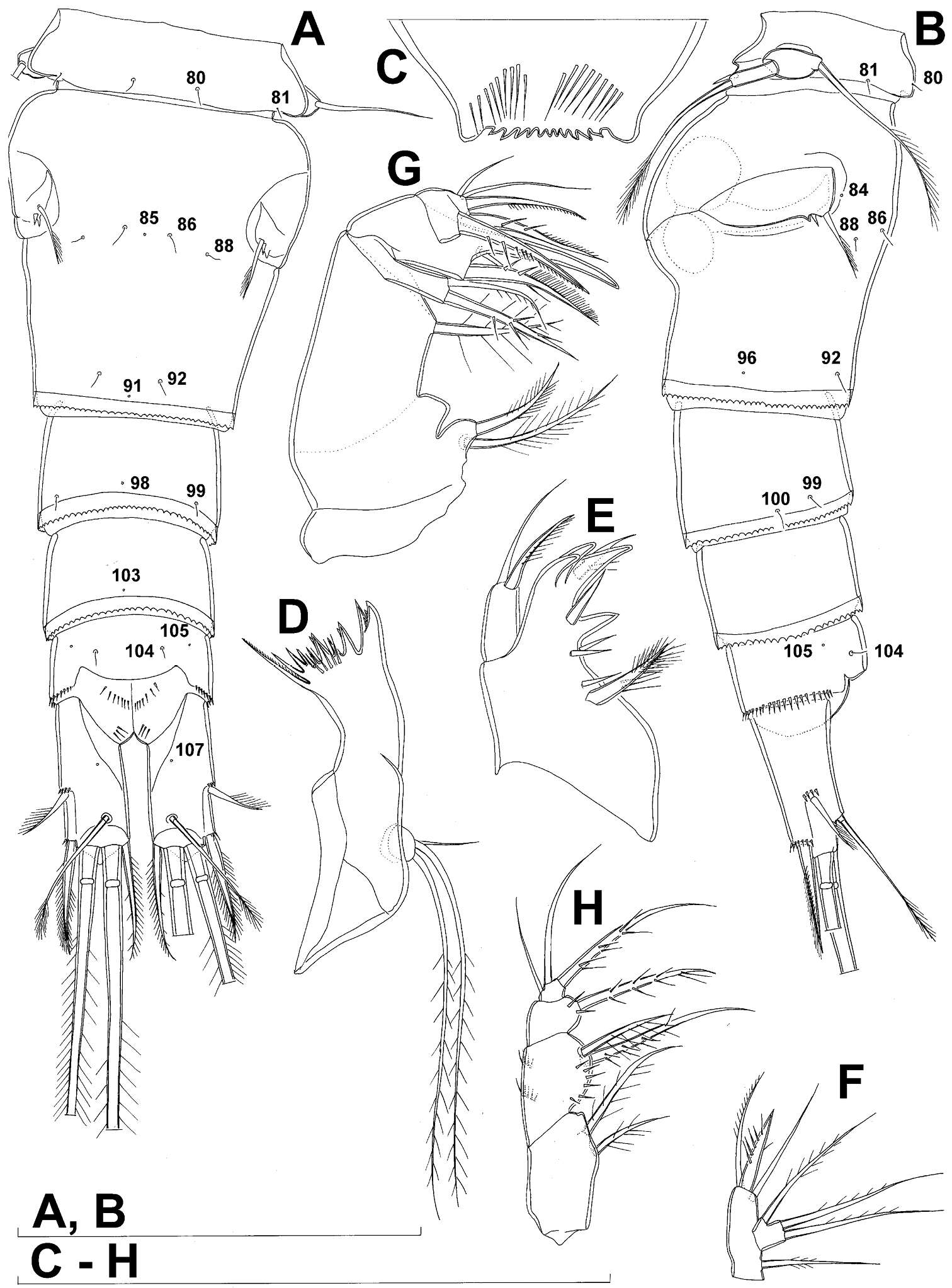
Figure 2.Diacyclops ishidai sp. n., holotype female: A urosome, dorsal view B urosome, lateral view C labrum, anterior view D mandibula, anterior view E maxillula, posterior view (palp armature omitted) F maxillular palp, anterior view G maxilla, posterior view H maxilliped, anterior view. Arabic numerals numbering sensilla and pores consecutively from anterior to posterior end of body, and from dorsal to ventral side (excluding appendages). Scale bars 100 μm.
-
Nancy F. Mercado-Salas, Eduardo Suárez-Morales, Alejandro M. Maeda-Martínez, Marcelo Silva-Briano
Zookeys
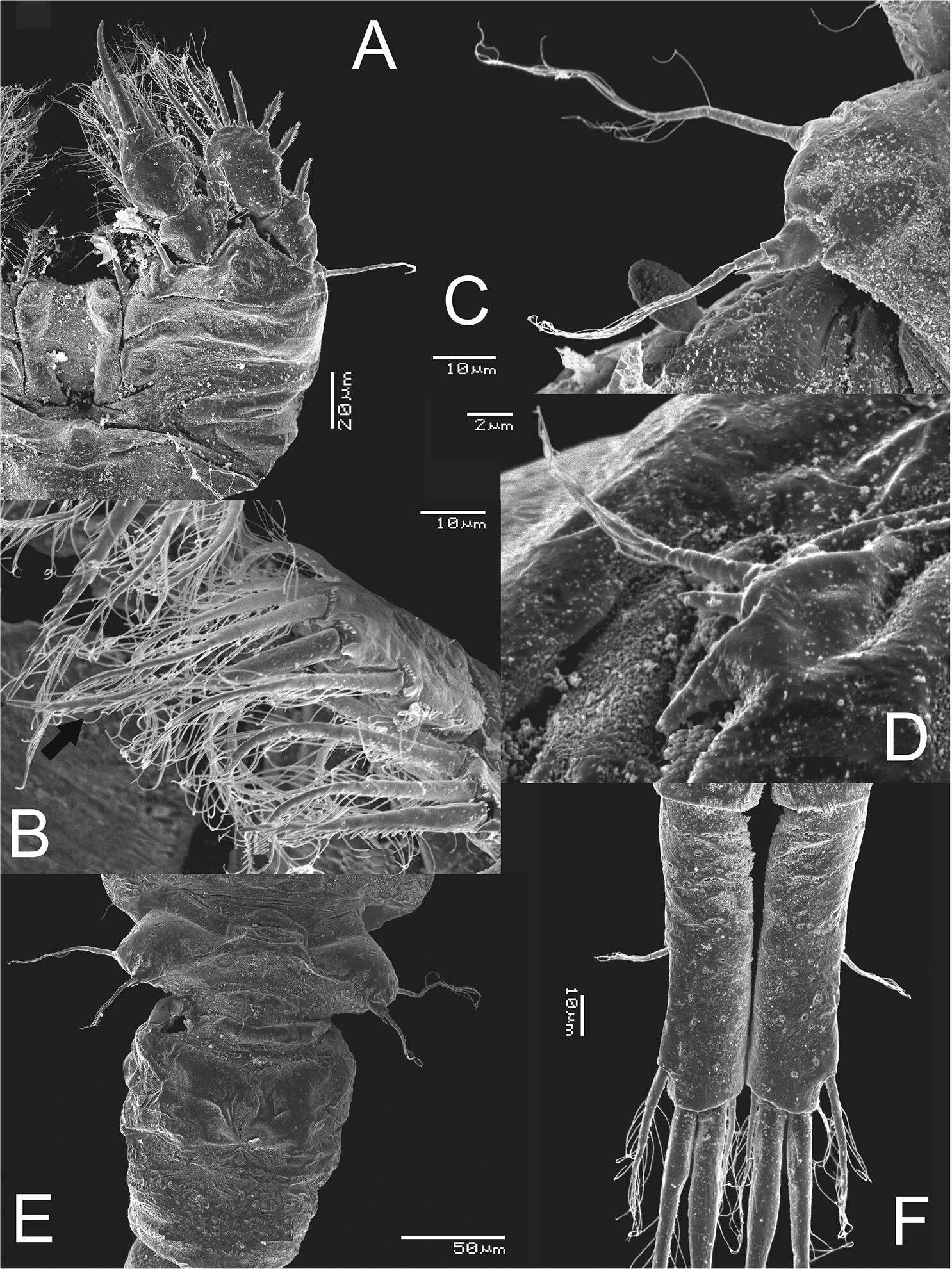
Figure 5.Metacyclops deserticus sp. n., SEM-processed female from Coahuila, México. A leg 1 B endopodite 2 leg 4 C leg 5 D leg 6 E genital double somite, ventral view F caudal ramus, ventral.
-
Terue C. Kihara, Carlos E. F. Rocha
Zookeys
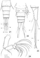
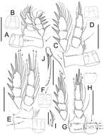
Figures 21–24.Clausidium rodriguesi sp. n. Male: 21 urosome lacking somite bearing P5, dorsal 22 urosome, ventral 23 caudal ramus, dorsal 24 antenna. Scale bars: 21–23 = 20 μm; 24 = 25 μm.
-
Martha Angélica Gutiérrez-Aguirre, Nancy Fabiola Mercado-Salas, Adrián Cervantes-Martínez
Zookeys
Figure 4.Eucyclops tziscao sp. n. A–B paratype C–G allotype from Laguna Tziscao, Chiapas. A Habitus, dorsal B P5, and P6 C Antennule, segments 1–14 D Antennule, segments 15–16 E Antenna, frontal F Antenna, caudal G P4, caudal. Scales bars: B–G = 50 µm; A = 100 µm.
-
Daisuke Uyeno, Kazuya Nagasawa
Zookeys
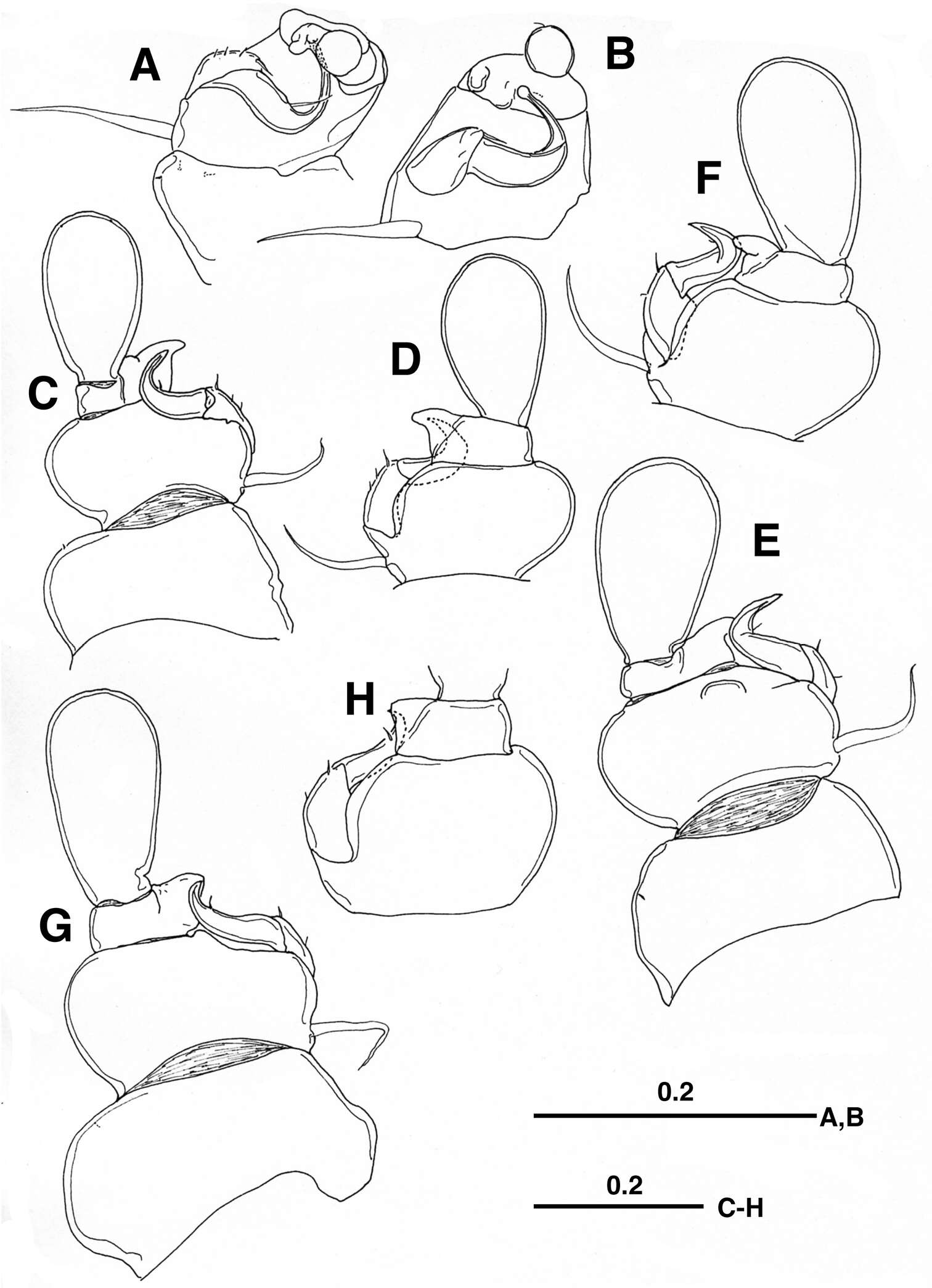
Figure 3.Ceratosomicola japonica sp. n., female, holotype NSMT–Cr 22240 (A–G), female, paratype NSMT–Cr 22243 (H), female, paratype NSMT–Cr 22242 (I). A cephalosome, anterior portion. ventral B antennule, anterior C antenna D mandible E maxilla F leg 1 G leg 2 H, leg 2 (drawn from a paratype, NSMT–Cr 22243) I leg 3. Scale bars = 100 μm in A; 20 μm in B; 30 μm in C, G, H; 10 μm in D, I; 50 μm in E, F.
-
Tomislav Karanovic, Mark J. Grygier, Wonchoel Lee
Zookeys
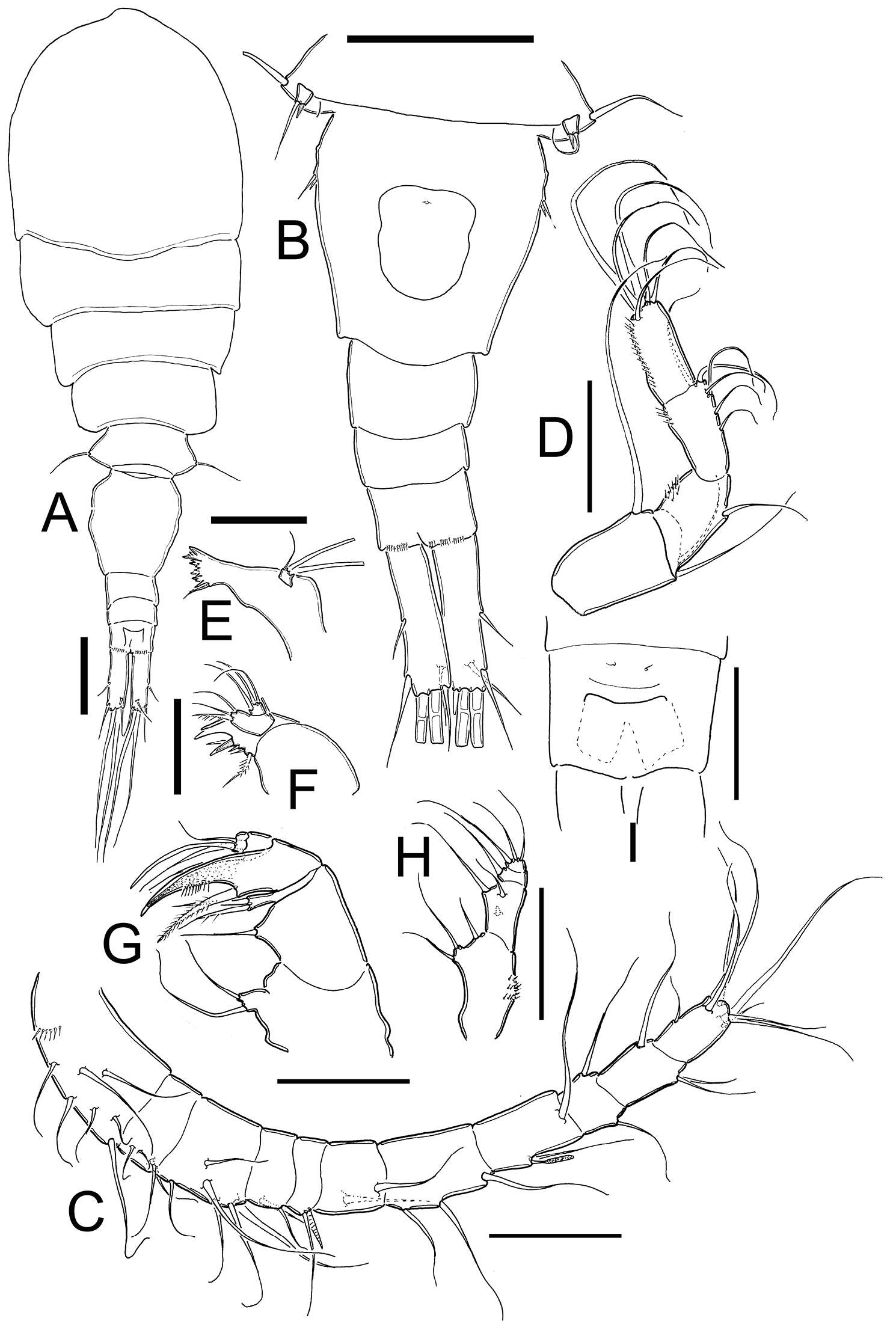
Figure 3.Diacyclops ishidai sp. n., A–E holotype female F paratype female A first swimming leg, anterior view B second swimming leg, anterior view C endopod of third swimming leg, anterior view D fourth swimming leg, anterior view E fifth leg, anterior view F genital double-somite, ventral view. Arabic numerals numbering sensilla and pores consecutively from anterior to posterior end of body, and from dorsal to ventral side (excluding appendages). Scale bars 100 μm.
-
Terue C. Kihara, Carlos E. F. Rocha
Zookeys
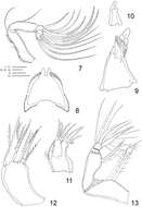
Figures 25–29.Clausidium rodriguesi sp. n. Male: 25 mandible 26 mandible, detail, ventral 27 mandible, detail, dorsal 28 P1, anterior 29 P2, anterior. Scale bars: 25–27 = 10 μm; 28, 29 = 20 μm.
-
Martha Angélica Gutiérrez-Aguirre, Nancy Fabiola Mercado-Salas, Adrián Cervantes-Martínez
Zookeys
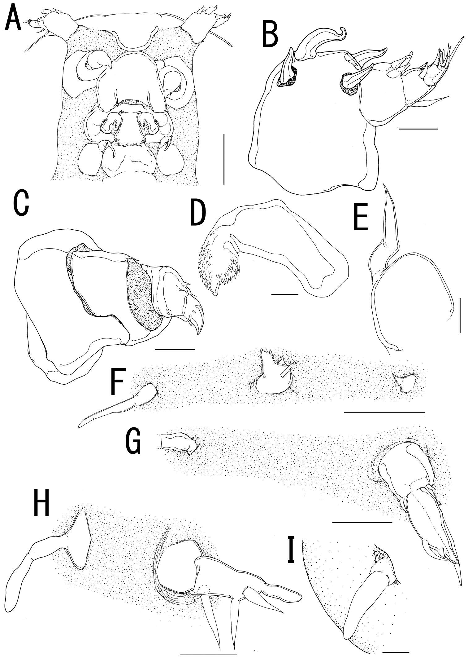
Figure 3.Eucyclops tziscao sp. n. Holotype from Laguna Tziscao, Chiapas. A P1, frontal B Intercoxal sclerite of P1, caudal C P2, frontal D Intercoxal sclerite of P2, caudal E P3, frontal, Exp and Enp separated F Intercoxal sclerite of P3, caudal G P4, caudal H Intercoxal sclerite of P4, frontal I Coxal spine P4 J P5. Scales bars: I= 25µm, J= 50 µm; A–H = 100 µm.
-
Daisuke Uyeno, Kazuya Nagasawa
Zookeys
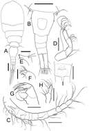
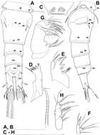
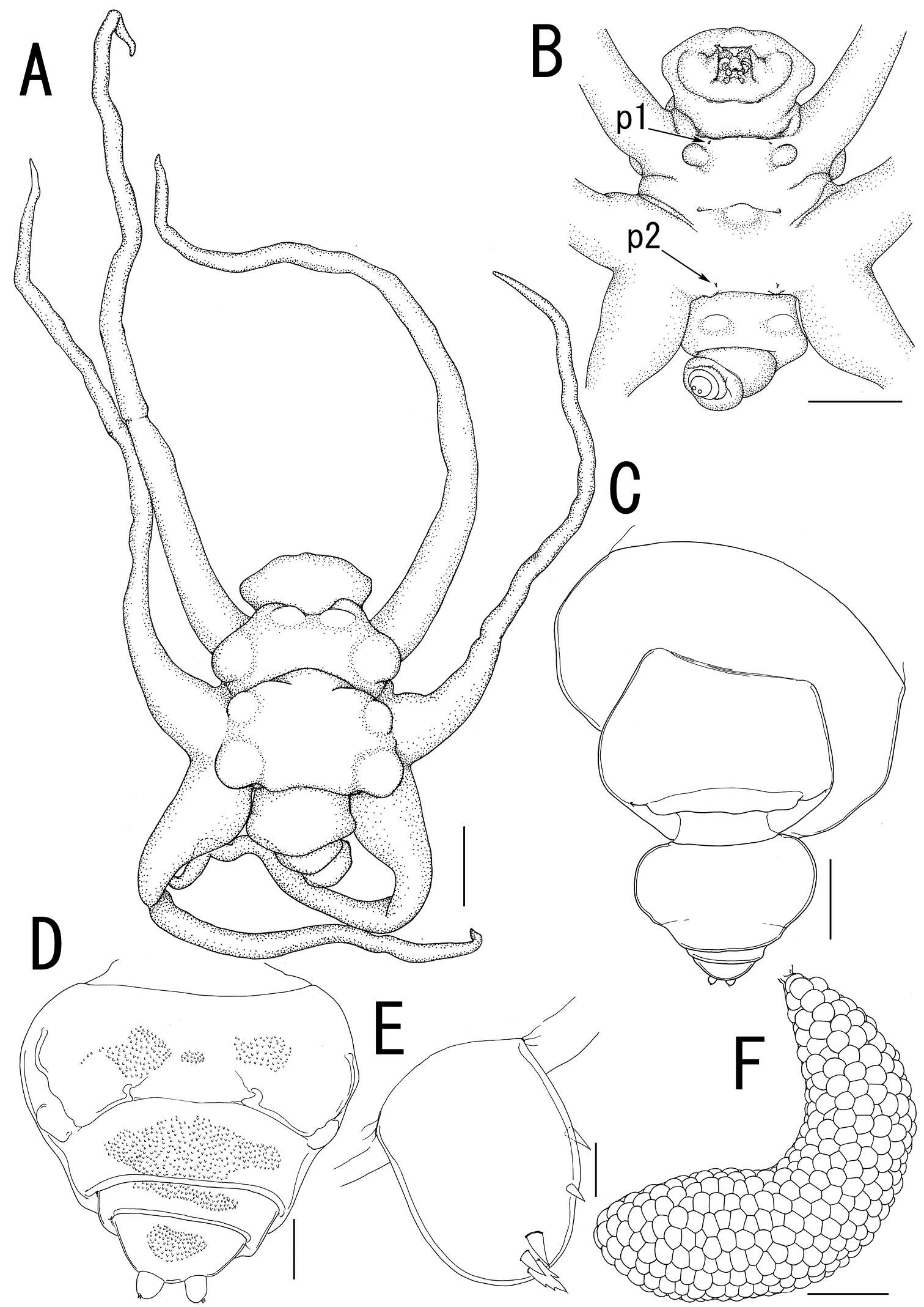
Figure 4.Ceratosomicola japonica sp. n., male, allotype NSMT–Cr 22241. A habitus, dorsal B habitus, ventral, p1 = leg 1, p2 = leg 2 C habitus, lateral D free thoracic somites and abdomen, ventral, p3 = leg 3 E caudal ramus, dorsal F maxilla G leg 1 H leg 2 I leg 3. Scale bars = 500 μm in A, B, C; 200 μm in D; 10 μm in E, I; 20 μm in F, G; 30 μm in H.
-
Tomislav Karanovic, Mark J. Grygier, Wonchoel Lee
Zookeys
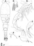
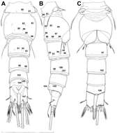
Figure 4.Diacyclops ishidai sp. n., allotype male: A habitus, dorsal view B antennula, flattened and slightly uncoiled, ventral view C middle part of antennula, flattened and uncoiled, dorsal view D third endopodal segment of fourth swimming leg, anterior view E fifth leg, anterior view F sixth leg, ventro-lateral view. Arabic numerals numbering sensilla and pores consecutively from anterior to posterior end of body, and from dorsal to ventral side (excluding appendages). Scale bars 100 μm.
-
Terue C. Kihara, Carlos E. F. Rocha
Zookeys
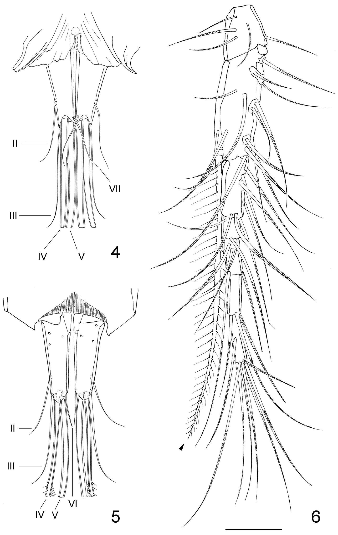
Figures 4–6.Clausidium rodriguesi sp. n. Female: 4 caudal rami, dorsal 5 caudal rami, ventral 6 antennule (arrow head indicating bipinnate seta). Scale bar = 50 μm.
-
Martha Angélica Gutiérrez-Aguirre, Nancy Fabiola Mercado-Salas, Adrián Cervantes-Martínez
Zookeys
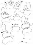
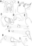
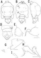
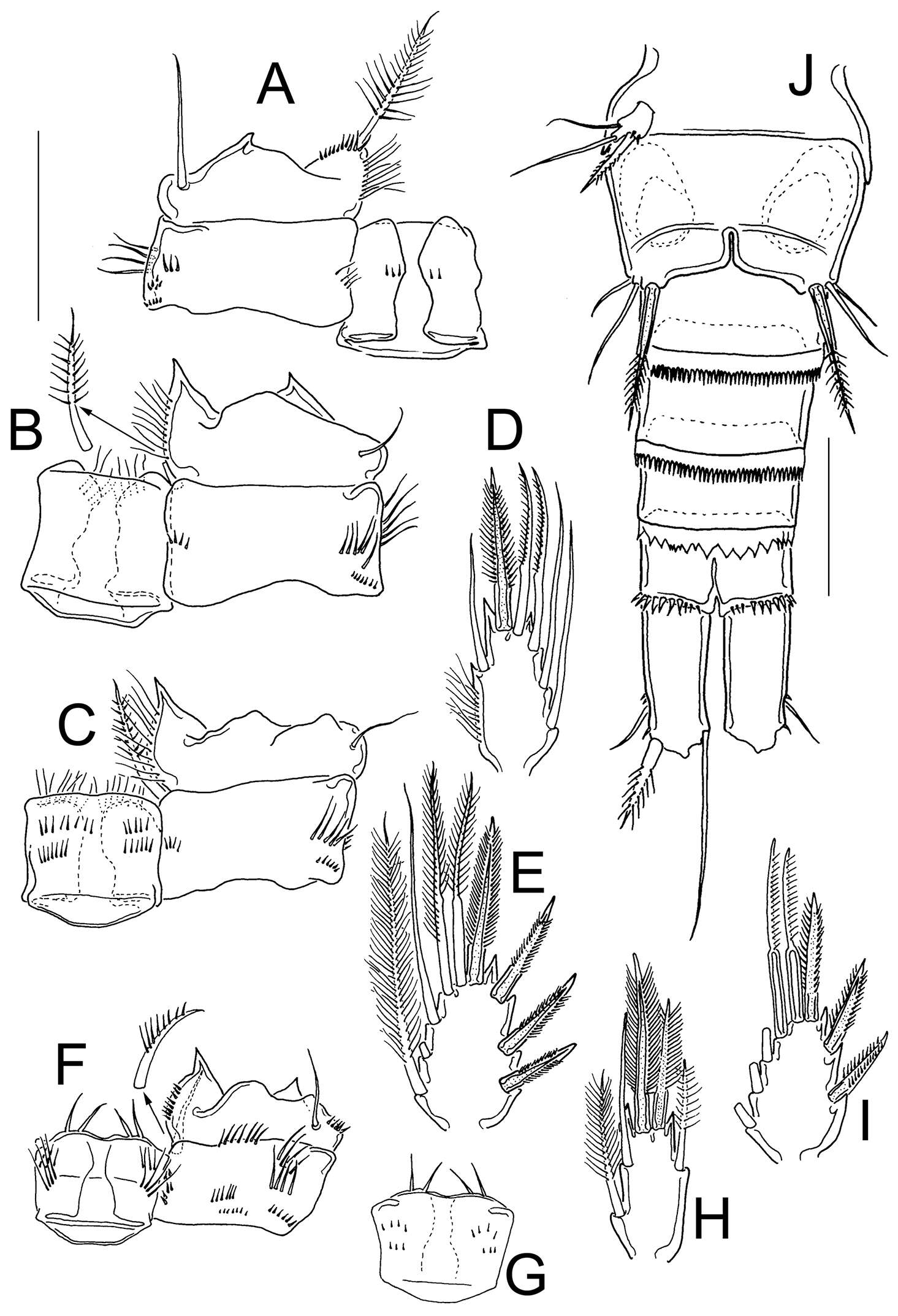
Figure 5.Eucyclops angeli sp. n. A–C paratype D–F holotype from grassland in San Cristóbal de las Casas, Chiapas. A Habitus, dorsal B Second to fourth prosomites, dorsal C Third and fourth prosomites, lateral D Urosome, ventral E Anal somite and one caudal ramus, dorsal F P5. Scale bars 50 µm.
-
Daisuke Uyeno, Kazuya Nagasawa
Zookeys
Figure 1.Live coloration of the host nudibranchs and the splanchnotrophids. A Hypselodoris festiva infected by an ovigerous specimenof Certosomicola japonica sp. n. B an egg sac of Ceratosomicola japonica sp. n. and the gill circle of Hypselodoris festiva with the mantle malformed into an elongate tube C Thecacera pennigera infected by an ovigerous specimen of Splanchnotrophus helianthus sp. n. D Trapania pennigera with the mantle removed to show a female specimen of Splanchnotrophus helianthus on the visceral sac E Trapania miltabrancha infected by an ovigerous specimen of Splanchnotrophus imagawai sp. n (photo by K. Imagawa) F gill circle of Trapania miltabrancha with egg sacs of Splanchnotrophus imagawai sp. n. (photo by K. Imagawa) G Roboastra luteolineata infected by an ovigerous specimen of Majimun shirakawai gen. et sp. n. (photo by N. Shirakawa) H female Majimun shirakawai gen. et sp. n. with dwarf male attached to the posterior part of the body. Scale bars = 5 mm in A; 1 mm in B, D, H.
-
Tomislav Karanovic, Mark J. Grygier, Wonchoel Lee
Zookeys
Figure 5.Diacyclops ishidai sp. n., allotype male: A urosome, dorsal view B urosome, lateral view C urosome, ventral view. Arabic numerals numbering sensilla and pores consecutively from anterior to posterior end of body, and from dorsal to ventral side (excluding appendages). Scale bar 100 μm.
-
Terue C. Kihara, Carlos E. F. Rocha
Zookeys
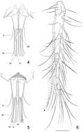
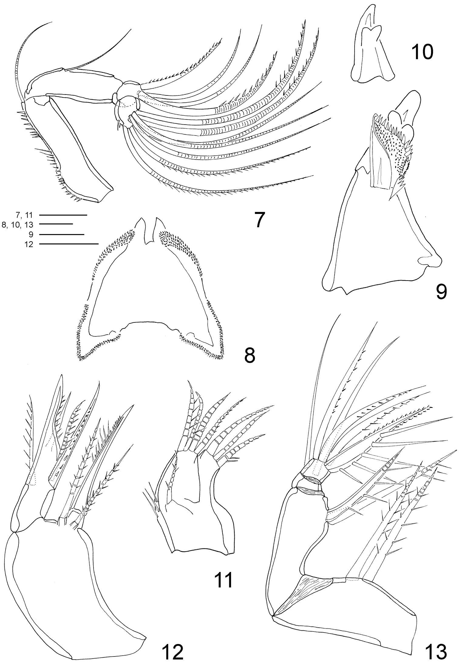
Figures 7–13.Clausidium rodriguesi sp. n. Female: 7 antenna 8 labrum 9 mandible 10 Detail of mandible tooth 11 maxillule 12 maxilla 13 maxilliped. Scale bars: 7 = 50 μm; 8 = 10 μm; 9, 10 = 25 μm; 11–13 = 20 μm.
-
Martha Angélica Gutiérrez-Aguirre, Nancy Fabiola Mercado-Salas, Adrián Cervantes-Martínez
Zookeys
Figure 9.Eucyclops angeli sp. n. Allotype from grassland in San Cristóbal de las Casas, Chiapas. A Coxa, basis, and intercoxal sclerite of P1, caudal B Coxa, basis, and intercoxal sclerite of P2, caudal C Coxa, basis, and intercoxal sclerite of P3, caudal D Enp3P3 E Exp3P3 F Coxa, basis, and intercoxal sclerite of P4, caudal G Intercoxal sclerite of P4, frontal H Enp3P4 I Exp3P4 J Urosome, ventral. Scale bars 50 µm.