-
Daisuke Uyeno, Kazuya Nagasawa
Zookeys
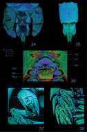
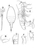
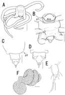
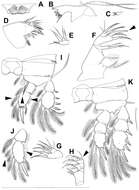
Figure 5.Splanchnotrophus helianthus sp. n., female, holotype NSMT–Cr 22244. A habitus, dorsal A’ enlarged view of egg sac B habitus, ventral, p1 = leg 1, p2 = leg 2 C posterior portion of body, ventral, p3 = leg 3 D fourth pedigerous somite and genito-abdomen, ventral E caudal ramus, dorsal F cephalosome, ventral. Scale bars = 1 mm in A; 100 μm in A’; 500 μm in B; 200 μm in C, F; 100 μm in D; 20 μm in E.
-
Tomislav Karanovic, Mark J. Grygier, Wonchoel Lee
Zookeys
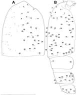
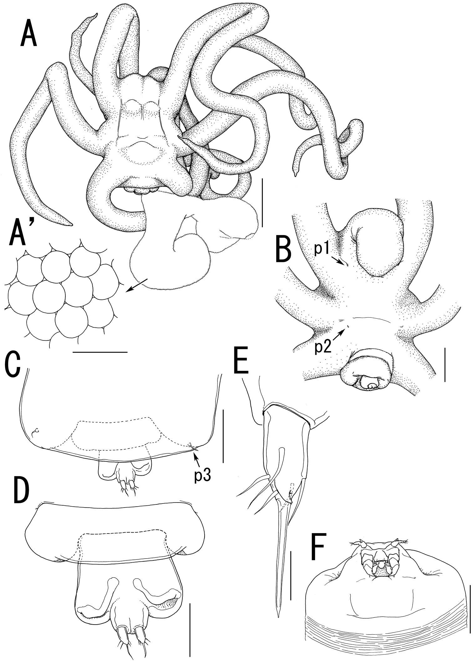
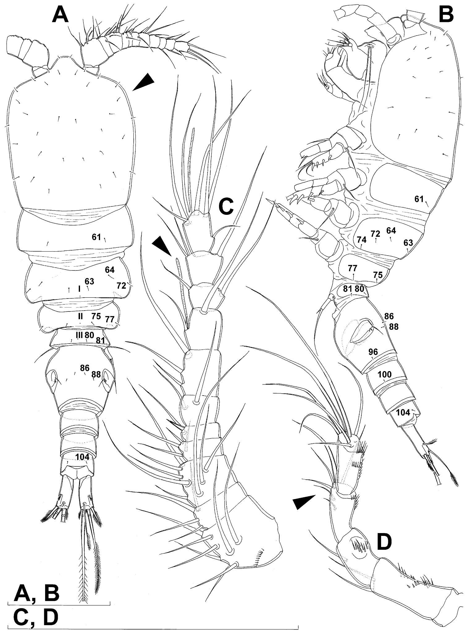
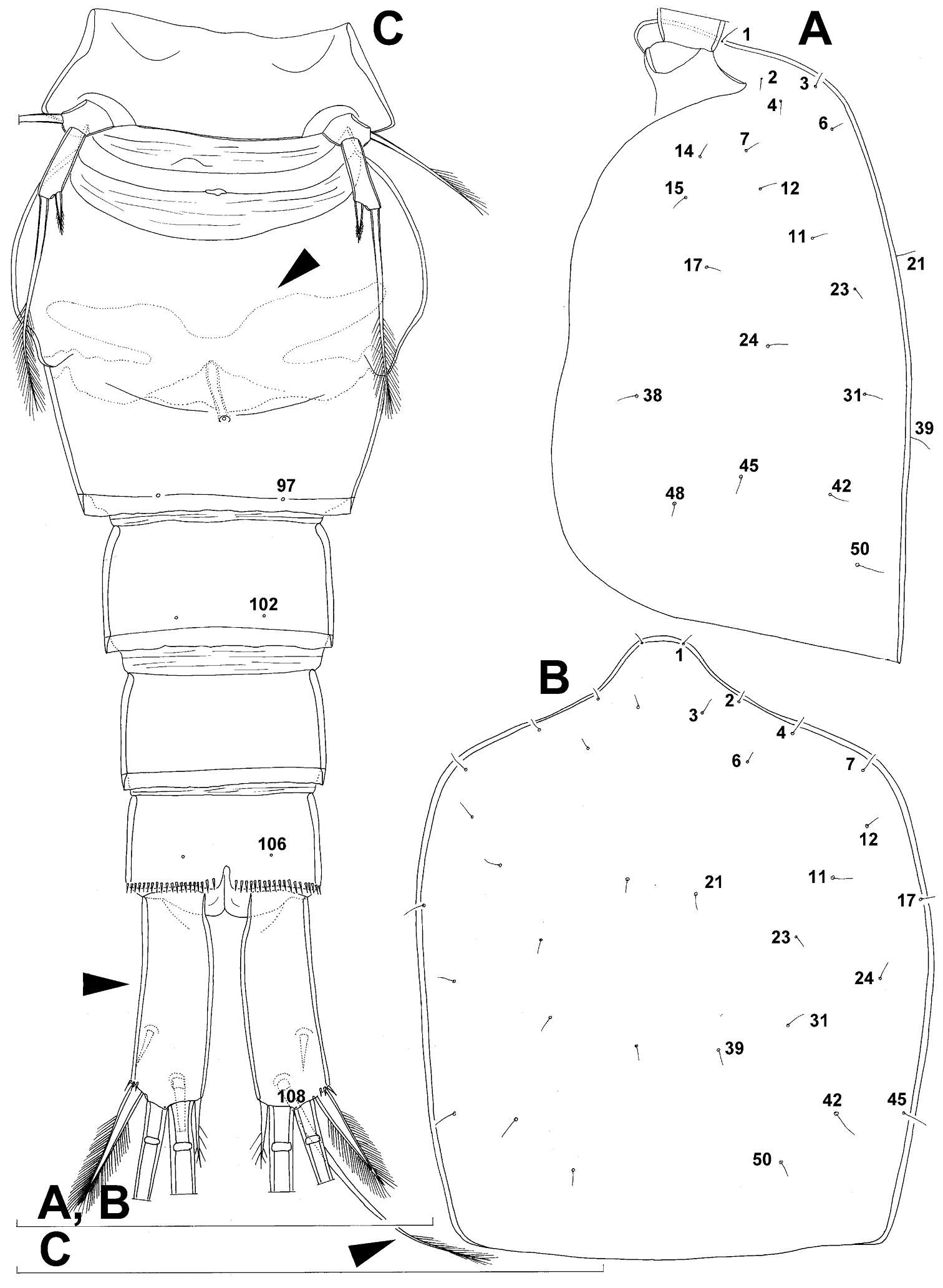
Figure 6.Diacyclops ishidai sp. n., allotype male: A cephalothorax, dorsal view B cephalothoracic shield and pleurons of free prosomites, lateral view. Arabic numerals numbering sensilla and pores consecutively from anterior to posterior end of body, and from dorsal to ventral side (excluding appendages). Scale bar 100 μm.
-
Terue C. Kihara, Carlos E. F. Rocha
Zookeys
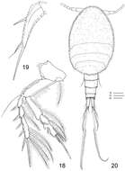
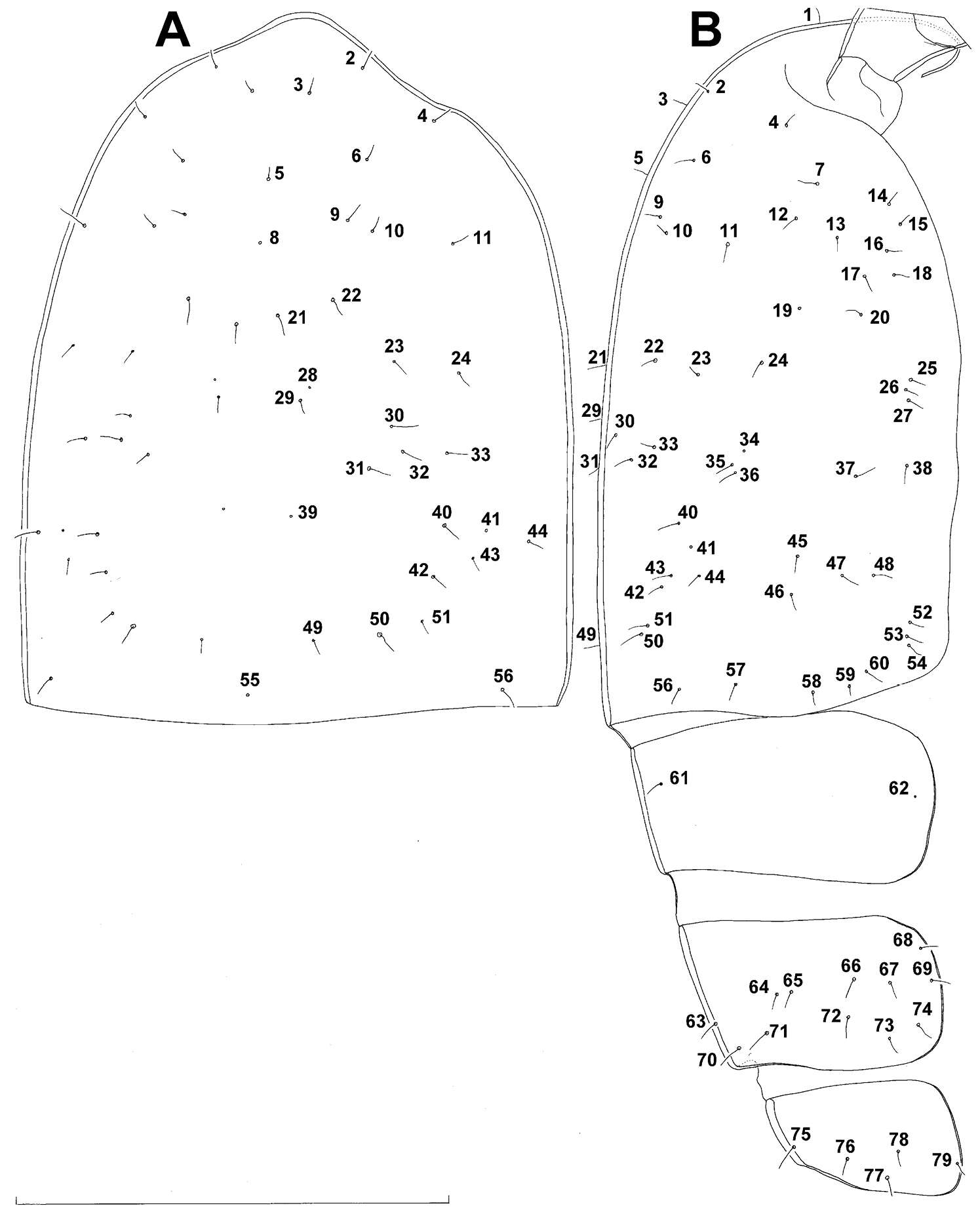
Figures 18–20.Clausidium rodriguesi sp. n. Female: 18 P4, anterior 19 P5, anterior. Male: 20 habitus, dorsal. Scale bars: 18 = 20 μm; 19 = 50 μm; 20 = 100 μm.
-
Martha Angélica Gutiérrez-Aguirre, Nancy Fabiola Mercado-Salas, Adrián Cervantes-Martínez
Zookeys
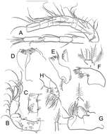
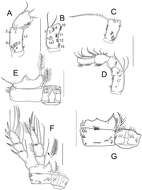
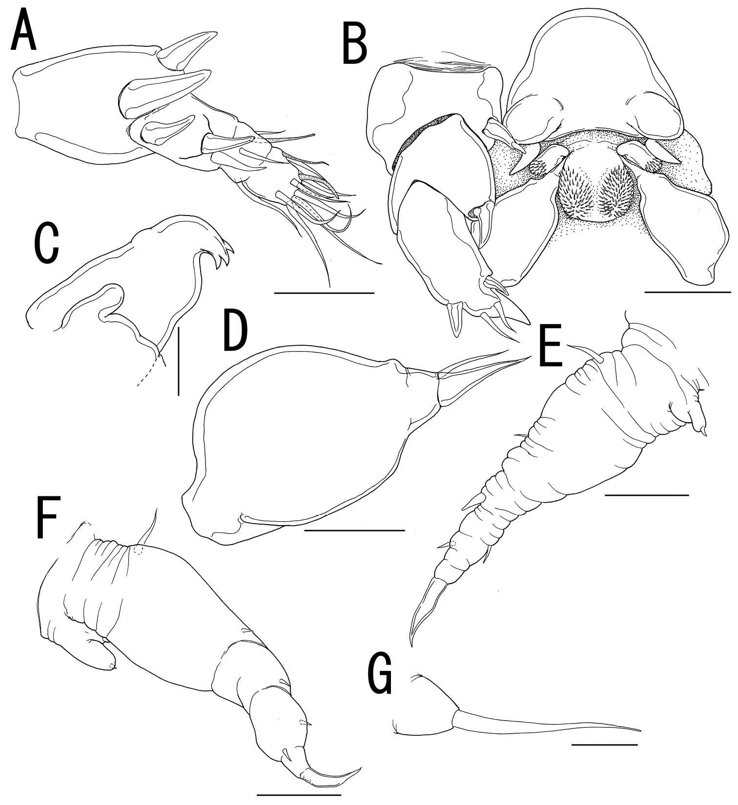
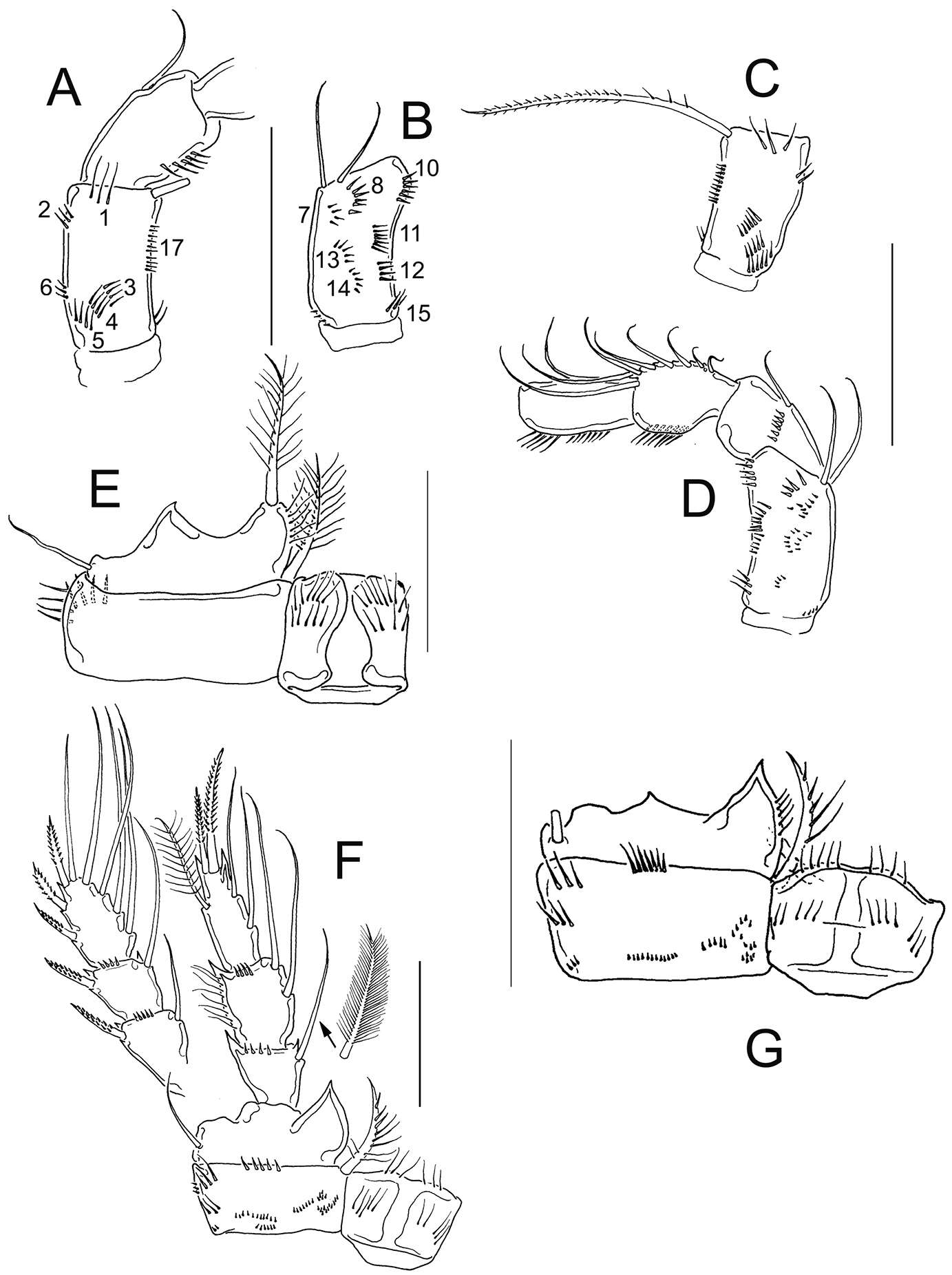
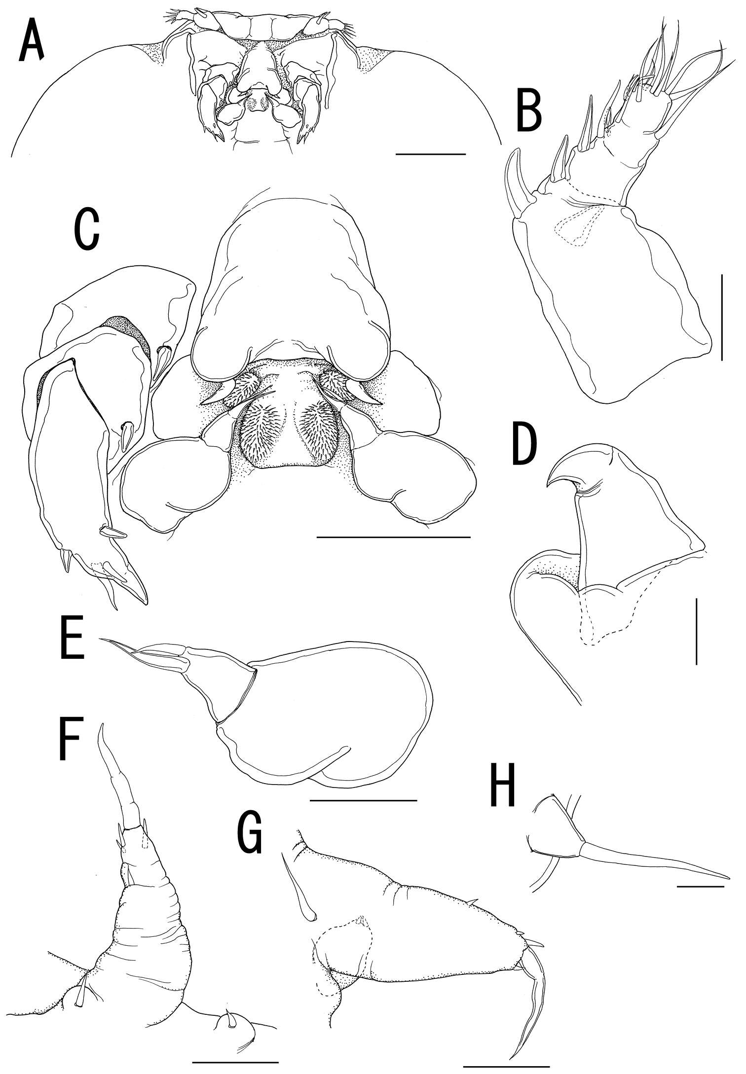
Figure 6.Eucyclops angeli sp. n. Holotype from grassland in San Cristóbal de las Casas, Chiapas. A Antennule B Antenna, caudal C Antenna, frontal D Labrum E Mandible F Maxillule, palp separated G Maxilla, proximal and distal endites of the coxa, separated H Maxilliped, frontal. Scale bar 50 µm.
-
Daisuke Uyeno, Kazuya Nagasawa
Zookeys
Figure 6.Splanchnotrophus helianthus sp. n., female, holotype NSMT–Cr 22244. A antennule, anterior B oral area C mandible, posterior D maxilla E leg 1 F leg 2 G leg 3. Scale bars = 20 μm in A, E, F; 30 μm in B; 10 μm in C, D, G.
-
Tomislav Karanovic, Mark J. Grygier, Wonchoel Lee
Zookeys
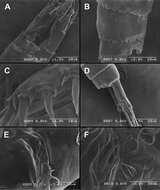
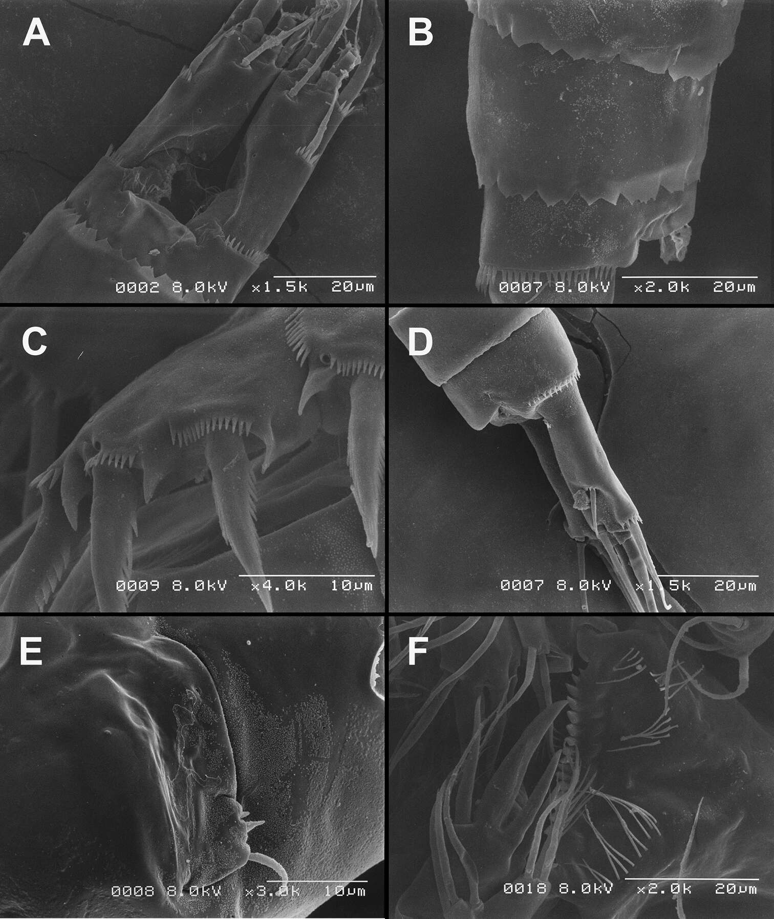
Figure 26.Scanning electron micrographs, A–C Diacyclops ishidai sp. n. D–E Diacyclops parasuoensis sp. n. F Diacyclops suoensis Ito, 1954: A anal somite and caudal rami, dorsal view, paratype female 1 B preanal and anal somites, lateral view, paratype female 2 C last two exopodal segments of second swimming legs, lateral view, paratype female 2 D anal somite and caudal rami, lateral view, paratype female E sixth leg, lateral view, paratype female F labrum and maxillulae, ventral view. Scale bars 20 μm (A, B, D, F) and 10 μm (C, E).
-
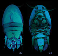
Terue C. Kihara, Carlos E. F. Rocha
Zookeys
Figures 32–33.Clausidium rodriguesi sp. n. Female: Confocal laser scanning microscopy maximum projections33 habitus, dorsal 34 habitus, ventral. Scale bars: 100 μm.
-
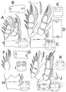
Martha Angélica Gutiérrez-Aguirre, Nancy Fabiola Mercado-Salas, Adrián Cervantes-Martínez
Zookeys
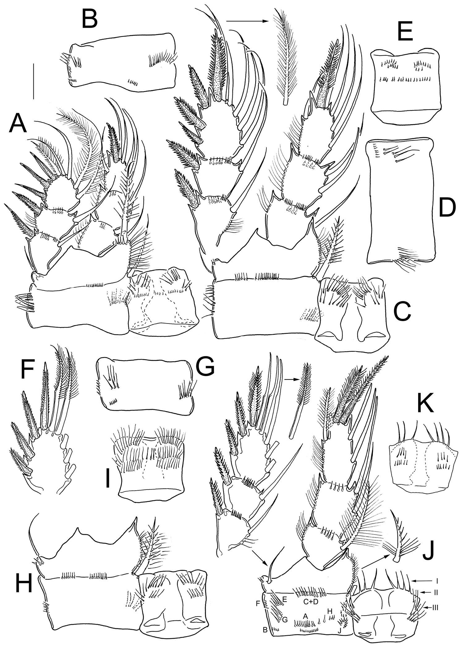
Figure 7.Eucyclops angeli sp. n. Holotype from grassland in San Cristóbal de las Casas, Chiapas. A P1, frontal B Coxa of P1, caudal C P2, frontal, Exp separated D Coxa of P2, caudal E Intercoxal sclerite of P2, caudal F Exp3P3 G Coxa of P3, caudal H Coxa, basis, and intercoxal sclerite of P3, frontal I Intercoxal sclerite P3, caudal J P4, caudal; exopod and coxal spine separated K Intercoxal sclerite of P4, frontal. Scale bar 50 µm.
-
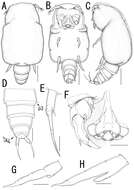
Daisuke Uyeno, Kazuya Nagasawa
Zookeys
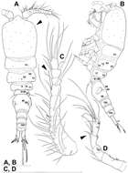
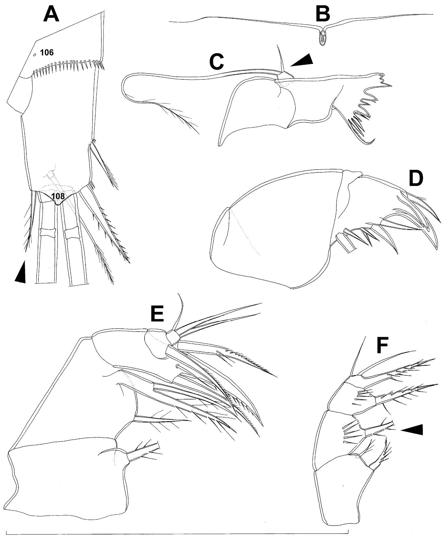
Figure 7.Splanchnotrophus helianthus sp. n., male, allotype NSMT–Cr 22245. A habitus, dorsal B habitus, ventral C habitus, lateral D free thoracic somites and abdomen, ventral E caudal ramus, ventral F oral area G leg 1 H leg 2. Scale bars = 100 μm in A, B, C; 50 μm in D; 20 μm in E.
-
Tomislav Karanovic, Mark J. Grygier, Wonchoel Lee
Zookeys
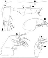
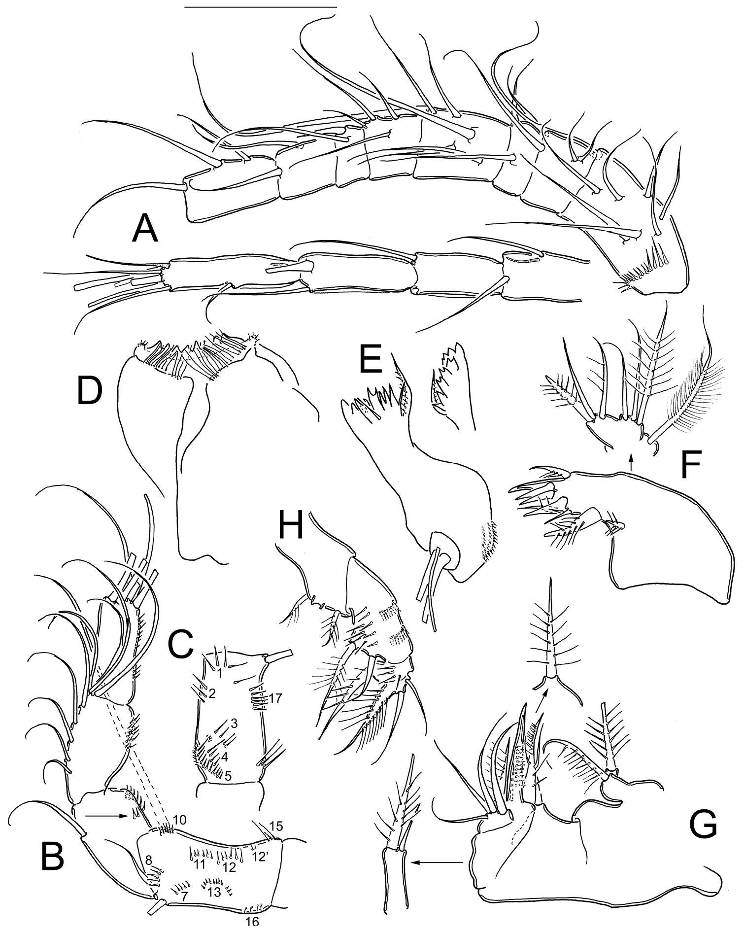
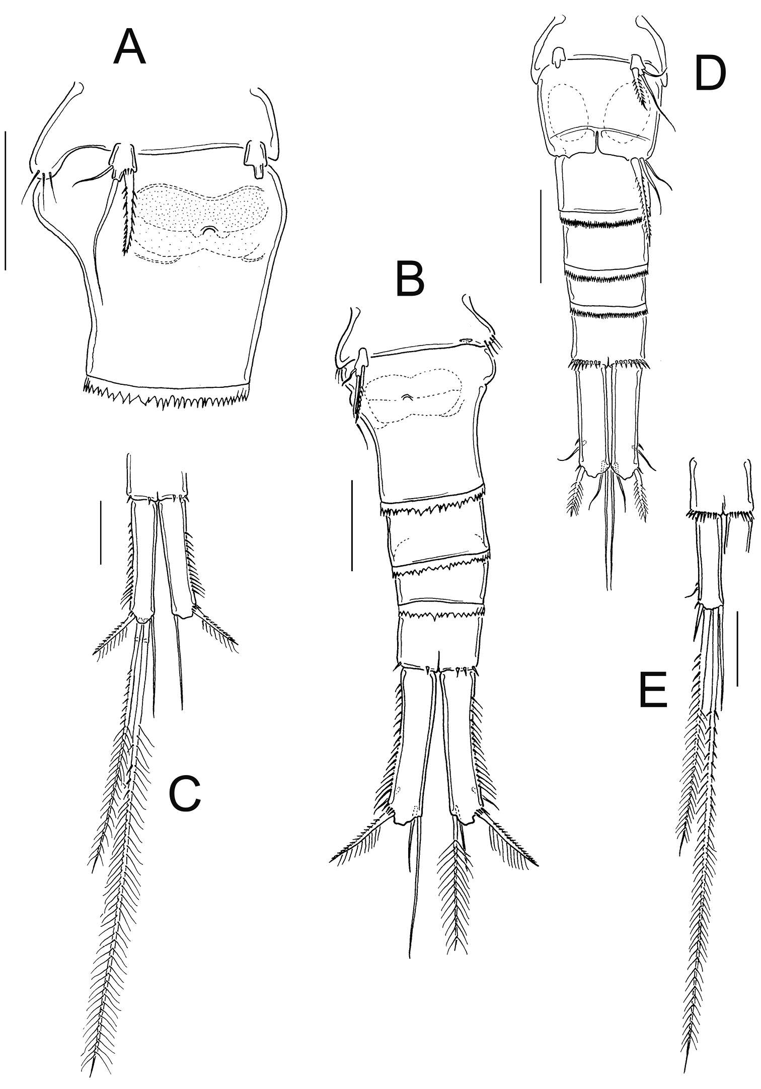
Figure 7.Diacyclops brevifurcus Ishida, 2006, holotype female: A left caudal ramus, ventral view B copulatory pore, ventral view C mandibula, posterior view D maxillula, posterior view (palp broken off) E maxilla, anterior view F maxilliped, anterior view. Arabic numerals indicating sensilla and pores presumably homologous to those in Diacyclops ishidai sp. n. Arrows pointing most prominent specific features. Scale bar 100 μm.
-
Terue C. Kihara, Carlos E. F. Rocha
Zookeys
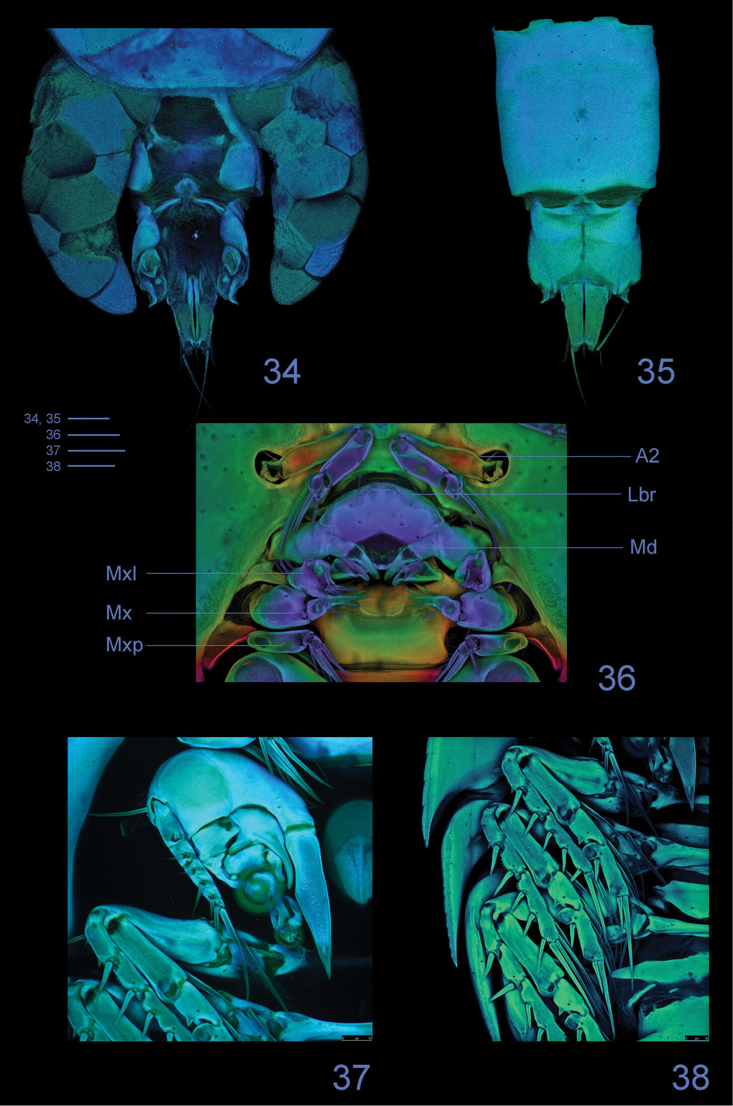
Figures 34–38.Clausidium rodriguesi sp. n. Female: Confocal laser scanning microscopy maximum projections 34 urosome, dorsal 35 urosome lacking somite bearing P5, ventral 36 antenna and oral region 37 P1, anterior 38 P2-P4, anterior. Scale bars: 50 μm.
-
Martha Angélica Gutiérrez-Aguirre, Nancy Fabiola Mercado-Salas, Adrián Cervantes-Martínez
Zookeys
Figure 8.Eucyclops angeli sp. n. A–B paratype C–F allotype from grassland in San Cristóbal de las Casas, Chiapas. A Habitus, dorsal B Third, and fourth prosomites, lateral C First to fourth urosomites, lateral D Anal somite and caudal ramus, dorsal E Antennule, last two segments separated F Antenna, caudal G Antenna, frontal. Scale bars 50 µm.
-
Daisuke Uyeno, Kazuya Nagasawa
Zookeys
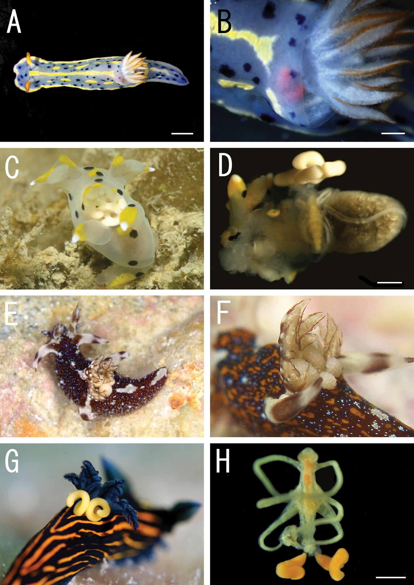
Figure 1.Live coloration of the host nudibranchs and the splanchnotrophids. A Hypselodoris festiva infected by an ovigerous specimenof Certosomicola japonica sp. n. B an egg sac of Ceratosomicola japonica sp. n. and the gill circle of Hypselodoris festiva with the mantle malformed into an elongate tube C Thecacera pennigera infected by an ovigerous specimen of Splanchnotrophus helianthus sp. n. D Trapania pennigera with the mantle removed to show a female specimen of Splanchnotrophus helianthus on the visceral sac E Trapania miltabrancha infected by an ovigerous specimen of Splanchnotrophus imagawai sp. n (photo by K. Imagawa) F gill circle of Trapania miltabrancha with egg sacs of Splanchnotrophus imagawai sp. n. (photo by K. Imagawa) G Roboastra luteolineata infected by an ovigerous specimen of Majimun shirakawai gen. et sp. n. (photo by N. Shirakawa) H female Majimun shirakawai gen. et sp. n. with dwarf male attached to the posterior part of the body. Scale bars = 5 mm in A; 1 mm in B, D, H.
-
Tomislav Karanovic, Mark J. Grygier, Wonchoel Lee
Zookeys
Figure 8.Diacyclops brevifurcus Ishida, 2006, holotype female: A third swimming leg, posterior view B fourth swimming leg, posterior view C fifth leg, anterior view. Arrows pointing most prominent specific features. Scale bars 100 μm.
-
Terue C. Kihara, Carlos E. F. Rocha
Zookeys
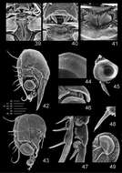
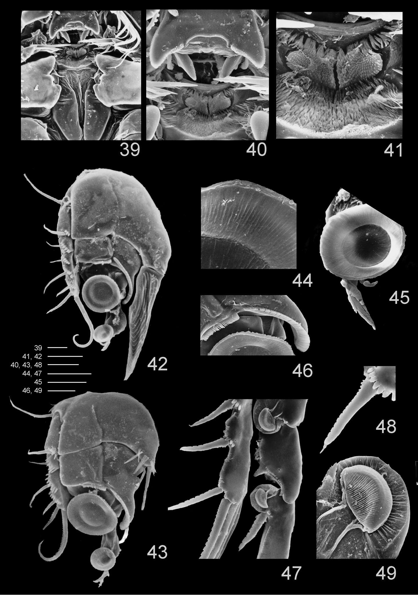
Figures 39–49.Clausidium rodriguesi sp. n.: Scanning electron microscopy photos 39 metastomal area, male 40 detail of metastomal area, male 41 detail of metastomal area, male 42 P1, anterior, female 43 P1, anterior, male 44 detail of sucking disc of P1, male 45 detail of lobe with serrate margin and distal sucking disc of enp-3 of P1, female 46 detail of P1 enp-1 adhesive fringe, male 47 sucking discs of P2, female 48 detail of serrate spine with apical flagellum of P2, female 49 detail of sucking disc of P2, female. Scale bars: 39, 40, 47 = 25 μm; 41, 44–46 = 10 μm; 42 = 35 μm; 43 = 20 μm; 48 = 12.5 μm; 49 = 4 μm.
-
Martha Angélica Gutiérrez-Aguirre, Nancy Fabiola Mercado-Salas, Adrián Cervantes-Martínez
Zookeys
Figure 10.Eucyclops festivus Lindberg, 1955; from pond 3 to Laguna Montebello, Chiapas. A First urosomite, and genital double-somite, ventral B Urosome, ventral C Anal somite and caudal ramus, ventral D Urosome, ventral E Anal somite and caudal ramus, ventral. Scale bars 50 µm. A–C female; D–E male.
-
Daisuke Uyeno, Kazuya Nagasawa
Zookeys
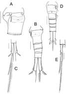
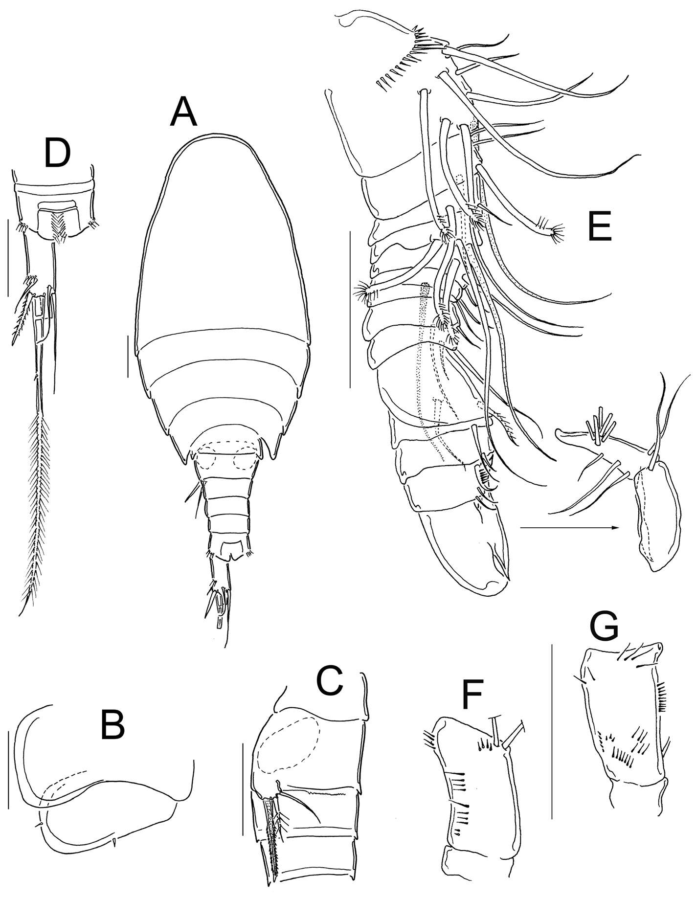
Figure 8.Splanchnotrophus imagawai sp. n., female, holotype NSMT–Cr 22249. A habitus, dorsal B habitus, ventral, p1 = leg 1, p2 = leg 2 C posterior portion of body, ventral, p3 = leg 3 D fourth pedigerous somite and genito-abdomen, ventral E caudal ramus, ventral F egg sac. Scale bars = 1 mm in A; 500 μm in B, F; 200 μm in C; 100 μm in D; 20 μm in E.
-
Tomislav Karanovic, Mark J. Grygier, Wonchoel Lee
Zookeys
Figure 9.Diacyclops parasuoensis sp. n., holotype female: A habitus, dorsal view B habitus, lateral view C antennula, ventral view D antenna, dorsal view. Arabic numerals indicating sensilla and pores presumably homologous to those in Diacyclops ishidai sp. n. Roman numerals indicating pores not present in Diacyclops ishidai sp. n. Arrows pointing most prominent specific features. Scale bars 100 μm.
-
Terue C. Kihara, Carlos E. F. Rocha
Zookeys
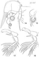
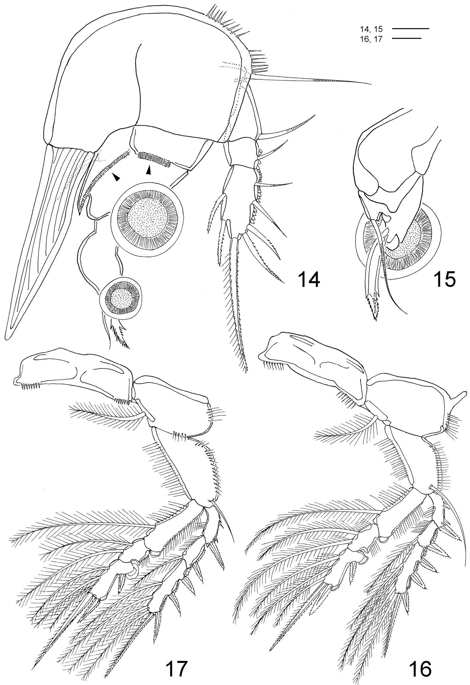
Figures 14–17.Clausidium rodriguesi sp. n. Female: 14 P1, anterior (arrows indicating adhesive fringe) 15 detail of distal area of P1 endopod, posterior 16 P2, anterior 17 P3, anterior. Scale bars: 14 = 20 μm; 15 = 10 μm; 16, 17 = 50 μm.
-
Martha Angélica Gutiérrez-Aguirre, Nancy Fabiola Mercado-Salas, Adrián Cervantes-Martínez
Zookeys
Figure 11.Eucyclops festivus Lindberg, 1955; from pond 3 to Laguna Montebello, Chiapas. A Antenna, frontal B Antenna, caudal C Antenna, frontal D Antenna, caudal E Coxa, basis, and intercoxal sclerite of P1, frontal F P4, caudal, Exp, and one inner seta separated G Coxa, basis, and intercoxal sclerite of P4, caudal. Scale bars 50 µm. A, B, E, F female; C, D, G male.
-
Daisuke Uyeno, Kazuya Nagasawa
Zookeys
Figure 9.Splanchnotrophus imagawai sp. n., female, holotype NSMT–Cr 22249. A anterior portion of cephalosome B antennule, ventral C oral area D mandible, posterior E maxilla F leg 1 G leg 2 H leg 3. Scale bars = 100 μm in A; 50 μm in B, C; 10 μm in D, H; 20 μm in E, F, G.
-
Tomislav Karanovic, Mark J. Grygier, Wonchoel Lee
Zookeys
Figure 10.Diacyclops parasuoensis sp. n., holotype female: A cephalothoracic shield, lateral view B cephalothorax, dorsal view C urosome, ventral view. Arabic numerals indicating sensilla and pores presumably homologous to those in Diacyclops ishidai sp. n. Arrows pointing most prominent specific features. Scale bars 100 μm.
-
Terue C. Kihara, Carlos E. F. Rocha
Zookeys
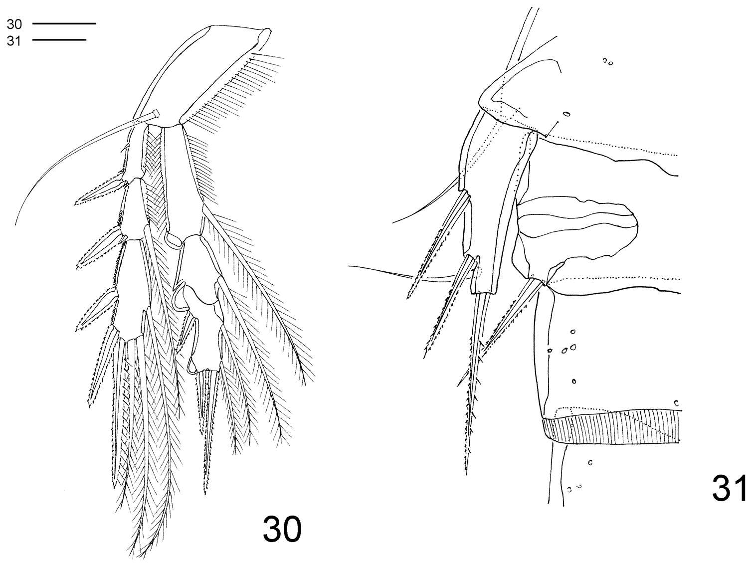
Figures 30–31.Clausidium rodriguesi sp. n. Male: 30 P4, anterior 31 P5 and P6. Scale bar: 20 μm.
-
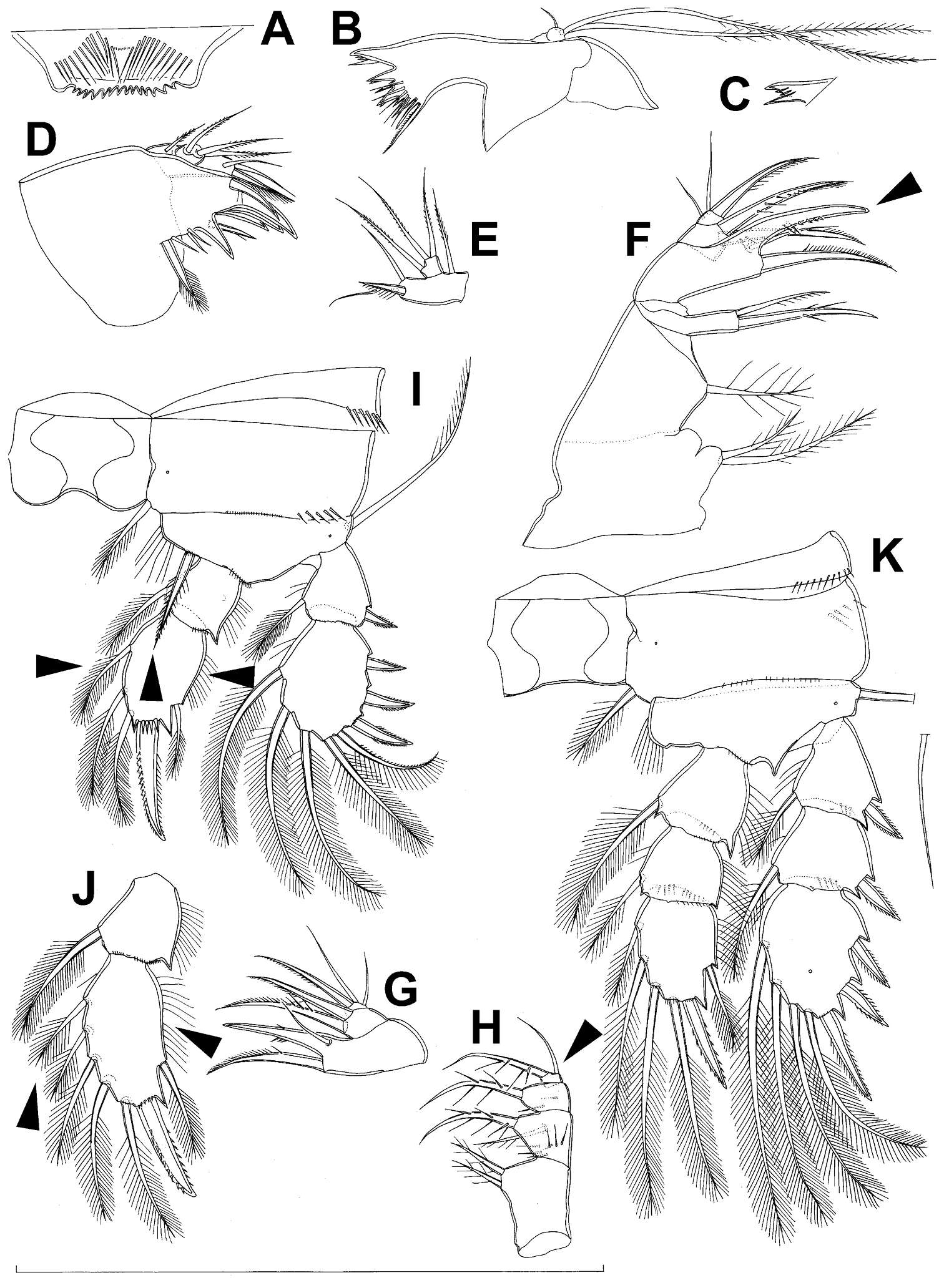
Tomislav Karanovic, Mark J. Grygier, Wonchoel Lee
Zookeys
Figure 11.Diacyclops parasuoensis sp. n., holotype female: A labrum, anterior view B mandibula, anterior view C quadricuspidate ventralmost tooth of mandibula, posterior view D maxillula, posterior view E maxillular palp, anterior view F maxilla, anterior view G basis and endopod of maxilla, posterior view H maxilliped, posterior view I first swimming leg, anterior view J endopod of second swimming leg, anterior view K third swimming leg, anterior view. Arrows pointing most prominent specific features. Scale bar 100 μm.