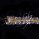
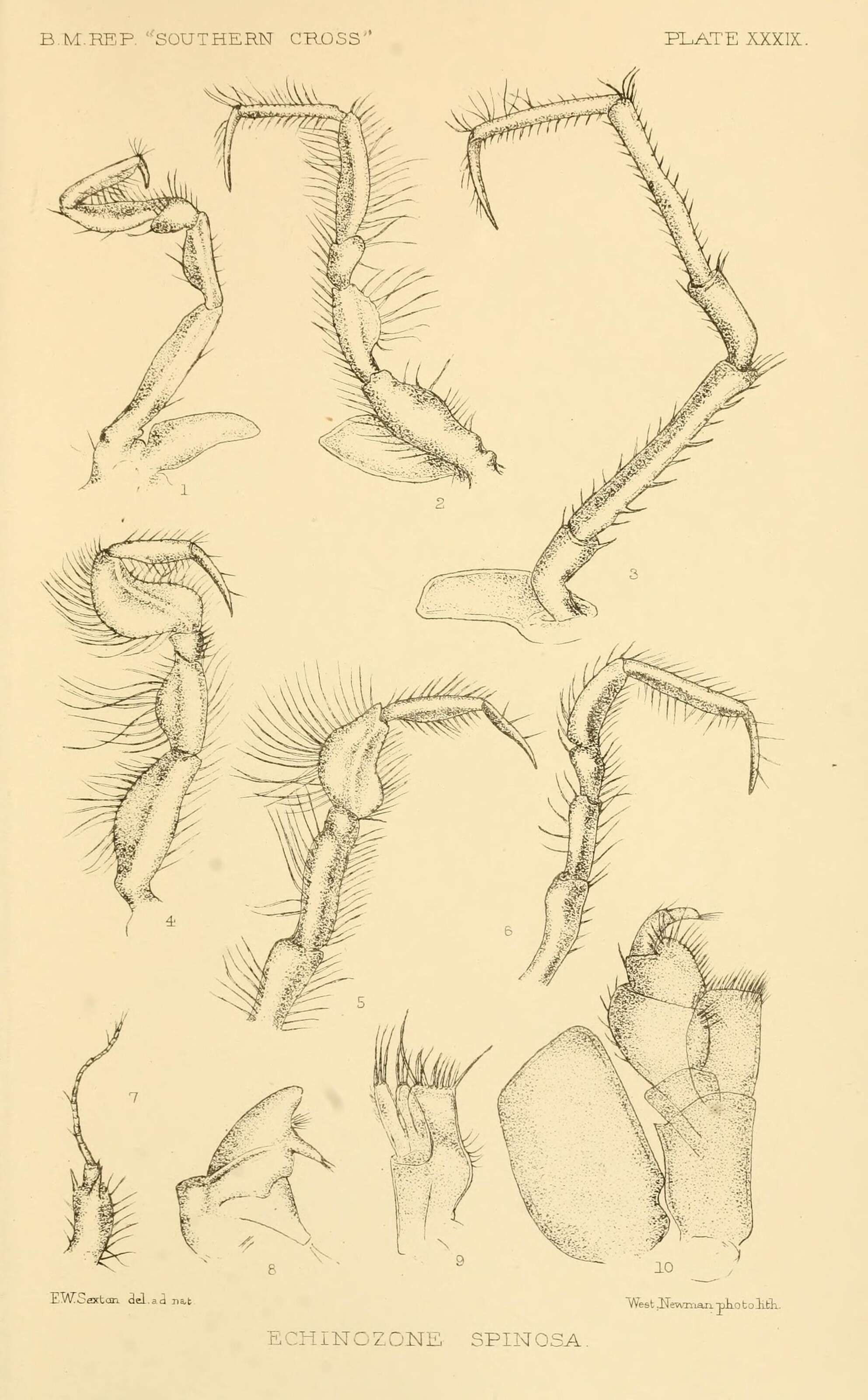
Report on the collections of natural history made in the Antarctic regions during the voyage of the "Southern Cross.".
London :Printed by order of the Trustees,1902..
biodiversitylibrary.org/page/12554348
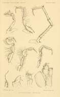
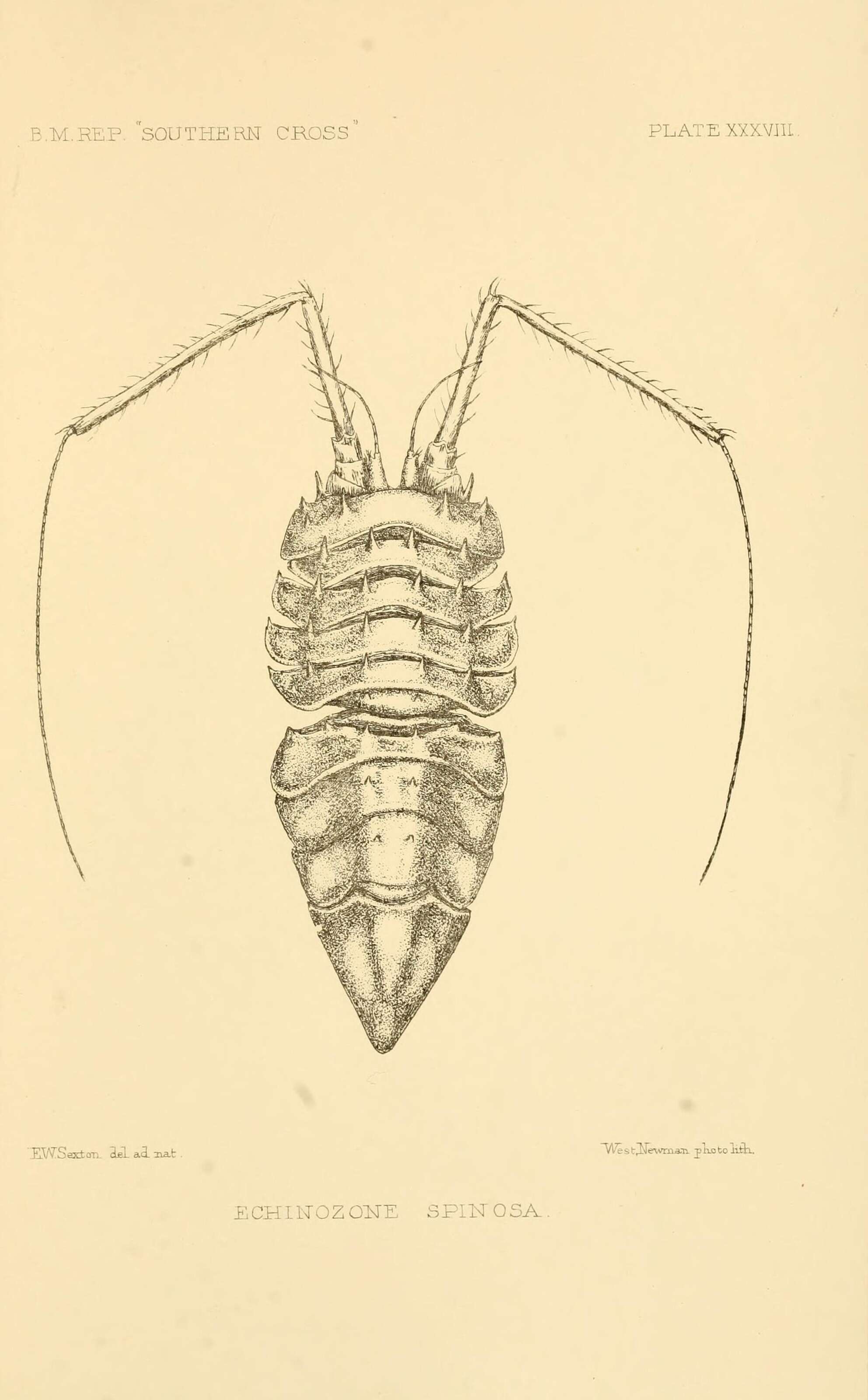
Report on the collections of natural history made in the Antarctic regions during the voyage of the "Southern Cross.".
London :Printed by order of the Trustees,1902..
biodiversitylibrary.org/page/12554346
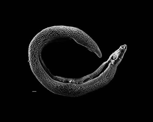
Description: English: Electron micrograph of an adult male Schistosoma parasite worm. The bar (bottom left) represents a magnification of 500 μm. Français : Photographie au micrographe électronique d'un ver parasite Schistosoma adulte mâle. La barre (en bas à gauche) représente une longueur de 500 μm. Date: 2 September 2009. Source: http://www.genome.gov/dmd/img.cfm?node=Photos/Animals/Trihinella (NHGRI-79094.jpg, formerly 20041-300.jpg) Transferred from en.wikipedia to Commons by User:Magnus Manske using CommonsHelper. Author: David Williams, Illinois State University. Permission(Reusing this file): Page states "This image is freely available and may be used without special permission." See also http://www.genome.gov/copyright.cfm.
No machine-readable author provided. Fvanhout assumed (based on copyright claims).
Wikimedia Commons
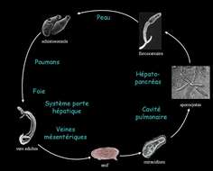
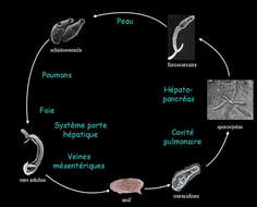
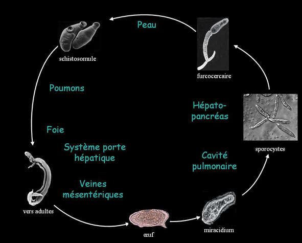
Description: Cycle de vie du schistosome. Date: 30 November 2006 (original upload date). Source: No machine-readable source provided. Own work assumed (based on copyright claims). Author: No machine-readable author provided. Fvanhout assumed (based on copyright claims).
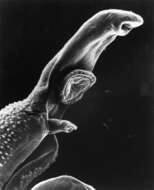
Description: Title: Pathology: SEM: Schistosome Parasite Description: This schistosome parasite enters the body through the skin of persons coming in contact with infested waters. The adult worm lives in the veins of its host. The parasite is magnified x256 in this photograph. Subjects (names): Topics/Categories: Pathology -- Scanning Electron Microscopy (SEM) Type: Black & White Print. Black & White Slide Source: National Cancer Institute Author: Bruce Wetzel (photographer). Harry Schaefer (phot AV Number: AV-0000-3692 Date Created: Unknown Date Entered: 1/1/2001 Access: Public. Source: http://visualsonline.cancer.gov/details.cfm?imageid=1762. Author: Bruce Wetzel (photographer). Harry Schaefer (phot. Permission(Reusing this file): PD.
No machine-readable author provided. Fvanhout assumed (based on copyright claims).
Wikimedia Commons
Description: Cycle de vie du schistosome Image perso. Date: 30 November 2006 (original upload date). Source: No machine-readable source provided. Own work assumed (based on copyright claims). Author: No machine-readable author provided. Fvanhout assumed (based on copyright claims).
Description: Micrografia eletrônica de um verme adulto masculino Schistosoma parasita. Date: 16 December 2013 (original upload date). Source: No machine-readable source provided. Own work assumed (based on copyright claims). Author: No machine-readable author provided. IcaroGustavo assumed (based on copyright claims).
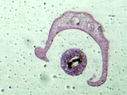
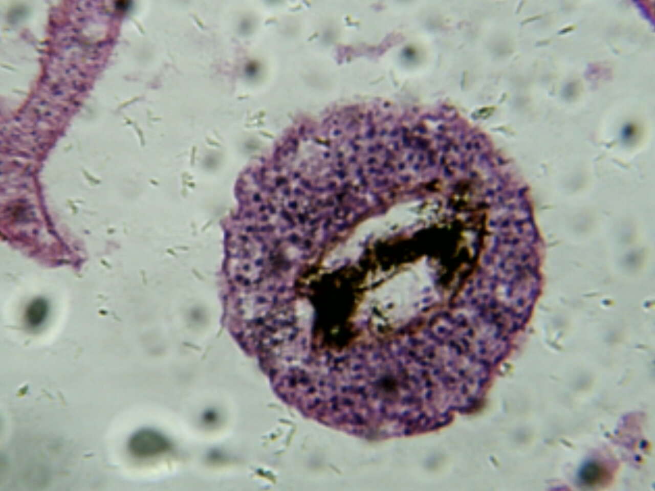
Description: Schistosoma - Male (in the centre) and Female - Cross Section. Date: 11 February 2010. Source: Own work. Author: Ciência e Saúde XXI. Permission (Reusing this file): Free to use. Other versions: Pt: Schistosoma - Macho (no centro) e fêmea - Secção transversal.
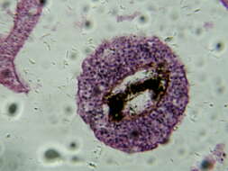
Description: Schistosoma - Male (Cross Section). Date: 11 February 2010. Source: Own work. Author: Ciência e Saúde XXI. Permission (Reusing this file): Free to use. Other versions: Pt: Schistosoma - Macho (Secção Transversal).
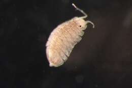
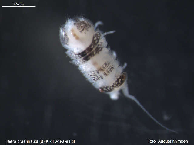
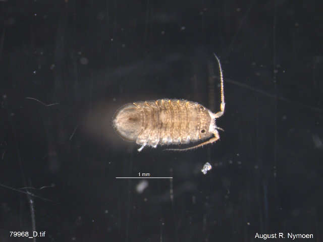
Description: English: A cluster of Munnid isopods (Munnidae sp.) in the Aquarium of the Bay's Octopus and Friends Gallery exhibit. They can be found on the top left corner of the filter-feeder tank’s viewing window. Date: 22 November 2021. Source: Own work. Author: Coughdrop12.
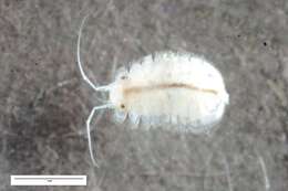
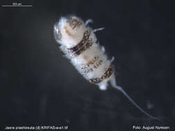
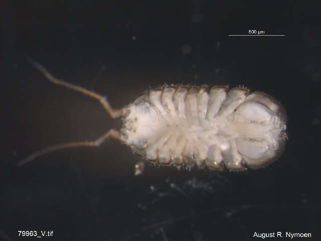
Luisa Borges. Universidade do Minho. Luisa Borges. Year: 2012. Contact: luisa.borges@bio.uminho.pt; luisaborges2000@yahoo.co.uk.
Barcode of Life Data Systems
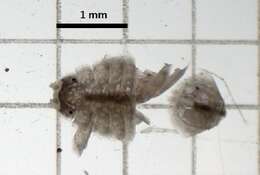
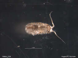
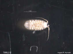
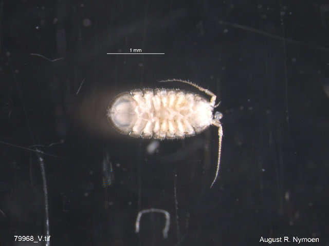
Full body. Specimen ID: 1122485. Field no.: 58-Bio_M2-081020-4-1. Taxon rep.: Jaera nordmanni. Image quality: 1. Aspect ratio: 1.347.
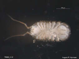
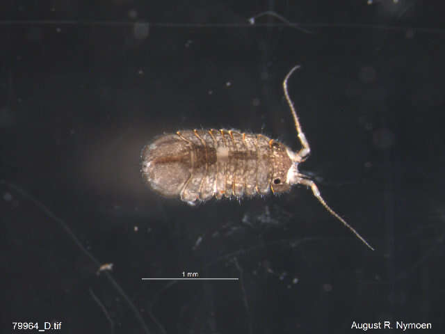
Zoologisches Forschungsmuseum Alexander Koenig. Year: 2018. Contact: Zoologisches Forschungsmuseum Alexander Koenig, Museumsmeile Bonn, Adenauerallee 160, D-53113 Bonn, Germany, barcoding@zfmk.de.
Barcode of Life Data Systems