-
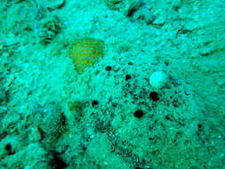
Coral Sea, Duration 15 seconds
-
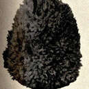
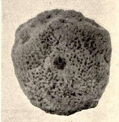
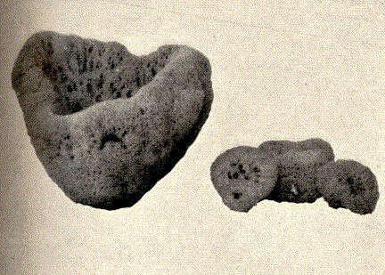
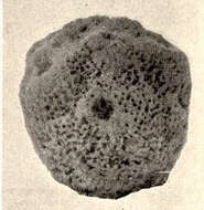
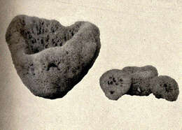
Hippospongia canaliculata, var. gossypina.
-
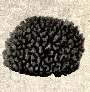
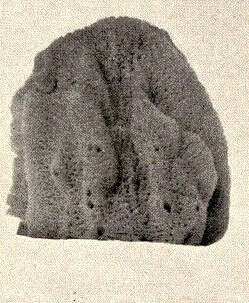
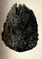
Hippospongia equina, var. meandriformis.
-
Renata Manconi, Barbara Cadeddu, Fabio Ledda, Roberto Pronzato
Zookeys
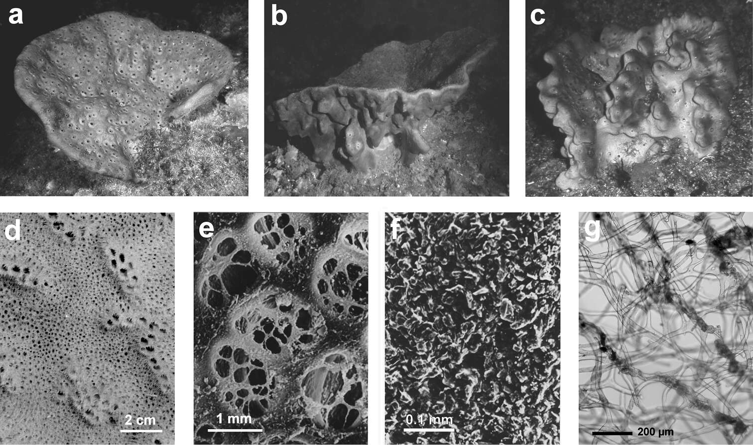
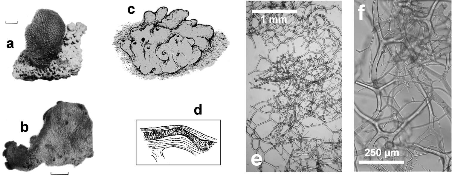
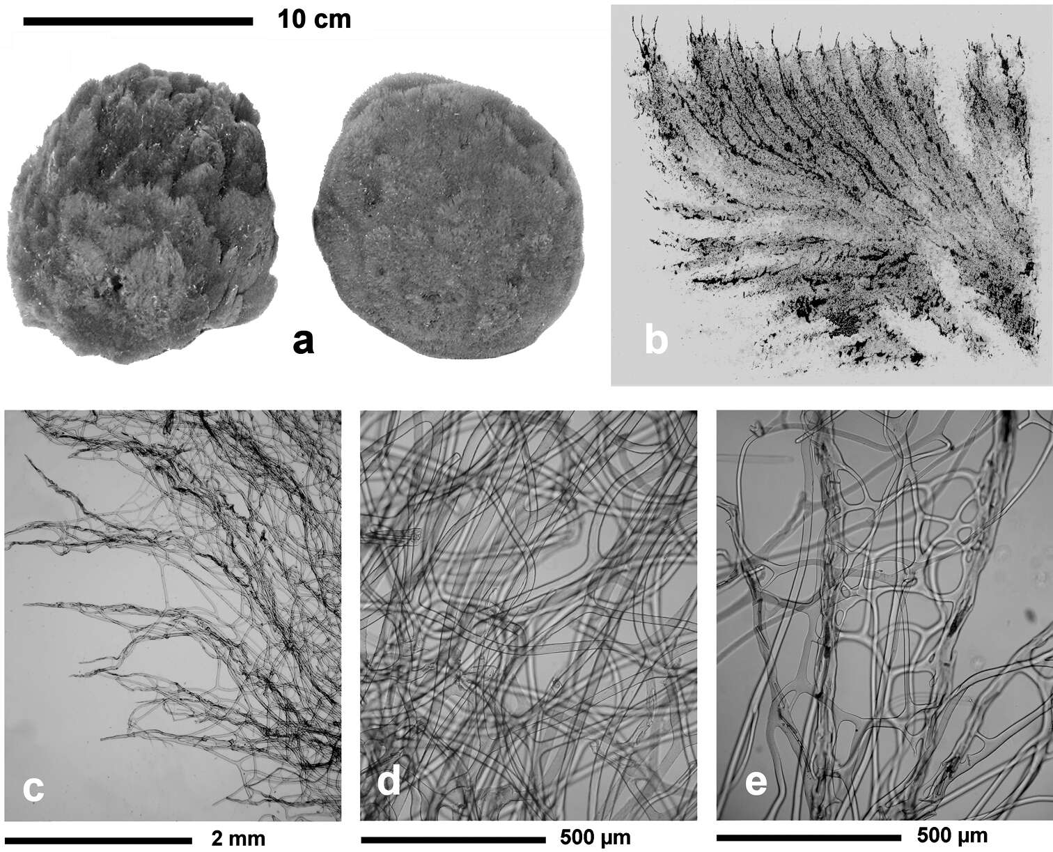
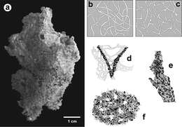
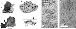
Figure 24Coscinoderma sporadense. a type specimen b, c network architecture of almost transparent secondary fibres d connections between primary and secondary fibres e primary fibre completely cored by inclusions f close-up of the sponge’s surface engulfing mineral grains and spicules. a–f modified from Voultsiadou-Koukoura et al. (1991).
-
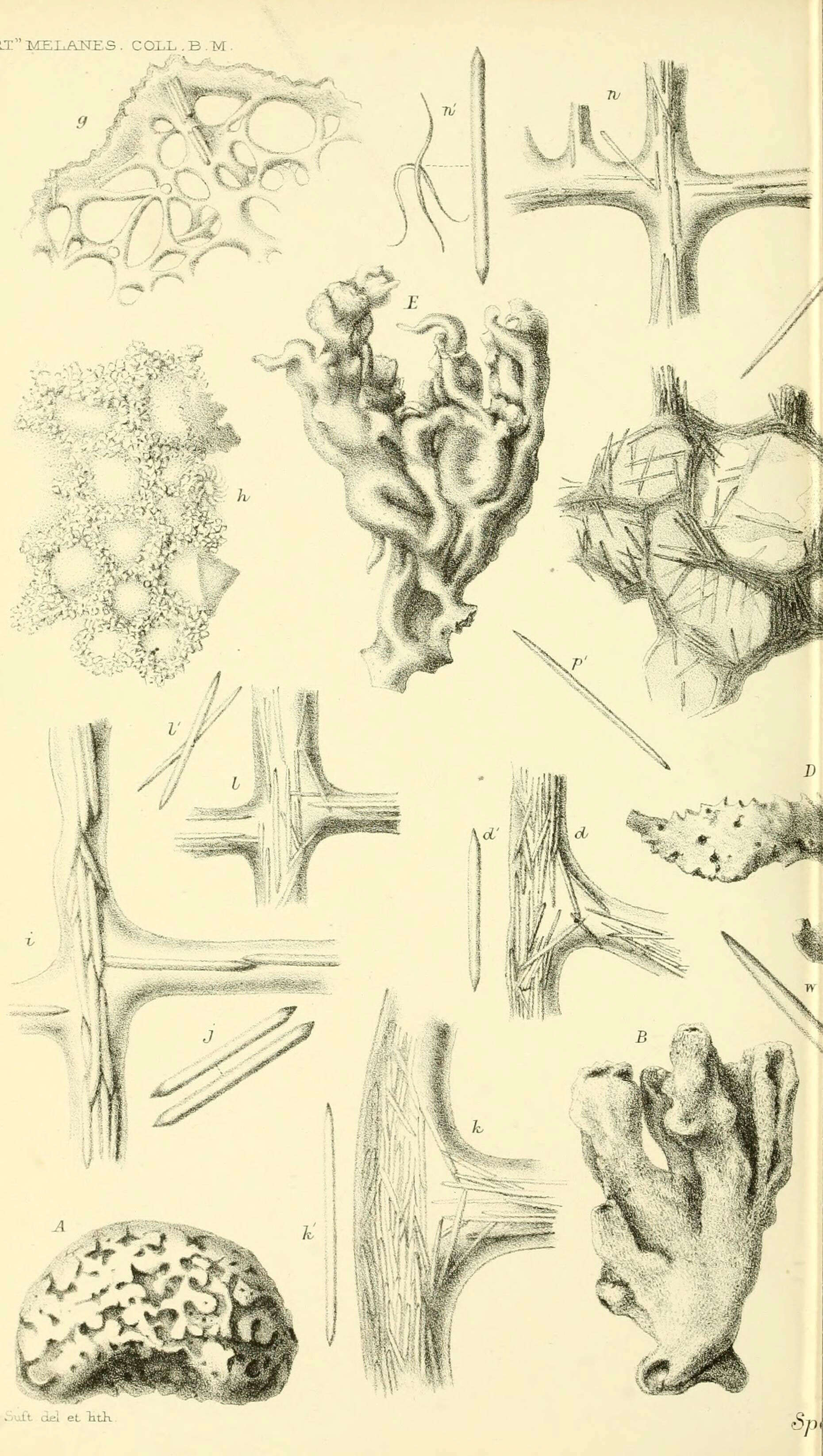
Report on the zoological collections made in the Indo-Pacific Ocean during the voyage of H.M.S. 'Alert' 1881-2.London :Printed by order of the Trustees,1884.
biodiversitylibrary.org/page/12067773
-
Coral Sea, Duration 13 seconds
-
Euspongia officinalis, var. tuba.
-
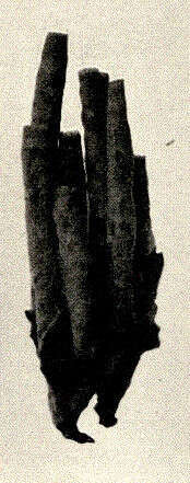
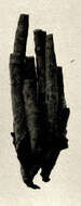
Hippospongia equina, var. elastica.
-
Renata Manconi, Barbara Cadeddu, Fabio Ledda, Roberto Pronzato
Zookeys
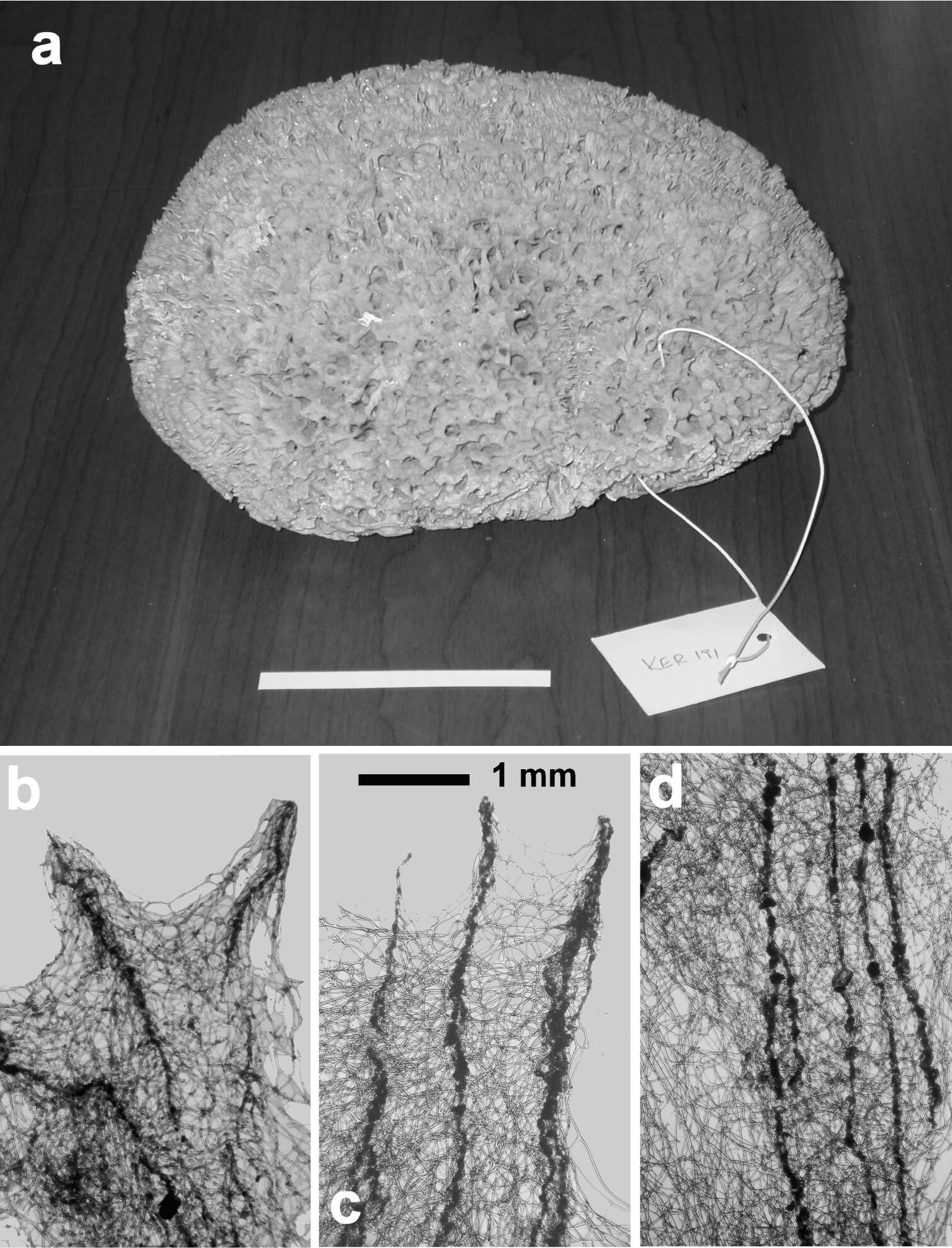
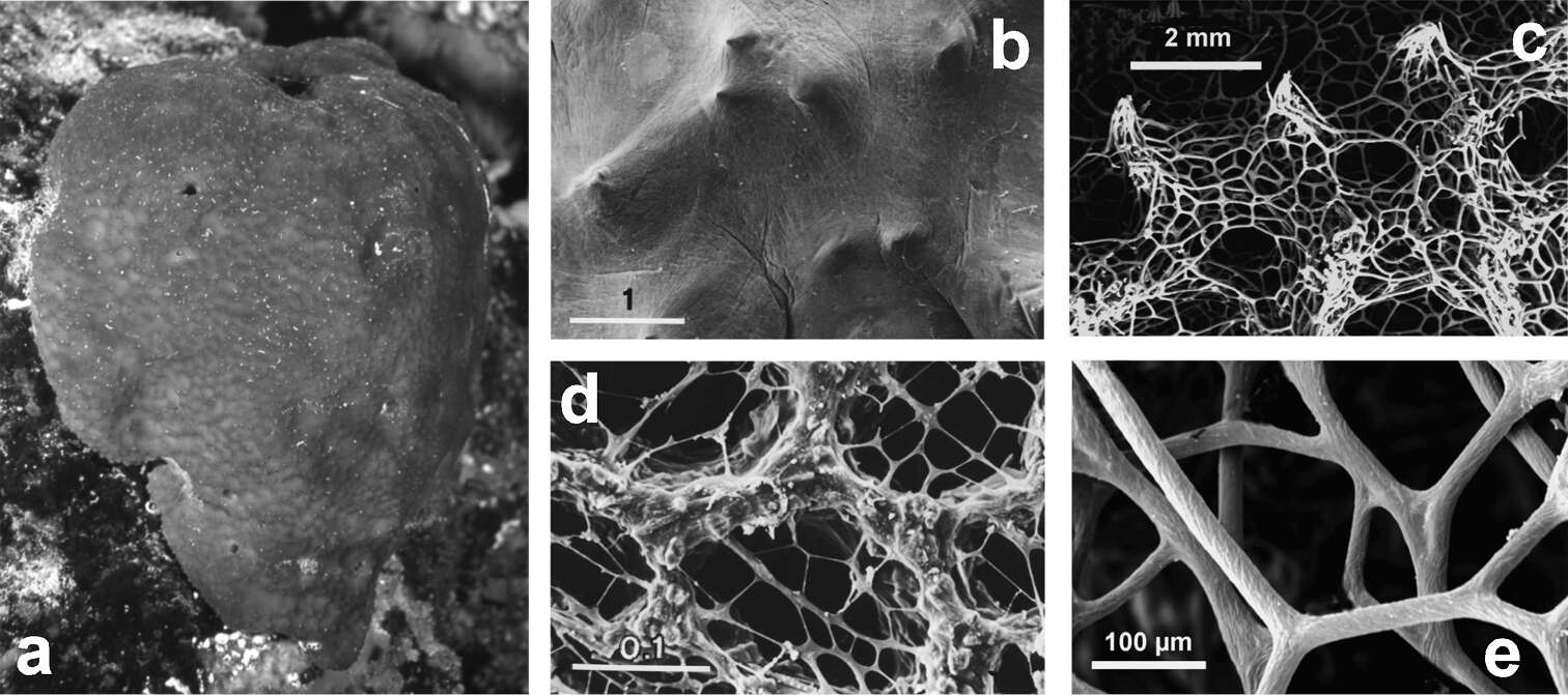
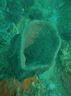
Figure 25Hippospongia communis. a a large, over 25 cm, specimen collected along the Libyan coast b, c skeletal network with tips of primary cored fibres supporting conules at the sponge surface d ascending tracts of primary fibres in the choanosome.
-
Coral Sea, Duration 15 seconds
-
Euspongia officinalis.
-
Renata Manconi, Barbara Cadeddu, Fabio Ledda, Roberto Pronzato
Zookeys
Figure 26Spongia lamella. a–c different growth forms d grouped oscules in the inner exhalant sponge surface e detail (SEM) of the inhalant apertures f detail of sponge surface with mineral grains enclosed in the slim collagenous layer g skeletal network of a lamina with abundant, cored primary fibres extended between the inner and outer surfaces, and inter-connected by a network of thinner secondary fibres without inclusions.
-
Coral Sea, Duration 15 seconds
-
Euspongia officinalis, var. adriatica.
-
Renata Manconi, Barbara Cadeddu, Fabio Ledda, Roberto Pronzato
Zookeys
Figure 27Spongia nitens. a, b dry specimens of the Schmidt’s collection preserved in the Landes Museum Joanneum of Graz c drawing of a living specimen d fibre showing an opaque narrow core e, f different magnification (LM) of the skeletal network, entirely free of mineral inclusions. a, b modified from Desqueyroux-Faundez and Stone (1992) c, d modified from Vacelet (1987) a, b scale bars = 1 cm.
-
Coral Sea, Duration 18 seconds
-
Renata Manconi, Barbara Cadeddu, Fabio Ledda, Roberto Pronzato
Zookeys
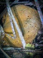
Figure 28Spongia officinalis. a massive large living specimen (ca. 25 cm) showing a finely conulose surface with scattered small oscula b close up of the conulose surface covered by a thin uncellularized collagenous layer (SEM) c magnifications of an inhalant cribrose basal area (SEM) d conules at the spongin skeleton surface (SEM) e twisted surface of secondary fibres (SEM). b, c modified from Pronzato et al. (1998) d, e modified from Pronzato & Manconi (2008) b, d scale bars in mm.
-
Renata Manconi, Barbara Cadeddu, Fabio Ledda, Roberto Pronzato
Zookeys
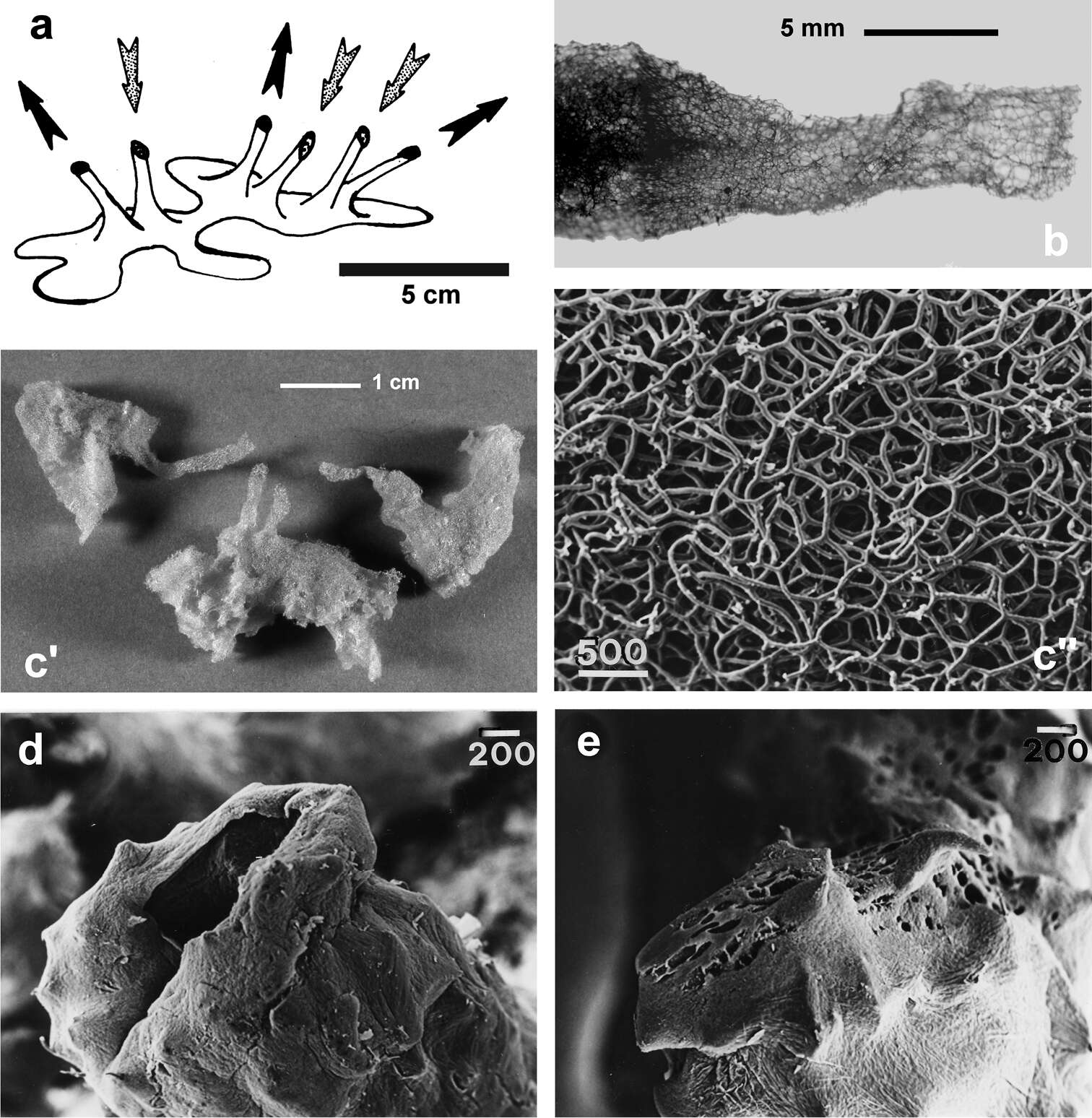
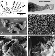
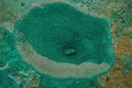
Figure 29Spongia virgultosa. a schematic drawing of the aquiferous system architecture and direction of incurrent and excurrent water flow b low magnification of the skeleton (LM) supporting a funnel c' spongin skeletons of some specimens showing the exhalant funnels (arrows) of the aquiferous system c'' blowup of skeleton skeleton characterised by the absence of cored primary fibres (LM) d exhalant funnel (SEM) e inhalant funnel (SEM). c-e) modified from Pronzato et al. (1998). d, e, f scale bars in µm.
-
Renata Manconi, Barbara Cadeddu, Fabio Ledda, Roberto Pronzato
Zookeys
Figure 30Spongia zimocca. a specimens from the sponge market (Djerba, Tunisia) b drawing of the skeletal network at the sponge surface c long and dense conules supported by tips of primary fibres at the sponge surface (LM) d network of uncored secondary fibres e cored primary fibres among uncored secondaries. b modified from Schulze (1879a).
-
-
-
-
-