Nudibranchs are shell-less marine slugs, so named for the “naked gills” that form feathery tufts on their dorsal side. More than 3,000 known species of nudibranchs exist, and they exhibit a wide variety of coloration, textures, sizes, feeding habits, and behaviors (Wägele and Klussmann-Kolb, 2005). Aeolidioidea is a subgroup of the Order Nudibranchia – however, the exact taxonomic level of Aeolidioidea is conflicting among the literature, with some classifying it as a suborder, superfamily, family, or subfamily. Aeolidoidea (commonly known as aeolid nudibranchs) are found worldwide, although their highest diversity is in the tropics (King & Valda, 2001). Many nudibranchs, primarily the aeolids, feed on organisms in the Phylum Cnidaria – the sea anemones, corals, hydroids, and jellyfish – which are notorious for their painful sting. Stinging cells, called cnidocytes, encapsulate a coiled, barbed, harpoon (nematocyst), which produces this sting (Hessinger and Lenhoff, 1988). Some aeolids nudibranchs have the ability to process ingested nematocysts without discharging them. Even more amazingly, some can sequester these nematocysts into projections of the digestive system in their cerata (tentacle-like projections along the nudibranch’s back, unique to the aeolids) in a process called foreign organellar retention. Foreign organellar retention involves the phagocytosis (engulfment) of nematocysts in the digestive system by cnidophage cells, which accumulate in a muscular capsule called the cnidosac in the cerata. This is most commonly a feature of the aeolids, but members of other families have this ability as well. These “stolen” nematocysts are termed “kleptocnidae,” and it is commonly thought that these are used in defense against the nudibranch’s own predators (Edbunds, 2009).
More than 3,000 known species of nudibranchs exist, and they exhibit a wide variety of coloration, textures, sizes, feeding habits, and behaviors (Wägele and Klussmann-Kolb, 2005). Aeolidioidea is a subgroup of the Order Nudibranchia – however, the exact taxonomic level of Aeolidioidea is conflicting among the literature, with some classifying it as a suborder, superfamily, family, or subfamily.
Aeolidoidea are found worldwide, although their highest diversity is in the tropics (King & Valda, 2001). They are found in the intertidal zones, but have also been found at great depths (King & Valda, 2001).
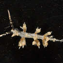
Nudibranchs are shell-less marine slugs. General characteristics of Nudibranchia include: a foot, which the nudibranch uses to crawl on the substrate. This is often covered in cilia (hair-like structures) or mucus, which facilitate mobility (King & Valda, 2001). They are named for their “naked gills,” which form tufts that encircle the anus on the external, dorsal side of the animal (King & Valda, 2001). However, in aeolids, these gills are often indistinct (Carmona et al., 2013).The nudibranch nervous system is concentrated around the esophagus, and there are no dorsal or ventral nerve cords (Denny & Gaines, 2007). Nudibranchs sense the world with rhinophores, chemosensory organs located on the head that look like two spiraled ear tufts (King & Valda, 2001). There are genital openings on the right side of the body (Denny & Gaines, 2007). Nudibranchs, like other gastropods, have radula, a serrated tongue with which to scrape their food (King & Valda, 2001). Aeolids have pectinate (comb-like) radular teeth (Carmona et al., 2013). Aeolids can be distinguished from other nudibranchs by their elongated, tapering bodies, which are covered in fingerlike projections along their dorsal side (Denny & Gaines, 2007). These projections are called cerata, and contain branches of the digestive tract (Carmona et al., 2013). Sacs at the tips of the cerata called cnidosacs store sequestered nematocysts in aeolids that have this ability (Carmona et al., 2013).
Nudibranchs are simultaneously hermaphroditic, meaning they have both male and female reproductive organs at the same time (Denny & Gaines, 2007). The reproductive system is therefore quite large, as it contains male and female gonads, glands for treating ova (a mature female reproductive cell), and receptacles for storing sperm (Denny & Gaines, 2007). However, they are unable to self fertilize, and therefore require a mate (King & Valda, 2001). When a nudibranch comes in contact with another, they line up, facing opposite directions, to align their reproductive organs that occur on the right side of their necks (King & Valda, 2001). They have simultaneous fertilization, with each exchanging sperm packets with the other (King & Valda, 2001). Fertilization is internal (Denny & Gaines, 2007), and nudibranchs can store sperm in their bodies until fertilization is required (King & Valda, 2001). Eggs are laid on substrate in a mucus-covered, ribbon-like matrix (King & Valda, 2001). Eggs hatch from these eggs as veliger larvae, which have two ciliated flaps for swimming and feeding, and are planktonic (King & Valda, 2001). This allows them to be carried by the water great distances. Most have a shell during the larval stage (King & Valda, 2001). Nudibranchs are estimated to live for a year or two, but very few studies have been able to satisfactorily determine life spans in the wild (King & Valda, 2001).
Nudibranchs can feed on a variety of foods, including sponges, soft corals, anemones, and hydroids (King & Valda, 2001). Aeolids are generally specialists, eating only one or two types of food, or even specializing on a specific species (Todd et al., 2001). Most aeolids feed on members of the Phylum Cnidaria (anemones, corals, jellyfish, hydroids, etc.) (Carmona et al., 2013). Many aeolids have the remarkable ability to sequester the stinging cells (nematocysts) of these organisms in their cnidosacs without discharging them (Carmona et al., 2013). These “stolen” nematocysts are termed “kleptocnidae,” and it is commonly thought that these are used in defense against the nudibranch’s own predators (Edbunds, 2009). Although many aeolids have the ability to sequester nematocysts, not all ingested nematocysts end up in the cerata. Many of them are passed intact through the alimentary canal and are excreted in feces (Schlesinger et al., 2009). Although dependent on a variety of external factors, an individual Spurilla neapolitana aeolid has been estimated to excrete 1.5×10⁵ intact nematocysts each day (Schlesinger et al., 2009).
But how do nudibranchs protect themselves from the dangerous sting of nematocysts while they consume them? One possibility is that the digestive epithelium of cnidaria-consuming aeolids contain chitinous, intracellular disks, known as spindles (Martin et al., 2007). In addition, the esophagus, radula pouch, and pharynx possess a chitinous coating (Martin et al. 2006). These features of the digestive tract are not seen in nudibranchs that do not consume cnidarians. The presence of chitin in nudibranchs is surprising, as chitin is a substance normally associated with organisms with hard exoskeletons, like arthropods, where it serves a protective function (Martin et al., 2007). Because of this, Martin et al. (2006) suggest that spindles help protect cnidaria-ingesting nudibranchs from the deleterious effects of nematocysts. Spindles are also found in the rhinophores, cerata, and sensory cilia, areas that frequently come in contact with nematocysts as nudibranchs graze for food in a cnidarian-populated habitat (Martin et al., 2006). In order to understand the mechanism by which spindles protect the nudibranch, Martin et al. (2006) examined epidermal (skin) spindles after contact with nematocysts. When discharged, nematocysts rupture the nudibranch’s epidermis. This liberates masses of spindles, which become entangled in the barbs of the nematocyst. As the spindles are released from the epidermis, they fall away, bringing the entangled nematocysts with them (Martin et al., 2007). The spindles in the epidermis and this intriguing detachment mechanism protect the deeper vital tissues from the injurious effects of the discharged nematocyst (Martin et al., 2007).
Other aeolids can sequester zooxanthellae from their prey (Carmona et al., 2013). Zooxanthellae are symbiotic protists that live in the tissues of some Cnidaria, and provide them with nutrients and waste cycling services (Carmona et al., 2013). Aeolids can sequester these protists and keep them alive in their tissues, using their photosynthetic products for their own nutrition, as did their former Cnidarian hosts (Carmona et al., 2013).
Predators may include crabs, fish, and asteroids (Miller & Byrne, 2000).