Haminoea bubble snails are marine mollusks with thin, fragile shells. These snails are herbivorous, apparently feeding mainly on diatoms (Malaquias et al. 2004, 2009).
There has been some confusion over the identity and correct name of the bubble snail described from Washington State, U.S.A., in 1989 as Haminoea callidegenita (Gibson and Chia 1989). This is now considered to be a junior (and thus invalid) synonym of the valid name H. japonica (Gosliner and Behrens 2006). (see Systematics and Taxonomy, below)
Haminoea japonica is unusual in that it produces some offspring that are swimming, non-feeding veliger larvae and others that metamorphose into crawling juveniles prior to hatching (Gibson and Chia 1989, 1995).
Haminoea japonica has poecilogonous development (i.e. it produces more than one type of offspring). Gibson and Chia (1989, 1995, as H. callidegenita) found that swimming non-feeding (lecithotrophic) veligers (typical mollusk larvae) and crawling juveniles were simultaneously released from nearly every egg mass observed, indicating that development mode varied not only within the population, but even among the offspring of single individuals. Development of all offspring from an egg mass appeared to be identical until just prior to hatching, when approximately half of the siblings metamorphosed and hatched as juveniles (these capsular metamorphic larvae underwent development, including a veliger stage, within the egg capsule, before hatching). The remaining siblings hatched as veligers and metamorphosed after a planktonic period ranging from 1 to 30 days. The percentage of juvenile hatchlings was highly variable among egg masses (ranging from 4 to 100%, with most egg masses having 50 to 70% of total hatchlings emerging as juveniles). Variability in hatchling type occurred among clutches, populations, and years. The percentage of juveniles released per egg mass was not influenced by a variety of factors tested, including physical egg mass characteristics, female reproductive traits (fecundity, female size, egg size), time of year, source of metamorphic inducer, and culture conditions. (Gibson and Chia 1995)

Haminoea japonica (=H. callidegenita) is apparently native to the northwestern Pacific, but has been introduced to (at least) the eastern Pacific, the Atlantic coast of Spain, and the Mediterranean (Gosliner and Behrens 2006 and references therein).
In Padilla Bay, Washington State, U.S.A., Haminoea japonica (=H. callidegenita) are found primarily in association with eelgrass (Zostera marina and Z. japonica) blades, surface sediments around Zostera roots, and mats of the green alga Ulva sp. (sea lettuce) in the upper intertidal zone (Gibson and Chia 1995).
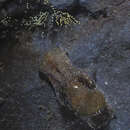
Gosliner and Behrens (2006) report that the preserved Haminoea japonica specimens they examined were up to 20 mm in length. In live specimens, the headshield is lobed and rounded anteriorly with elongate, rounded, deeply divided posterior lobes. The ground color is translucent white with scattered opaque white spots and a dense covering of dark brown spots that are most dense on the center of the headshield and on the short parapodia lobes, which partially envelop the yellowish, transparent shell (parapodia are the fleshy protrusions on the sides of the snail). A posterior mantle lobe with brown and opaque white bands partially covers the posterior end of the shell. A long, rounded posterior end of the foot extends posteriorly from the shell. Through the transparent shell, the translucent white mantle is covered with large brown spots and scattered bright orange spots. (Gosliner and Behrens 2006)
Spinella et al. (1998) identified alkylphenol compounds present in the external parts of Haminoea japonica (=H. callidegenita), but absent from internal organs; these may function as alarm pheromones.
In Washington State, U.S.A., Gibson and Chia (1989, as H. callidegenita) collected viable egg masses in all months except January and December. These were short, thick cylinders, ranging from almost ovate (10 X 5 X 5 mm) to sausage shaped (36 X 6 X 6 mm). A typical egg mass produced by a 26 mm adult was 15 X 5 X 5 mm and took approximately 20 minutes to be deposited. Egg masses were attached to the substrate along the length of one side, and were slightly curved in shape as a consequence of the adult turning slightly towards the mass during oviposition. Adults did not show any substrate preference for oviposition and deposited masses on any available solid surface, both in the field and under laboratory conditions. The bright orange-yellow eggs were individually encapsulated and arranged in a continuous string. This string spiralled through the periphery of the egg mass (irregularly deposited at a depth of approximately 1.5 mm), leaving the center and outer portion of the jelly free of embryos. Egg masses contained an average of 21 eggs/ mm3 of egg mass, for a total of 200 to 700 eggs per mass. Well developed veligers, with lengthened propodia, rhythmic contractions of the heart, and the ability to retract completely into the shell were visible in the egg masses 29 days after oviposition. Hatching began approximately 3 days later. Some individuals from each egg mass hatched as veliger larvae and others as juveniles. A few days before the onset of hatching, the jelly mass began to deteriorate and was colonized by diatoms and nematodes. Hatching generally occurred over a period lasting from 3 to 11 days. At hatching, capsule walls softened (i.e., became flexible and readily distorted) and were eventually split by the larval propodium, allowing the individual to escape. This process took approximately 1.5 to 2 minutes after the first visible softening of the capsule. After the larva emerged from the capsule, it slowly worked its way out of the jelly mass. Veligers would then swim away, while juveniles often remained on the deteriorating mass or crawled to filaments of the green alga Chaetomorpha if available. Hatching of veligers and juveniles occurred in the same way. (Gibson and Chia 1989)
In June 2005 at Crown Beach (San Francisco Bay) in Alameda, California (U.S.A.), over 90 people, primarily schoolchildren, reported rashes after wading in the water. The cause of this problem was apparently a schistosome (a parasitic trematode worm) in the genus Gigantobilharzia, carried by the recently introduced Japanese Bubble Snail, Haminoea japonica. (Cohen 2005) Although this parasite likely cannot complete its life cycle in humans or other mammals, requiring a bird host instead, accidental infection can cause an unpleasant dermatitis in humans.
In the original description of H. callidegenita based on specimens from commercial, non-native oyster beds in Washington State, U.S.A. (Gibson and Chia 1989), the authors noted the possibility that this species might have been introduced and not native to the temperate eastern Pacific. It has subsequently been reported from northwestern Spain and Venice, Italy, where the snails were associated with the introduced Manila Clam (Ruditapes philippinarum) (Alvarez et al. 1993, cited in Gosliner and Behrens 2006). Subsequent analysis has indeed indicated that these specimens from both the west coast of the United States and from Europe are introductions from the northwestern Pacific. Gosliner and Behrens (2006) examined putative Haminoea callidegenita specimens fron San Francisco Bay, California, U.S.A., and compared these with both the original description and type specimens of H. callidegenita from Washington State, U.S.A. (Gibson and Chia 1989) and with specimens of H. japonica from Japan. They concluded that Haminoea callidegenita is a junior synonym of H. japonica (i.e., H. callidegenita and H. japonica actually refer to the same species, and because H. japonica is an older name, this is the valid name). Gosliner and Behrens also note that H. japonica was originally described as merely a variety of H. binotata, but has been treated as a distinct species since at least 1961. They also note that in the past, H. rotundata, a species originally described from the Phillipines, may have been been considered to be the same species as H. japonica of Japan, but that H. japonica has now long been recognized as a distinct species. (Gosliner and Behrens 2006 and references therein; D. Behrens in litt. April 2010)
Haminoea japonica is herbivorous, apparently feeding mainly on diatoms (Malaquias et al. 2004, 2009). Gibson and Chia (1989, as H. callidegenita) reported that this species fed by grazing diatoms and detritus from any available surface, including the macroalgae Chaetomorpha linum and Ulva sp., the seagrass Phyllospadix scouleri, and the walls of glass aquaria. Examination of stomach contents and fecal pellets revealed sand, diatoms (both fragmented and, more often, entire and still pigmented), and clumps of Ulva cells (recognizable Chaetomorpha cells were not observed). Individuals secreted a mucous tube when crawling and other individuals seemed to follow these tubes. (Gibson and Chia 1989)
Haminoea japonica is een slakkensoort uit de familie van de Haminoeidae.[1] De wetenschappelijke naam van de soort is voor het eerst geldig gepubliceerd in 1895 door Pilsbry.
Bronnen, noten en/of referenties日本月华螺(学名:Haminoea japonica),又名日本泡螺,为海洋真後鰓類支序頭楯目腹足綱軟體動物[1]。舊屬阿地螺科月华螺属[1]的,今屬長葡萄螺科長葡萄螺屬[2]。
分布于日本[1][3]、朝鲜[1]、中国大陆的山东[1]、香港[3]、泰國及菲律宾[1]。在地中海[2]及美國加州的三藩市灣及華盛頓州也有發現[4]。属于温带性种类。其一般生活于潮间带石头下、海藻间。[1]
壳长11mm,壳宽8mm[5]。
本物種的寄生蟲包括曾在美國加州三藩市灣引致人類絛蟲皮炎(human cercarial dermatitis(英语:cercarial dermatitis))的一種禽類血吸蟲[4]。