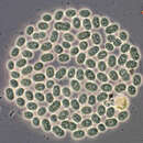
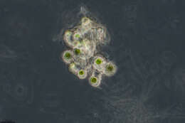
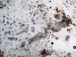
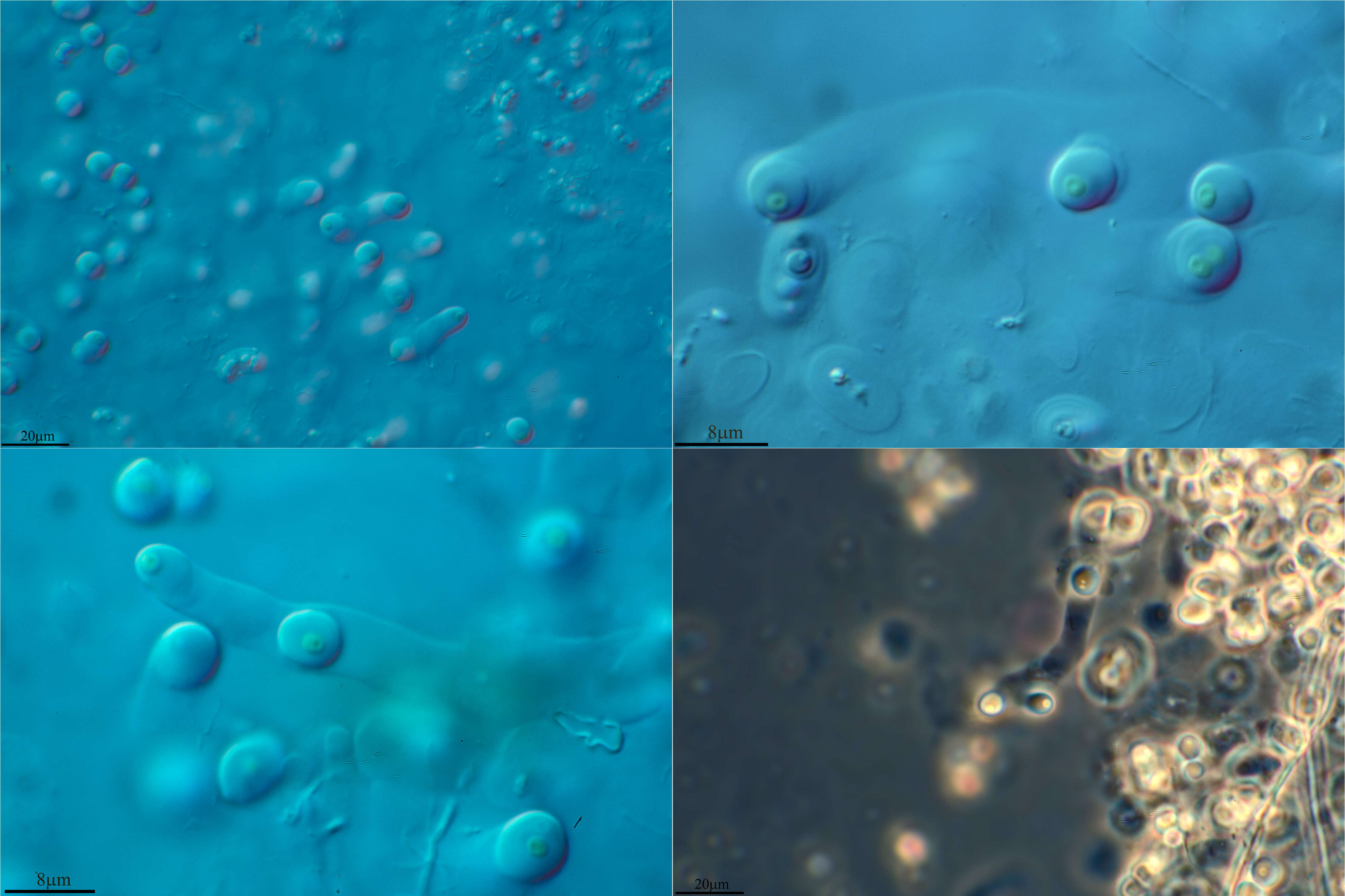
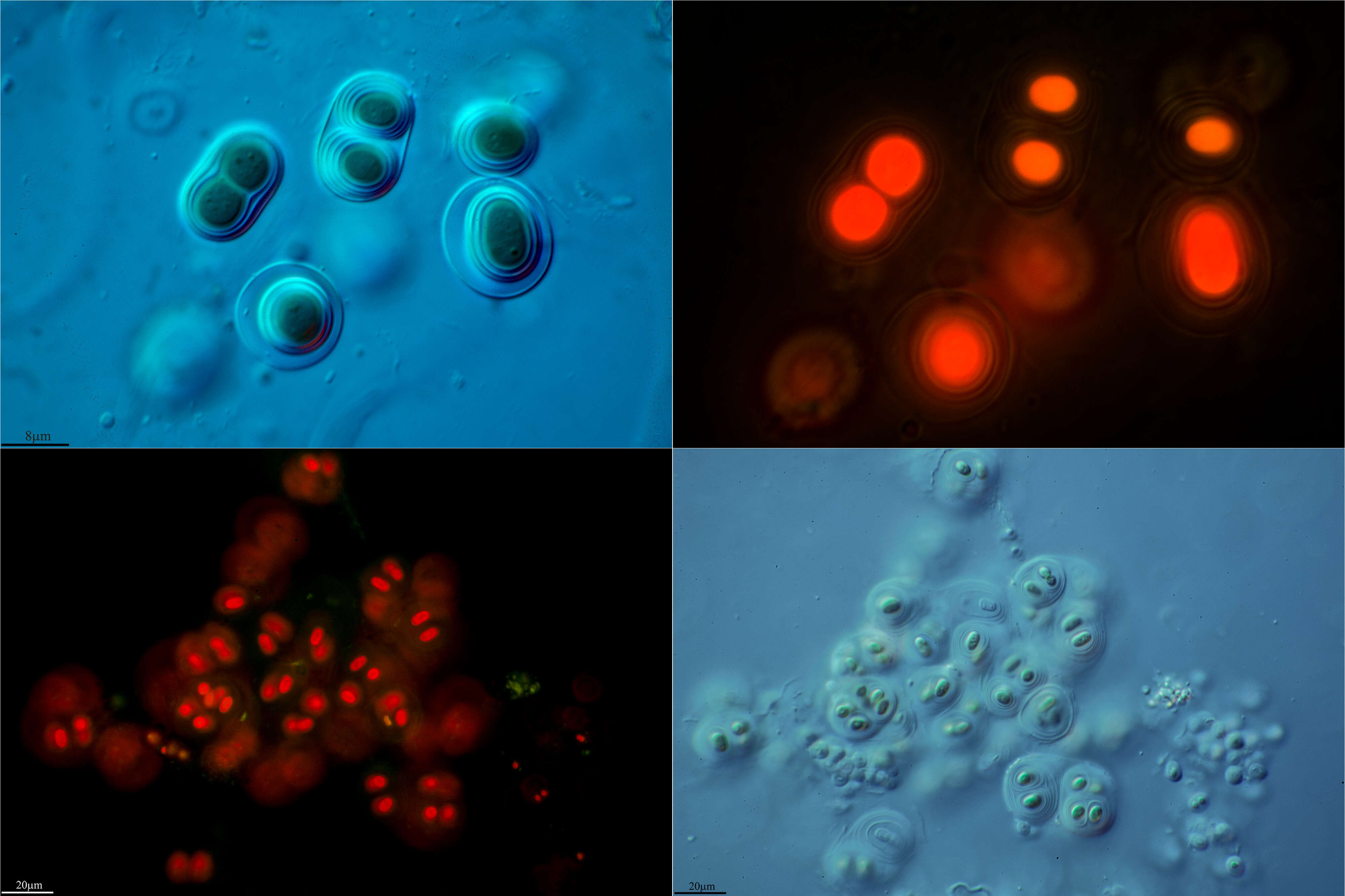
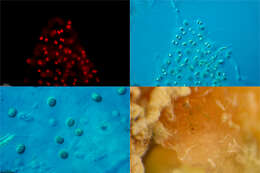
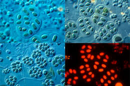
Aphanothece (a-fan-owe-theek-ee) blue green alga in which many coccoid or cylindrical cells share a common mucus sheath. Differential interference contrast.
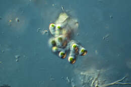
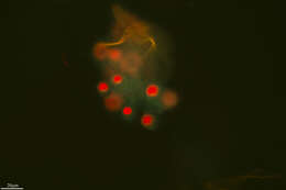
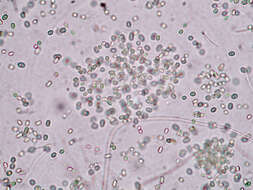
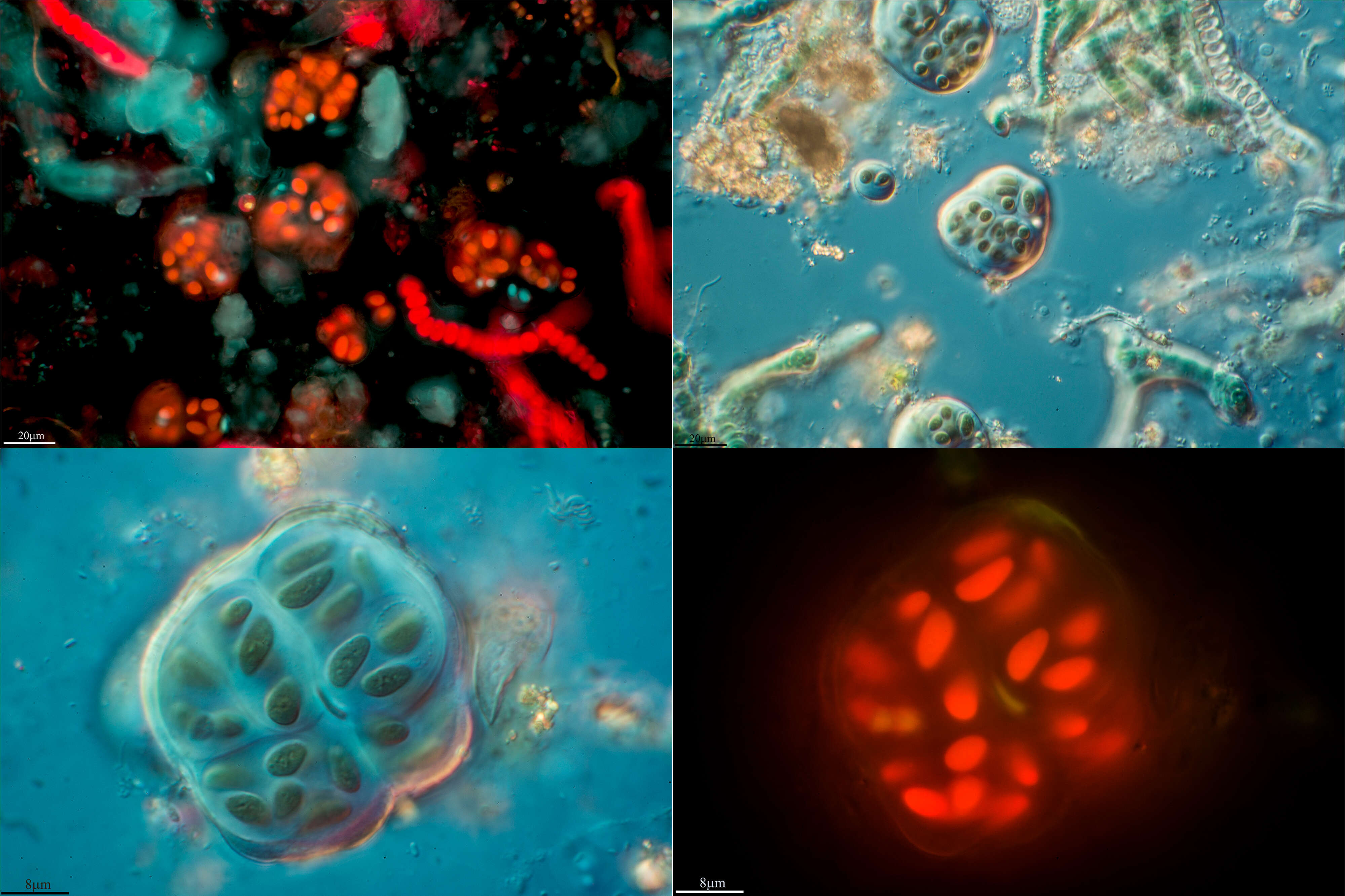
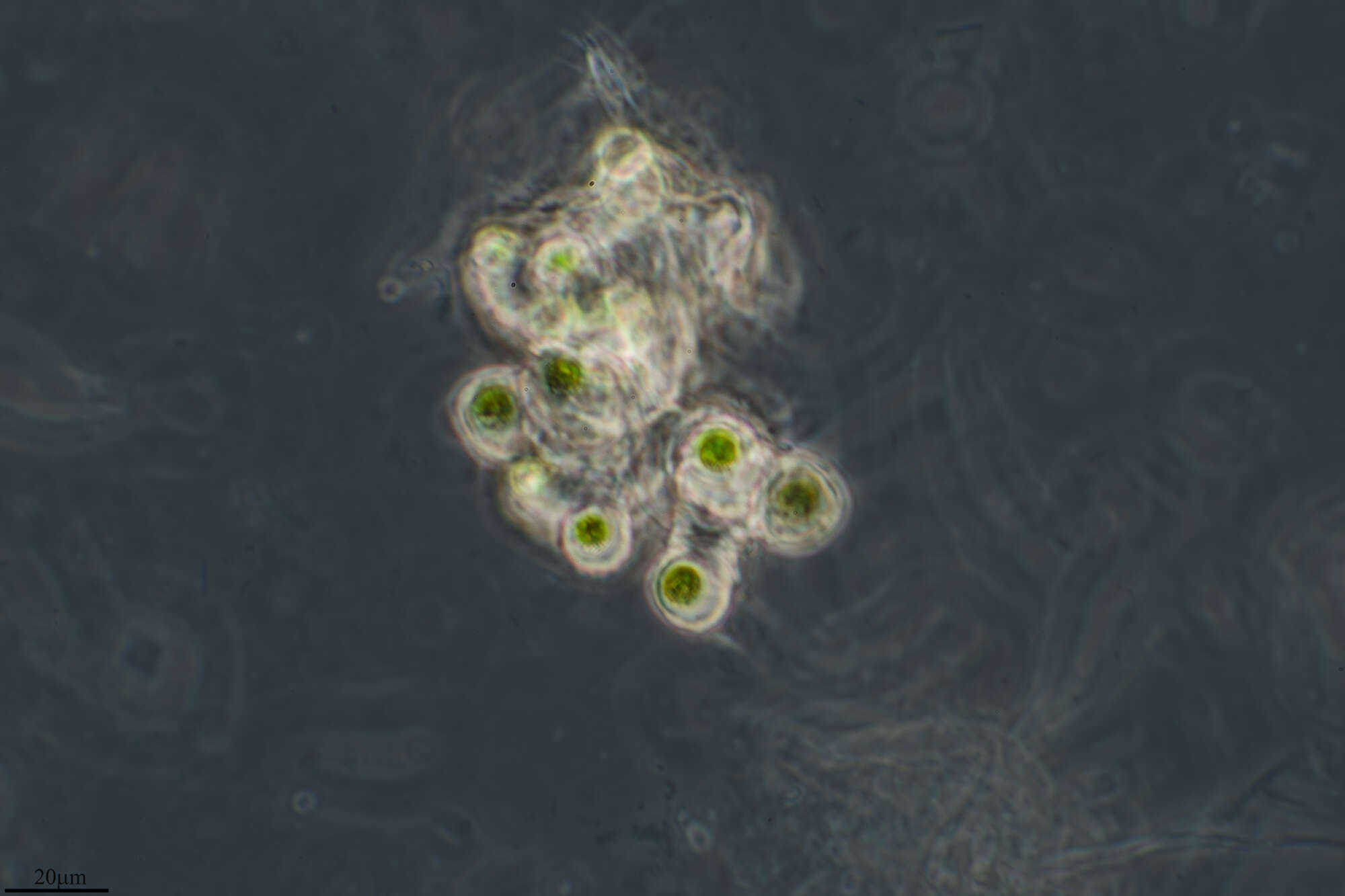
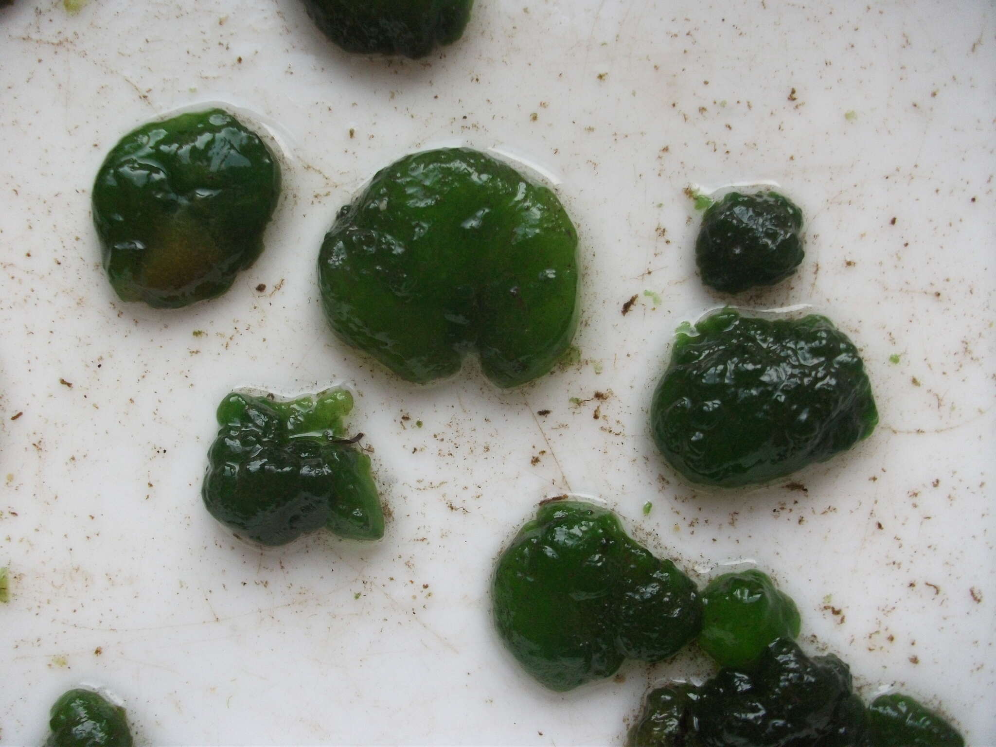
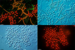
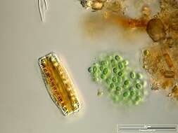
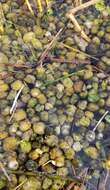
Colony accompanied by Epithemia adnata. Scale bar indicates 50 µm. Sample from a wetland at the Pillersee (Tyrol, Austria). The image was built up using several photomicrographic frames with manual stacking technique. Images were taken using Zeiss Universal with Olympus C7070 CCD camera.Image under Creative Commons License V 3.0 (CC BY-NC-SA).
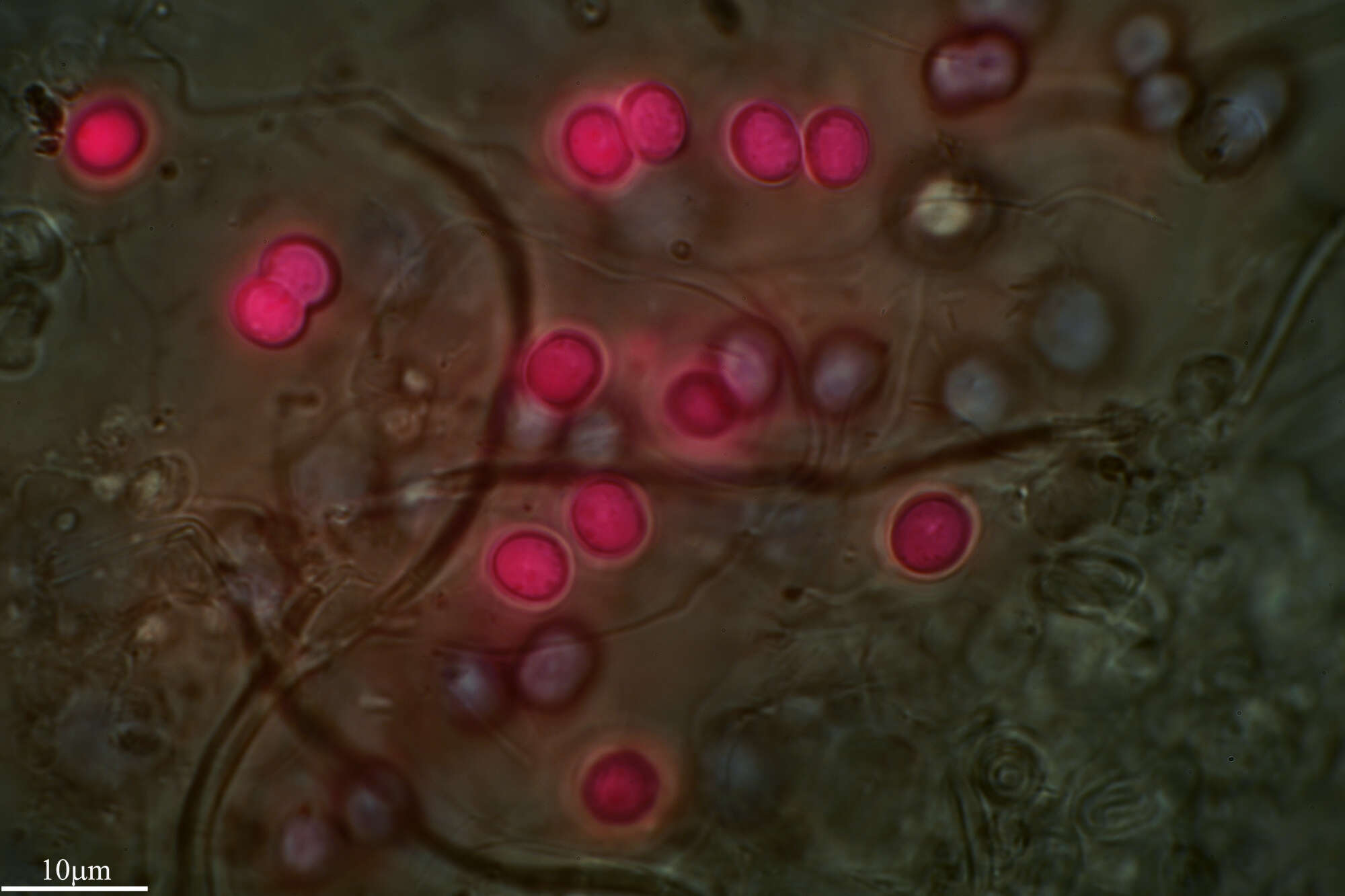
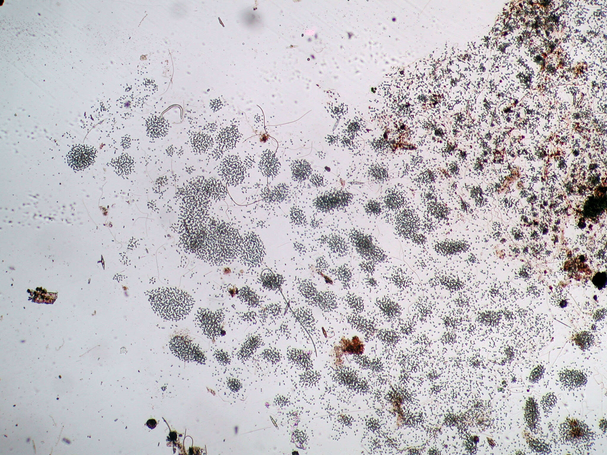
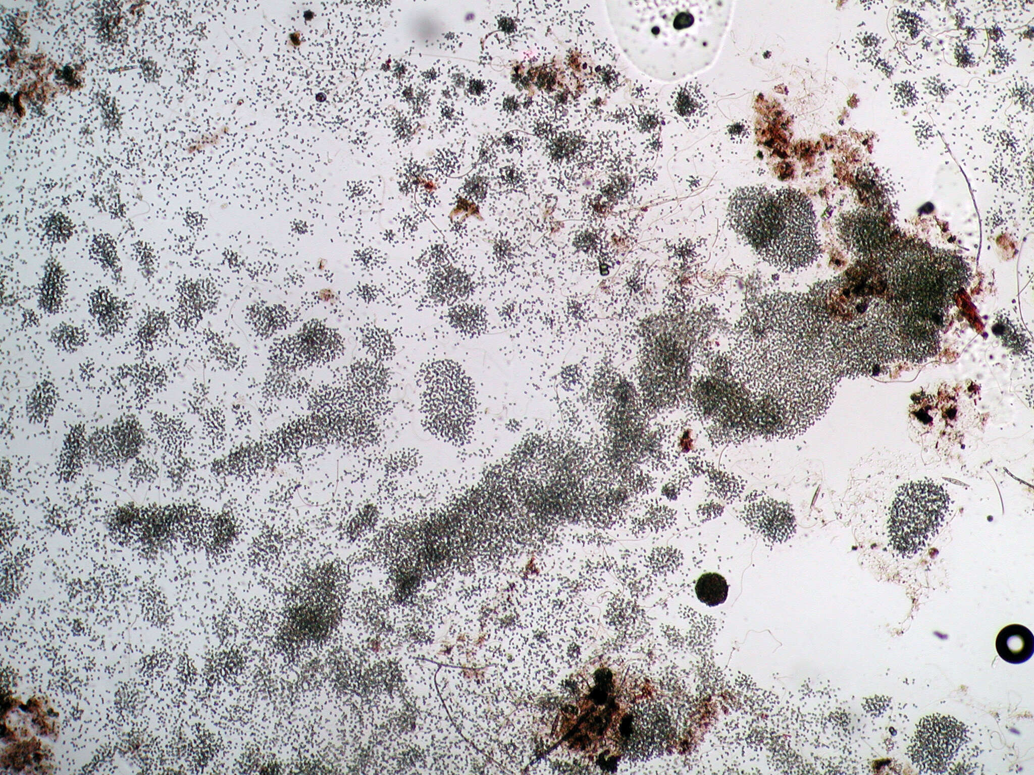
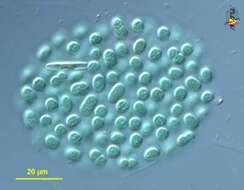
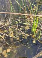
Scale bar indicates 100 µm. Sample from the pond Hegne Moor situated in the vicinity of Lake Constance. Images were taken using Zeiss Universal with Olympus C7070 CCD camera.Image under Creative Commons License V 3.0 (CC BY-NC-SA).
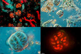
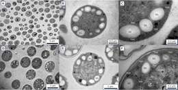
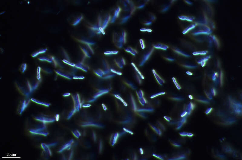
Inomura K, Deutsch C, Wilson ST, Masuda T, Lawrenz E, Lenka B, Sobotka R, Gauglitz JM, Saito MA, Prášil O, Follows MJ
Wikimedia Commons
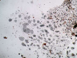
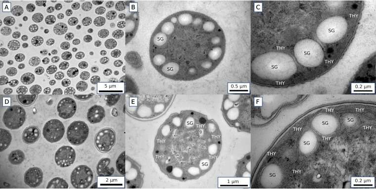
Description: English: Transmission electron micrographs of Crocosphaera cells harvested at the 6-h time point during the light period (A to C) and at the 6-h time point during the dark (D to F). Starch granules (SG) and thylakoid membranes (THY) are observed mostly on the edge of the cytosol. More-detailed images (C and F) show that SG are observed mostly between THY. Date: 11 December 2019. Source: Fig. 6 at https://europepmc.org/article/PMC/PMC6908418 Quantifying Oxygen Management and Temperature and Light Dependencies of Nitrogen Fixation by Crocosphaera watsonii. In: Msphere 4(6), doi:10.1128/msphere.00531-19, PMID 31826967, PMC 6908418. Author: Inomura K, Deutsch C, Wilson ST, Masuda T, Lawrenz E, Lenka B, Sobotka R, Gauglitz JM, Saito MA, Prášil O, Follows MJ.