-
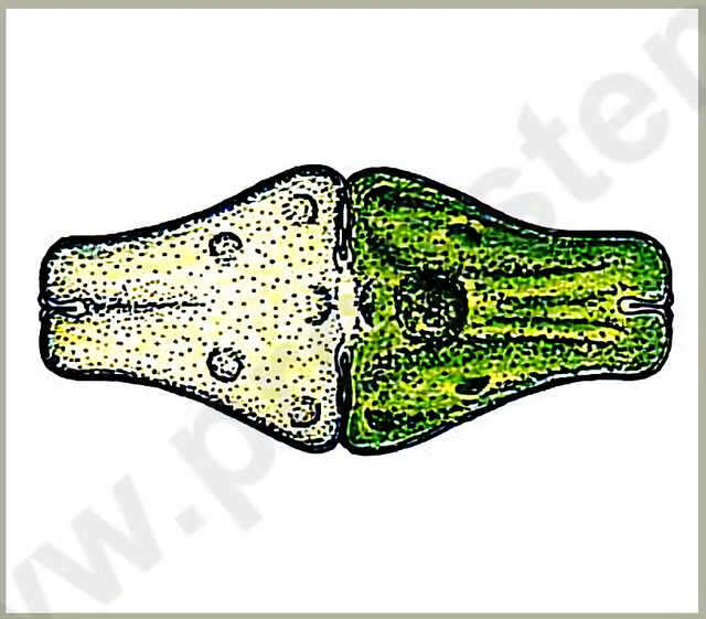
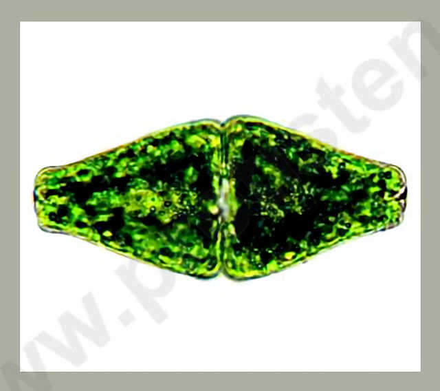
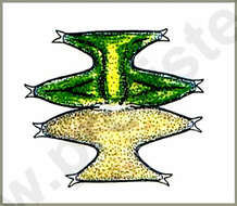
Euastrum ansatum RALFS var. ansatum Dimension: Length 80 90 µm, width 35 45 µm.Widely spread both in lowland and in middle altitude in moderate acidic waters.Copyright by Prof. Rupert Lenzenweger, Ried im Innkreis, Austria. Place name: n. a. Latitude: 0 Longitude: 0 Dimensionen: Länge 80 90 µm, Breite 35 45 µm.Sowohl im Flachland als auch in mittleren Höhenlagen in mäßig sauren Gewässern verbreitet. Copyright by Prof. Rupert Lenzenweger, Ried im Innkreis, Österreich. For permission to use of (high-resolution) images please contact postmaster@protisten.de.
-
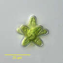
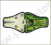
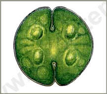
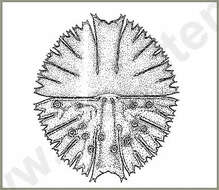
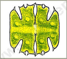
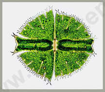
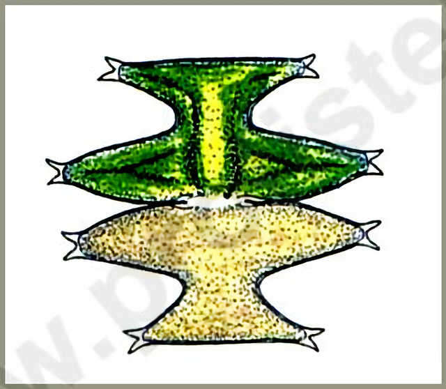
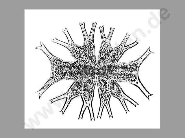
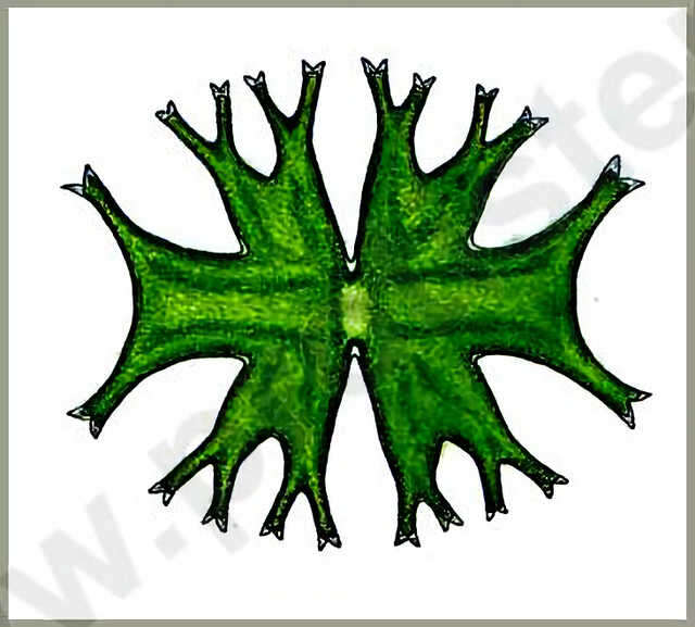
Micrasterias pinnatifida (KÜTZ.) RALFS The cells are only a little wider than long. The cell halves form three lobes, the lateral lobes are tapered peripherally and have a few denticles at the blunted ends. The lateral lobes are short and are strongly widened with straight ends. The central cut ist deep and strongly widened peripherally. Dimension: Length 60 - 70 µm, width 65 75 µm.Ecology: Common in moderate acidic to neutral waters of fens and litoral zones.Occurrence: Ubiquitous.Copyright by Prof. Rupert Lenzenweger, Ried im Innkreis, Austria. Place name: n. a. Latitude: 0 Longitude: 0 Die Zellen sind nur wenig breiter als lang. Die Zellhälften sind drei-lappig, die Seitenlappen sind nach außen konisch verschmälert mit einem Paar Zähnchen an den abgestumpften Enden. Die Scheitellappen sind kurz, stark verbreitert mit geraden Enden. Die Mitteleinschnitte sind tief, nach außen stark erweitert. Dimensionen: Länge 60 70 µm, Breite 65-75 µm.Ökologie: In schwach sauren bis neutralen Gewässern von Mooren und Uferzonen allgemein verbreitet.Verbreitung: Weltweit. Copyright by Prof. Rupert Lenzenweger, Ried im Innkreis, Österreich. For permission to use of (high-resolution) images please contact postmaster@protisten.de.
-
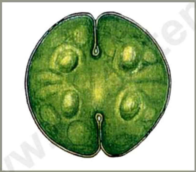
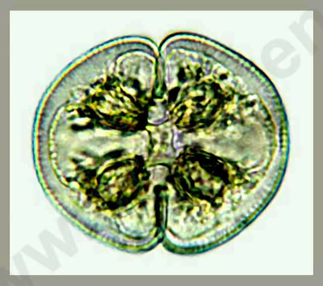
Cosmarium circulare REINSCH Dimension: Length 70 - 100 µm, width 50 - 90 µm.Occurrence: .Copyright by Prof. Rupert Lenzenweger, Ried im Innkreis, Austria. Place name: n. a. Latitude: 0 Longitude: 0 Dimensionen: Länge 70 100 µm, Breite 50 90 µm.Vorkommen: Azidophile Alge, eher selten. Copyright by Prof. Rupert Lenzenweger, Ried im Innkreis, Österreich. For permission to use of (high-resolution) images please contact postmaster@protisten.de.
-
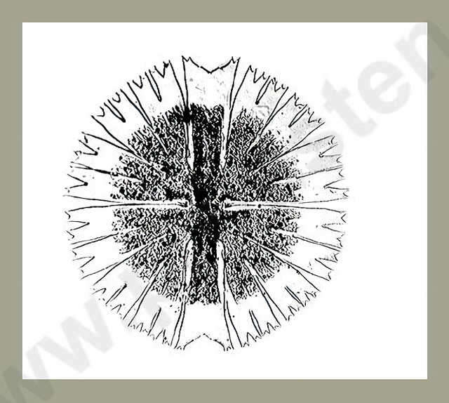
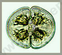
Micrasterias rotata (GREV.) RALFS The cells are 1.08 to 1.15 times longer than wide, the shape seems almost circular or wide elliptical. The cell is devided into lobes due to deep cuts, the terminations of lobes are denticulated. The central lobe is broadened evenly at the end. The termination is formed concavely and is lightly arched upwards at both sides. The lateral angles of the central lobe are little denticulated. The cut in the middle of the cell (sinus) is very deep and peripherally a little widened. The cellwall is densly punctuated by tiny pores. The Chromatophores have several scattered pyrenoids with varying sizes. Dimension: Length 200 - 300 µm, width 200 270 µm.This specie is very tolerant concerning living conditions. Therefore the species is widely spread in all altitudes, in forestal ditches and lowland fens sometimes abundant.Copyright by Prof. Rupert Lenzenweger, Ried im Innkreis, Austria. Place name: n. a. Latitude: 0 Longitude: 0 Die Zellen sind 1,08 bis 1.15mal länger als breit und im groben Umriss fast kreisrund bis breit oval. Die Zellseiten sind durch unterschiedlich tiefe Einschnitte in Lappen geteilt, deren Enden mit Zähnchen besetzt sind. Der Mittellappen ist gegen das Ende zu gleichmäßig verbreitert, dieses ist in der Mitte konkav abgerundet, beiderseits flach aufgewölbt. Die seitlichen Winkel der Mittellappen (Apikalwinkel) sind jeweils mit einem Paar Zähnchen besetzt. Der Mitteleinschnitt (Sinus) ist tief eingeschnitten und nach außen etwas erweitert. Die Zellwand ist mit kleinen Poren dicht besetzt. In die Chromatophoren sind zerstreut mehreren, unterschiedlich großen Pyrenoiden eingelagert. Dimensionen: Länge 200 300 µm, Breite 200 270 µm.Eine sehr milieutolerante Alge, daher In allen Höhenlagen verbreitet, im Tiefland in Waldgräben und Moorgebieten mitunter massenhaft. Copyright by Prof. Rupert Lenzenweger, Ried im Innkreis, Österreich. For permission to use of (high-resolution) images please contact postmaster@protisten.de.
-
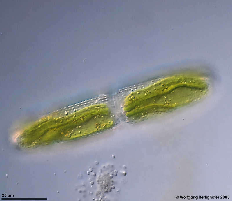
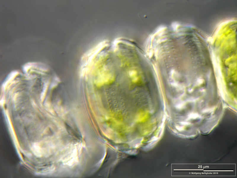
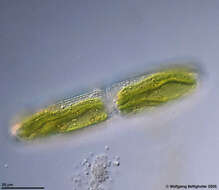
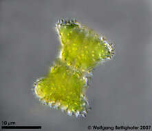
Tetmemorus brebissonii var. brebissonii Non-filamentous desmids have the ability to move slowly by means of directed secretion of mucilage. Depht of focus approach can show cell surface together with folded chloroplasts and cell contour. Picture generated from 5 shots using CombineZ by Alan Hadley. Sample from spagnum pond Dosenmoor near Neumuenster (Schleswig- Holstein, Germany). Images were taken using Zeiss Universal with Olympus C7070 CCD camera.Image under Creative Commons License V 3.0 (CC BY-NC-SA). Place name: Bog Dosenmoor near Neumuenster (Schleswig-Holstein, Germany) Latitude: 54.136219 Longitude: 10.026433 Einzellige Desmidiaceen haben die Fähigkeit, sich mittels gerichteter Sekretion von Schleim langsam zu bewegen. Die Multiebenenabbildung zeigt die Zelloberfläche zusammen mit dem gefalteten Chloroplasten und dem Zellumriss. Probe aus dem Dosenmoor in der Nähe von Neumünster. Mikrotechnik: Zeiss Universal, Kamera: Olympus C7070. Creative Commons License V 3.0 (CC BY-NC-SA). For permission to use of (high-resolution) images please contact postmaster@protisten.de.
-
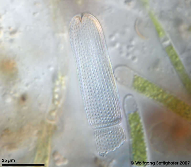
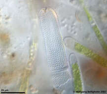
Tetmemorus brebissonii var. brebissonii Empty cell wall from Tetmemorus spec. The surface texture of cell wall is clearly visible. Sample from sphagnum pond Dosenmoor near Neumuenster (Schleswig-Holstein, Germany). Images were taken using Zeiss Universal with Olympus C7070 CCD camera.Image under Creative Commons License V 3.0 (CC BY-NC-SA). Place name: Bog Dosenmoor near Neumuenster (Schleswig-Holstein, Germany) Latitude: 54.136219 Longitude: 10.026433 Leere Zellwand von Tetmemorus spec. Das Zellwandmuster ist deutlich sichtbar. Probe aus dem Dosenmoor in der Nähe von Neumünster. Mikrotechnik: Zeiss Universal, Kamera: Olympus C7070. Creative Commons License V 3.0 (CC BY-NC-SA). For permission to use of (high-resolution) images please contact postmaster@protisten.de.
-
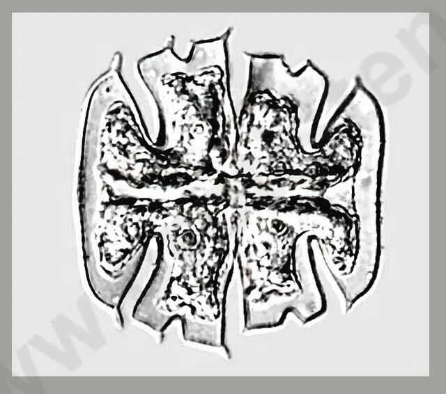
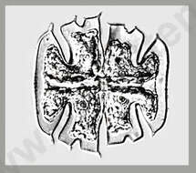
Micrasterias crux-melitensis Scale bar indicates 50 µm.Sample from a wetland at the Pillersee (Tyrol, Austria). The image was built up using several photomicrographic frames with manual stacking technique. Images were taken using Zeiss Universal with Olympus C7070 CCD camera.Image under Creative Commons License V 3.0 (CC BY-NC-SA). Place name: Wetland at the Pillersee (Tyrol, Austria) Latitude: 47.531785 Longitude: 12.573095 Multiebenen-Abbildung, manuell gestapelt. Der Messbalken markiert eine Länge von 50 µm. Probe aus einer Feuchtwiese beim Pillersee in Tirol. Mikrotechnik: Zeiss Universal, Kamera: Olympus C7070. Creative Commons License V 3.0 (CC BY-NC-SA). For permission to use of (high-resolution) images please contact postmaster@protisten.de.
-
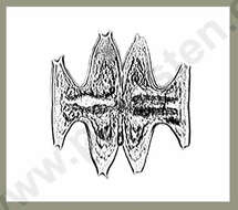
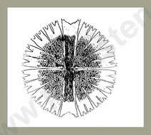
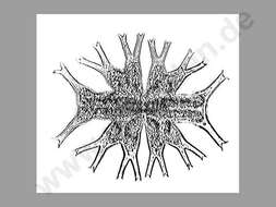
Micrasterias decemdentata (NÄG.) ARCH. Dimension: Length 55 - 60 µm, width 55 - 60 µm.Occurrence: .Copyright by Prof. Rupert Lenzenweger, Ried im Innkreis, Austria. Place name: n. a. Latitude: 0 Longitude: 0 Dimensionen: Länge 55 60 µm, Breite 55 60 µm.Vorkommen: Säureliebende Art in Torfmoos-schlenken, in Mitteleuropa recht selten. Copyright by Prof. Rupert Lenzenweger, Ried im Innkreis, Österreich. For permission to use of (high-resolution) images please contact postmaster@protisten.de.
-
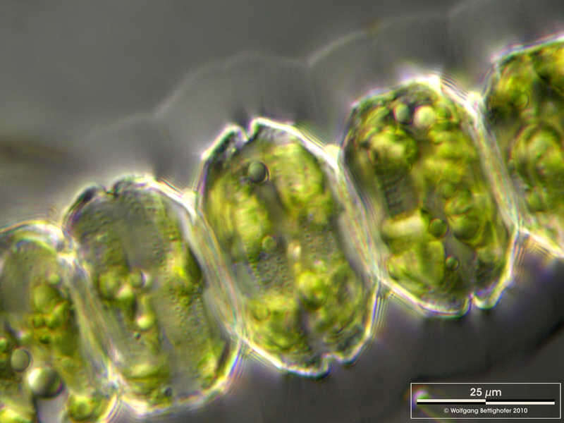
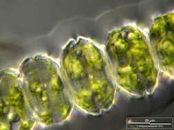
Desmidium grevillei The Multi-layer image shows the dotted pattern of the cell wall surface. Scale bar indicates 25 µm. Sample from a small wetland near Schladming (northern alpine region of Austria near Salzburg). Images were taken using Zeiss Universal with Olympus C7070 CCD camera.Image under Creative Commons License V 3.0 (CC BY-NC-SA). Place name: Wetland near Schladming (Austria) Latitude: 47.37386 Longitude: 13.823016 Die Multiebenenabbildung zeigt das Punktmuster der Zellwandoberfläche. Der Messbalken markiert eine Länge von 25 µm. Probe aus einer Wiesenvernässung nahe Schladming/Österreich. Mikrotechnik: Zeiss Universal, Kamera: Olympus C7070. Creative Commons License V 3.0 (CC BY-NC-SA). For permission to use of (high-resolution) images please contact postmaster@protisten.de.
-
Desmidium grevillei Centrical optical section showing nucleus. Scale bar indicates 25 µm. Sample from a small wetland near Schladming (northern alpine region of Austria near Salzburg). Images were taken using Zeiss Universal with Olympus C7070 CCD camera.Image under Creative Commons License V 3.0 (CC BY-NC-SA). Place name: Wetland near Schladming (Austria) Latitude: 47.37386 Longitude: 13.823016 Die mittige optischen Schnitt zeigt den Zellkern. Der Messbalken markiert eine Länge von 25 µm. Probe aus einer Wiesenvernässung nahe Schladming/Österreich. Mikrotechnik: Zeiss Universal, Kamera: Olympus C7070. Creative Commons License V 3.0 (CC BY-NC-SA). For permission to use of (high-resolution) images please contact postmaster@protisten.de.
-
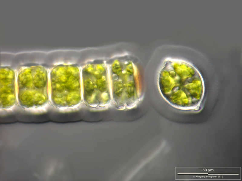
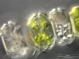
Desmidium grevillei Filamentous colony of Desmidium grevillii with its mucilaginous sheath. One cell was separated from the filament and is shown in optical cross section. Scale bar indicates 50 µm. Sample from a small wetland near Schladming (northern alpine region of Austria near Salzburg). Images were taken using Zeiss Universal with Olympus C7070 CCD camera.Image under Creative Commons License V 3.0 (CC BY-NC-SA). Place name: Bog Waasenmoos Pass Thurn near Mittersil (Tyrol, Austria) Latitude: 47.30234117 Longitude: 12.41751194 Fädige Kolonie von Desmidium grevillii mit seiner Gallerthülle. Eine Zelle hat sich vom Faden getrennt und ist im optischen Querschnitt dargestellt. Der Messbalken markiert eine Länge von 50 µm. Probe aus einer Wiesenvernässung nahe Schladming/Österreich. Mikrotechnik: Zeiss Universal, Kamera: Olympus C7070. Creative Commons License V 3.0 (CC BY-NC-SA). For permission to use of (high-resolution) images please contact postmaster@protisten.de.
-
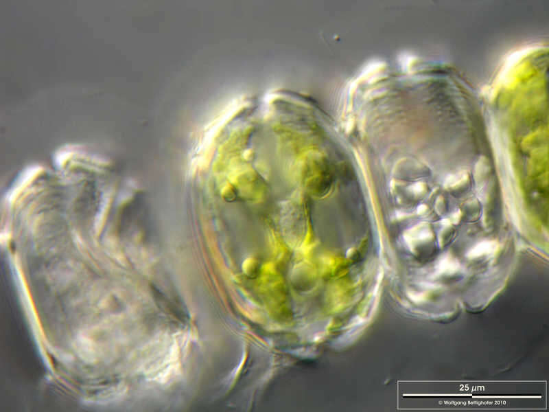
Desmidium grevillei Cell in optical cross section. Scale bar indicates 25 µm. Sample from a small wetland near Schladming (northern alpine region of Austria near Salzburg). Images were taken using Zeiss Universal with Olympus C7070 CCD camera.Image under Creative Commons License V 3.0 (CC BY-NC-SA). Place name: Wetland near Schladming (Austria) Latitude: 47.37386 Longitude: 13.823016 Zelle im optischen Querschnitt. Der Messbalken markiert eine Länge von 25 µm. Probe aus einer Wiesenvernässung nahe Schladming/Österreich. Mikrotechnik: Zeiss Universal, Kamera: Olympus C7070. Creative Commons License V 3.0 (CC BY-NC-SA). For permission to use of (high-resolution) images please contact postmaster@protisten.de.
-
Desmidium grevillei Filamentous colony of Desmidium grevillii with its mucilaginous sheath. One cell was separated from the filament and is shown in optical cross section. Scale bar indicates 50 µm. Sample from a small wetland near Schladming (northern alpine region of Austria near Salzburg). Images were taken using Zeiss Universal with Olympus C7070 CCD camera.Image under Creative Commons License V 3.0 (CC BY-NC-SA). Place name: Wetland near Schladming (Austria) Latitude: 47.37386 Longitude: 13.823016 Fädige Kolonie von Desmidium grevillii mit seiner Gallerthülle. Eine Zelle hat sich vom Faden getrennt und ist im optischen Querschnitt dargestellt. Der Messbalken markiert eine Länge von 50 µm. Probe aus einer Wiesenvernässung nahe Schladming/Österreich. Mikrotechnik: Zeiss Universal, Kamera: Olympus C7070. Creative Commons License V 3.0 (CC BY-NC-SA). For permission to use of (high-resolution) images please contact postmaster@protisten.de.
-
Desmidium grevillei Filamentous colony of Desmidium grevillii with its mucilaginous sheath. One cell was separated from the filament and is shown in optical cross section. Scale bar indicates 50 µm. Sample from a small wetland near Schladming (northern alpine region of Austria near Salzburg). Images were taken using Zeiss Universal with Olympus C7070 CCD camera.Image under Creative Commons License V 3.0 (CC BY-NC-SA). Place name: Bog Waasenmoos Pass Thurn near Mittersil (Tyrol, Austria) Latitude: 47.30234117 Longitude: 12.41751194 Fädige Kolonie von Desmidium grevillii mit seiner Gallerthülle. Eine Zelle hat sich vom Faden getrennt und ist im optischen Querschnitt dargestellt. Der Messbalken markiert eine Länge von 50 µm. Probe aus einer Wiesenvernässung nahe Schladming/Österreich. Mikrotechnik: Zeiss Universal, Kamera: Olympus C7070. Creative Commons License V 3.0 (CC BY-NC-SA). For permission to use of (high-resolution) images please contact postmaster@protisten.de.
-
Desmidium grevillei The Multi-layer image shows the dotted pattern of the cell wall surface. Scale bar indicates 25 µm. Sample from a small wetland near Schladming (northern alpine region of Austria near Salzburg). Images were taken using Zeiss Universal with Olympus C7070 CCD camera.Image under Creative Commons License V 3.0 (CC BY-NC-SA). Place name: Wetland near Schladming (Austria) Latitude: 47.37386 Longitude: 13.823016 Die Multiebenenabbildung zeigt das Punktmuster der Zellwandoberfläche. Der Messbalken markiert eine Länge von 25 µm. Probe aus einer Wiesenvernässung nahe Schladming/Österreich. Mikrotechnik: Zeiss Universal, Kamera: Olympus C7070. Creative Commons License V 3.0 (CC BY-NC-SA). For permission to use of (high-resolution) images please contact postmaster@protisten.de.
-

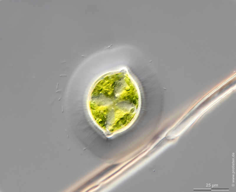
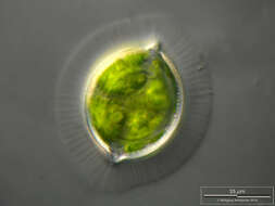
Staurastrum capitulum Staurastrum capitulum is a smal desmid with an acanthous cell wall. With its rotational symmetry the surface structure is not easy to capture with photos. Depth of focus technique was used for the attempt. Sample from spagnum pond situated in the northern alpine region of Austria near Salzburg. Images were taken using Zeiss Universal with Olympus C7070 CCD camera.Image under Creative Commons License V 3.0 (CC BY-NC-SA). Place name: Bogs near Salzburg (Austria) Latitude: 48.068516 Longitude: 12.954134 Staurastrum capitulum ist eine kleine Zieralge mit einer stacheligen Zellwand. Mit ihrer Rotationssymmetrie ist es nicht leicht, die Oberflächenstruktur fotografisch darzustellen. Tiefenschärfe durch Multiebenenabbildung aus vielen Bildebenen, manuell gestapelt. Probe aus einem Moor in den nördlichen Kalkalpen von Österreich in der Nähe von Salzburg. Mikrotechnik: Zeiss Universal, Kamera: Olympus C7070. Creative Commons License V 3.0 (CC BY-NC-SA). For permission to use of (high-resolution) images please contact postmaster@protisten.de.
-
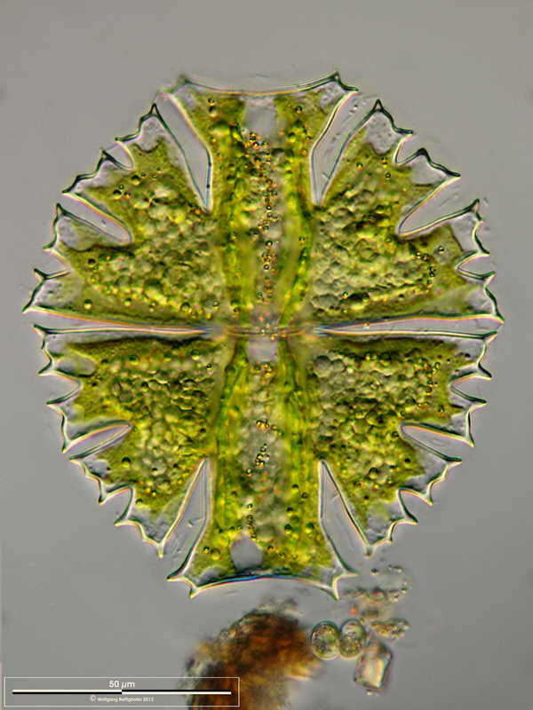
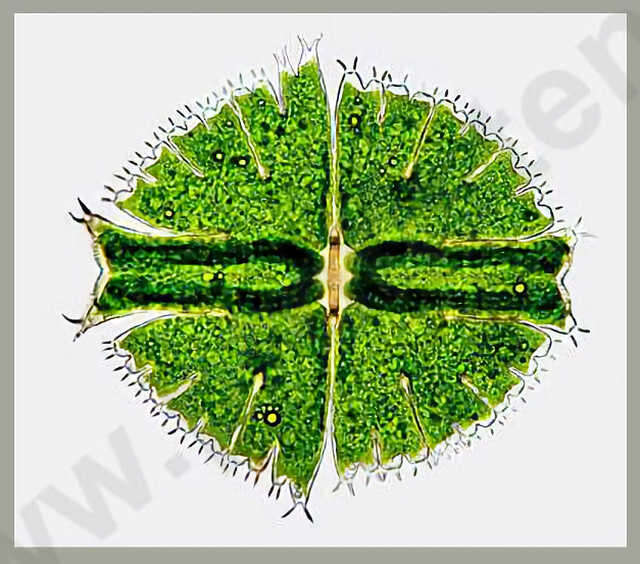
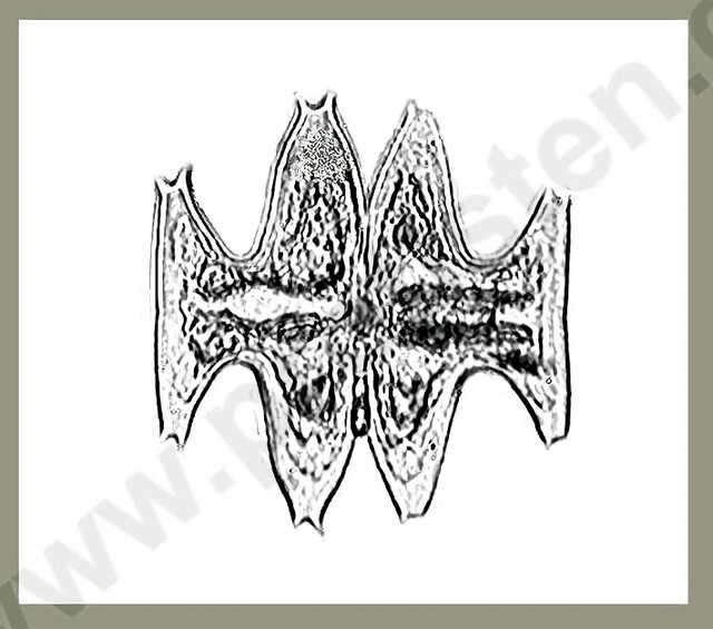
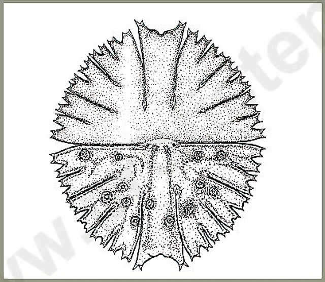
Micrasterias furcata RALFS The cells are about as long as wide and have a circular shape. The cell halves consist of three lobes. The lateral lobes are clearly separated. At the ends of the lateral lobes there are two outwards standing projections. Between the projections there is a concave area. The central cut is deep and peripherally strongly widened. Dimension: Length 150 - 180 µm, width 150 160 µm.Ecology: In acidic to moderate acidic fens and bogs.Occurrence: Ubiquitous, rather rare in Central Europe.Copyright by Prof. Rupert Lenzenweger, Ried im Innkreis, Austria. Place name: n. a. Latitude: 0 Longitude: 0 Die Zellen sind etwa so lang wie breit und im groben Umriss kreisrund. Die Zellhälften werden aus drei Lappen gebildet. Die Seitenlappen sind außen jeweils 4-fach gegabelt, die Einschnitte dazwischen diesen sind weit geöffnet. Die Scheitellappen sind deutlich abgesetzt, an den Enden mit zwei schräg nach außen abstehenden Fortsätzen zwischen diesen konkav. Der Mitteleinschnitt ist tief, nach außen stark erweitert. Dimensionen Länge 150 180 µm, Breite 150 160 µm.Ökologie: In sauren bis mäßig sauren Gewässern von Moortümpeln und Moorseen.Verbreitung: Weltweit, in Mitteleuropa eher selten. Copyright by Prof. Rupert Lenzenweger, Ried im Innkreis, Österreich. For permission to use of (high-resolution) images please contact postmaster@protisten.de.
-
Micrasterias fimbriata RALFS Dimension: Length 230 - 250 µm, width 200 - 230 µm.Occurrence: .Copyright by Prof. Rupert Lenzenweger, Ried im Innkreis, Austria. Place name: n. a. Latitude: 0 Longitude: 0 Dimensionen: Länge 230 250 µm, Breite 200 230 µm.Vorkommen: In mäßig sauren Gewässern von Nieder - und Zwischenmooren meist nur vereinzelt und wenig verbreitet. Copyright by Prof. Rupert Lenzenweger, Ried im Innkreis, Österreich. For permission to use of (high-resolution) images please contact postmaster@protisten.de.