-
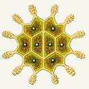
Pediastrum granulatum.
-
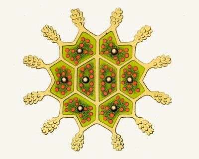
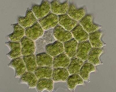
Portrait of Pediastrum duplex (Meyen, 1829 ).Collected from a freshwater pond near Boise, Idaho. Phase contrast.
-
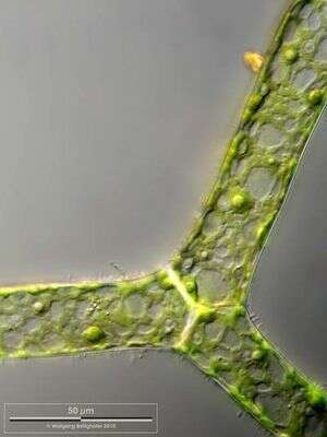
Scale bar indicates 50 µm. Sample from Lake Constance (Bodensee, Southern Germany) near Bodman. Images were taken using Zeiss Universal with Olympus C7070 CCD camera.
-
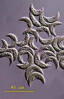
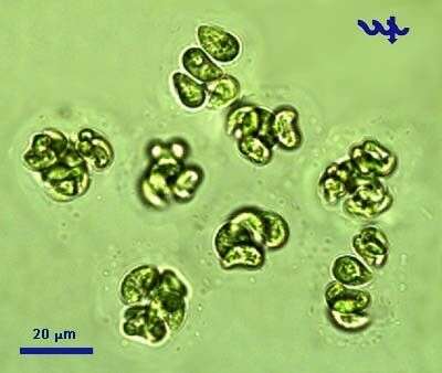
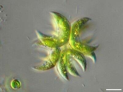
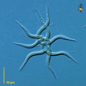
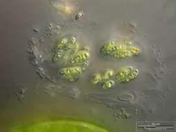
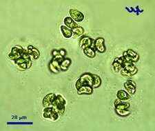
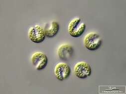
Kirchneriella (keyrsh-ner-ell-a) in which cells occur individually (particularly in cultures) or in colonies 4-16, rarely more, cells within gelatinous sheaths. Cells oval or oblong cylindrically, curved or screw-shaped curved. Both ends of the cells are rounded. A single chloroplast covers the inside of the cell and contains a single pyrenoid. The nucleus is located centrally near the concavity of the cell. Common in plankton of fresh water in lakes, ponds and rivers. This slightly squashed colony of Kirchneriella lunaris was collected in the plankton from Lake Constance, Germany. Cells average 16 - 19 (microns long. Cells are bent or moon shaped. Differential interference contrast.
-
Colony of green algae in thick mucilaginous envelope. Scale bar indicates 25 µm. Sample from sphagnum pond situated in the northern alpine region of Austria near Salzburg. Images were taken using Zeiss Universal with Olympus C7070 CCD camera.
-
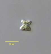
Portrait of the chlorellacean Tetraedron minimum var. minimum (A. Braun) Hansgirg. DIC.
-
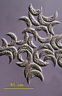
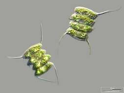
Portrait of Pediastrum duplex (Meyen, 1829 )demonstrating tufts of bristles protruding from the notch at the ends of the horns of peripheral cells (seen most clearly at one o'clock and 7 o'clock here). These bristles are thought to consist of proteinaceous microfilaments (see Scnepf,E. et al. J. Ultrastruct. Res. 72:367-379,1980). These nonmotile bristles may play a role in orienting the coenobia in the water column . DIC.
-
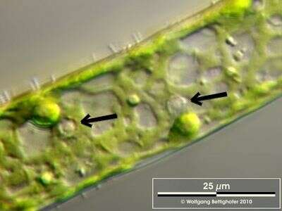
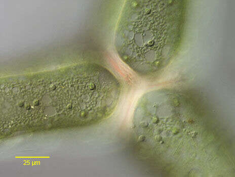
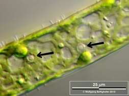
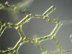
Cells of Hydrodictyon have several nuclei (arrows). Sample from Lake Constance (Bodensee, Southern Germany) near Bodman. Images were taken using Zeiss Universal with Olympus C7070 CCD camera.
-
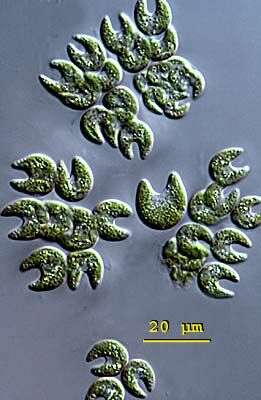
Kirchneriella (keyrsh-ner-ell-a) in which cells occur individually (particularly in cultures) or in colonies 4-16, rarely more. Cells live within gelatinous sheaths. Cells oval or oblong cylindrically, curved or screw-shaped curved. Both ends of the cells are rounded. A single chloroplast covers the inside of the cell and contains a single pyrenoid. The nucleus is located centrally near the concavity of the cell. Common in plankton of fresh water in lakes, ponds and rivers. This slightly squashed colony of Kirchneriella obesa was collected in the plankton from Lake Constance, Germany; cells measure 8 - 14 microns long. With an U-shaped incision. Differential interference contrast.
-
Scale bar indicates 25 µm.Sample from a wetland at the Pillersee (Tyrol, Austria). The image was built up using several photomicrographic frames with manual stacking technique. Images were taken using Zeiss Universal with Olympus C7070 CCD camera.Image under Creative Commons License V 3.0 (CC BY-NC-SA).
-
Portrait of the chlorellacean Tetraedron minimum var. minimum (A. Braun) Hansgirg. DIC .
-
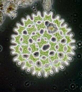
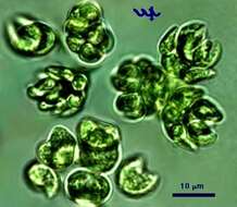
Pediastrum duplex is a common chlorophyte (Chlorococcales) of Lake Kinneret, relatively more abundant in winter, especially in âNo-Peridinium yearsâ. This specimen was collected at the shore by the Kinneret Limnological Laboratory in June 2006
-
Scale bar indicates 25 µm. Sample from the Lake Constance (vicinity of Bodman). The image was built up using several photomicrographic frames with manual stacking technique. Images were taken using Zeiss Universal with Olympus C7070 CCD camera.Image under Creative Commons License V 3.0 (CC BY-NC-SA).
-
-
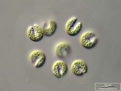
Scale bar indicates 10 µm. Sample from Lake Constance in the vicinity of Bodman. The image was built up using several photomicrographic frames with manual stacking technique. Images were taken using Zeiss Universal with Olympus C7070 CCD camera.Image under Creative Commons License V 3.0 (CC BY-NC-SA).
-
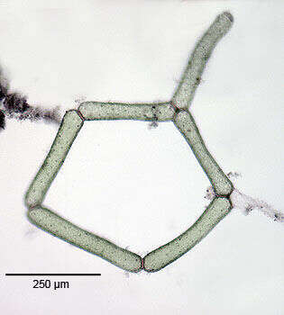
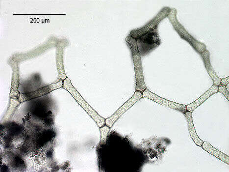
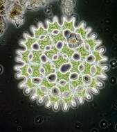
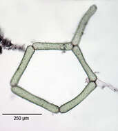
Portion of a coenobium of the chlorophycean, Hydrodictyon reticulatum (Lagerheim, 1894.The tubular coenobia have walls composed of a meshwork of cells whose interspaces are bordered by 5 or 6 cells (five in this case). Coenobia may form nets up to 2 feet in length.The cylindrical cells are uninucleate when toung and become multinucleate as they age. the plastid is peripheral and reticulate in older cells. there are many pyrenoids. Biflagellate zoospores released within the mother cell wall may may aggregate to form a mini meshwork that is ultimately released as a new coenobium.Collected from a temporary freshwater pond near Boise, Idaho. August 2005. Brightfield.
-
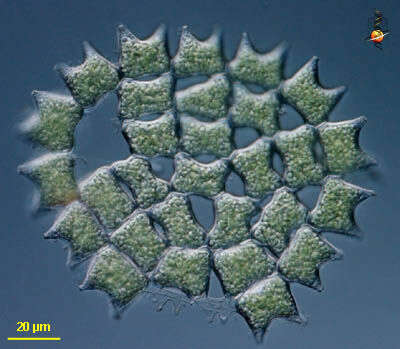
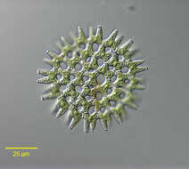
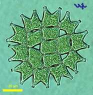
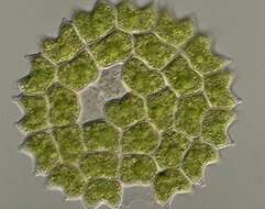
Pediastrum (pee-dee-ass-trum) a colonial green alga. cellulose cell walls, plastids with chlorophyll B, pyrenoids present. Colonies flat, and species distinguished by shape and arrangement of cells. Differential interference contrast.
-
-
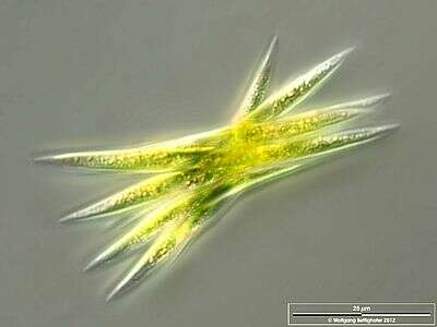
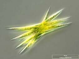
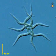
Aka Ankistrodesmus bibraianus. Scale bar indicates 10 µm.Sample from the pond Hegne Moor situated in the vicinity of Lake Constance. The image was built up using several photomicrographic frames with manual stacking technique. Images were taken using Zeiss Universal with Olympus C7070 CCD camera.Image under Creative Commons License V 3.0 (CC BY-NC-SA).
-
Portion of a coenobium of the chlorophycean, Hydrodictyon reticulatum (Lagerheim, 1894.The tubular coenobia have walls composed of a meshwork of cells whose interspaces are bordered by 5 or 6 cells (five in this case). Coenobia may form nets up to 2 feet in length.The cylindrical cells are uninucleate when toung and become multinucleate as they age. the plastid is peripheral and reticulate in older cells. there are many pyrenoids. Biflagellate zoospores released within the mother cell wall may may aggregate to form a mini meshwork that is ultimately released as a new coenobium.Collected from a temporary freshwater pond near Boise, Idaho. August 2005. Brightfield.
-
Pediastrum is a large, flat desmid. Many cells join together by their cellulose walls to form a regular, star-shaped array.
-
Scale bar indicates 10 µm. Sample from the Lake Constance (vicinity of Bodman). The image was built up using several photomicrographic frames with manual stacking technique. Images were taken using Zeiss Universal with Olympus C7070 CCD camera.Image under Creative Commons License V 3.0 (CC BY-NC-SA).
-
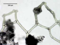
Detail of the junction of three cells of a coenobium of the chlorophycean, Hydrodictyon reticulatum (Lagerheim, 1894.The tubular coenobia have walls composed of a meshwork of cells whose interspaces are bordered by 5 or 6 cells (five in this case). Coenobia may form nets up to 2 feet in length.The cylindrical cells are uninucleate when toung and become multinucleate as they age. the plastid is peripheral and reticulate in older cells. there are many pyrenoids (seen here). Biflagellate zoospores released within the mother cell wall may may aggregate to form a mini meshwork that is ultimately released as a new coenobium.Collected from a temporary freshwater pond near Boise, Idaho. August 2005.DIC.
-
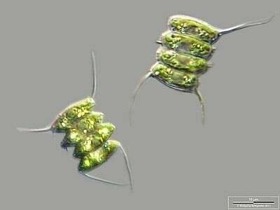
Ankistrodesmus (anne-kissed-ro-des-muss) is a green alga, small tangled clusters of twisted cells. Four cells in this tangle. Not uncommon in freshwater habitats. Differential interference contrast.