-
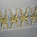
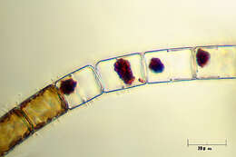
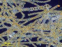
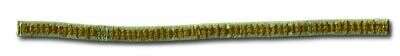
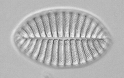
Filamentous diatom commonly found near the Tvarminne Zoological Station. Previously known as Achnantes taeniata.
-
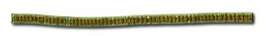
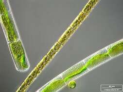
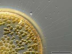
D. fragilissimus forms loosely connected chains. The cells bear processes on the valve end which fit into depressions on the adjacent cell. It is often found together with Leptocylindrus danicus and Guinardia delicatula
-
-
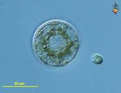
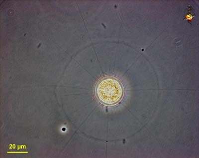
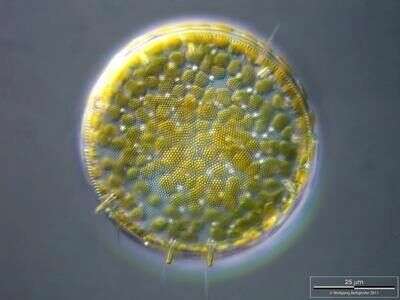
Cyclotella (sike-low-tell-a). Centric diatom, seen from valve view. The cell is surrounded by a sheath of mucus. Long thin organic spines project from the cell - and are believed to have a role in flotation. The pattern of pores in the frustule is used in identification. From a freshwater site. Phase contrast.
-
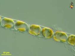
Eucampia (you-camp-ee-a) zoodiacus is a filament forming centric diatom (stramenochrome). Adjacent cells are attached by two interlocking apical elevations. Detail showing peripheral disc-shaped plastids and central nucleus. Differential interference microscopy.
data on this strain.
-
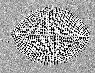
The oblique view exhibits short silicous spines, the so called occluded processes. On the lower left, lower right and central above chitinous spines are visible. Scale bar indicates 50 µm. The image was built up using several photomicrographic frames with manual stacking technique. Sample from North Sea near Heligoland (spring diatom bloom). Images were taken using Zeiss Universal with Olympus C7070 CCD camera.
-
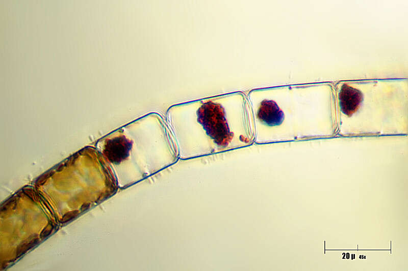
Detail showing plastids inside several cells at the end of a filament. Same filament as included in another picture in this collection.
-
-
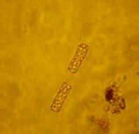
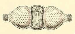
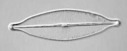
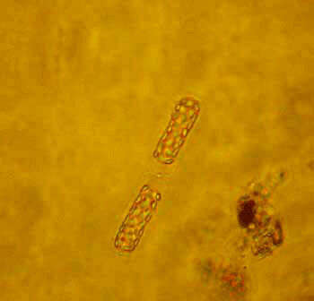
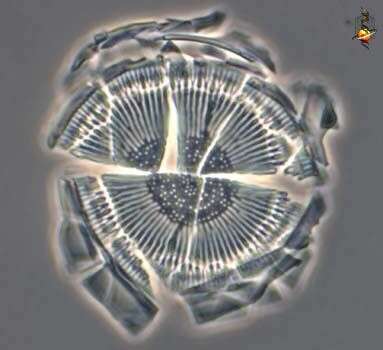
Biddulphia pulchella.
-
Melosira varians together with Mougeotia and Chlamydomonas. The scale bar indicates 50 µm. The specimen was gathered in the wetlands of Oderbruch (Oder valley 100 km north east of Berlin). The image was built up using several photomicrographic frames with manual stacking technique. Images were taken using Zeiss Universal with Olympus C7070 CCD camera.Image under Creative Commons License V 3.0 (CC BY-NC-SA).
-
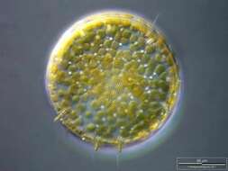
Cyclotella (sike-low-tell-a). Centric diatom, seen from valve view. With many small plastids containing chlorophylls a and c. From a freshwater site. Differential interference contrast.
-
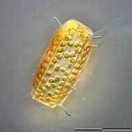
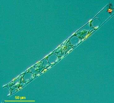
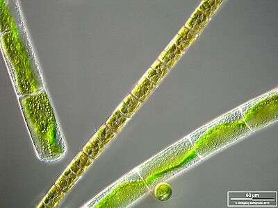
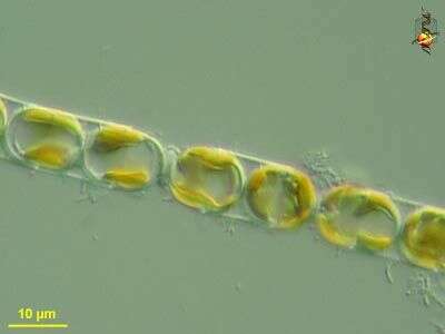
Melosira (mellow-sire-a) nummuloides, filament forming centric diatom, with multiple small plastids within the cell. Dark ground illumination. Leptosiropsis (leapt-owe-sire-op-sis) torulosa, green alga with organic wall that is produced in layers. Phase contrast microscopy.
data on this strain.
-
The oblique view exhibits short silicous spines, the so called occluded processes. Some chitinous spines protruding from the fultoportulae (also called strutted processes) along the dotted valve margin are also visible. Scale bar indicates 50 µm. The image was built up using several photomicrographic frames with manual stacking technique. Sample from North Sea near Heligoland (spring diatom bloom). Images were taken using Zeiss Universal with Olympus C7070 CCD camera.
-
Cell accompanied by epibiotic flagellates. Scale bar indicates 50 µm. Sample from the Federsee near Lake Constance. The image was built up using several photomicrographic frames with manual stacking technique. Images were taken using Zeiss Universal with Olympus C7070 CCD camera.Image under Creative Commons License V 3.0 (CC BY-NC-SA).
-
-
-
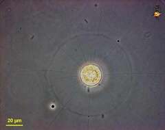
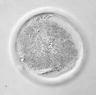
Cyclotella (sike-low-tell-a). Centric diatom, frustule only, seen from valve view, frustule broken from cover-slip pressure to show the brittle nature of the frustule. The pattern of pores in the frustule is used in identification. Phase contrast.
-
Melosira (mellow-sire-a) nummuloides, filament forming centric diatom, with multiple small plastids within the cell clearly shown in this micrograph. Differential interference microscopy.
data on this strain.
-
Silicious processes (the labiate and the occluded ones) are visible. Scale bar indicates 25 µm. The image was built up using several photomicrographic frames with manual stacking technique. Sample from North Sea near Heligoland (spring diatom bloom). Images were taken using Zeiss Universal with Olympus C7070 CCD camera.
-
-
-
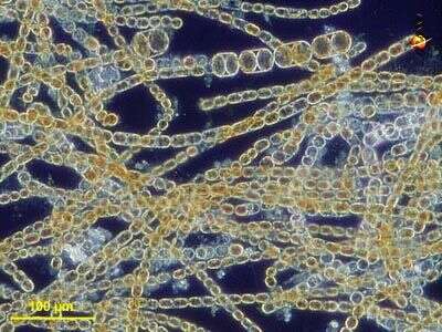
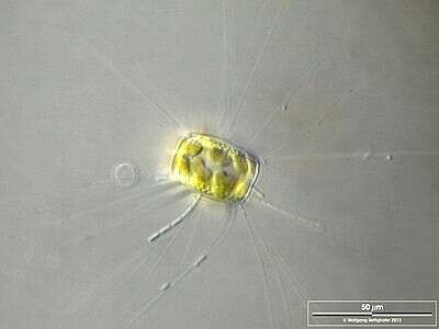
The thin, barely visible floating extensions are made of chitin. Furthermore, filamentous bacteria colonies are attached. Scale bar indicates 50 µm. Sample from the Lake Constance (vicinity of Bodman). The image was built up using several photomicrographic frames with manual stacking technique. Images were taken using Zeiss Universal with Olympus C7070 CCD camera.Image under Creative Commons License V 3.0 (CC BY-NC-SA).
-
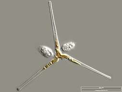
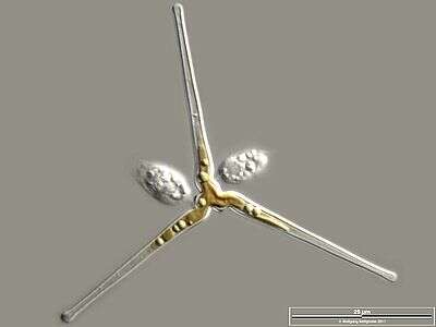
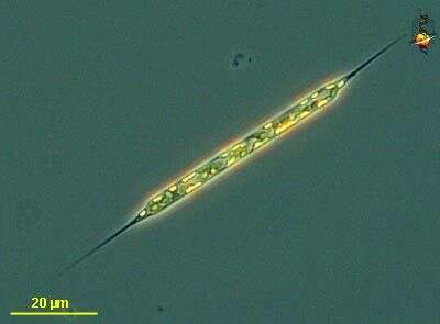
Rhizosolenia (rye-so-so-lean-ee-a) setigera, one of the common genera of marine phytoplantkonic diatoms, a centric diatom in which the valves, at the ends of the cells, are conical and give rise to spines. Much of the long cylindrical body is enclosed with hoop-shaped girdle bands. Phase contrast microscopy.
data on this strain.
-
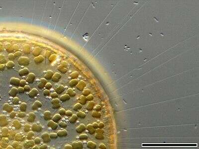
The image shows numerous chitinous spines which minimize their sedimentation speed. Scale bar indicates 25 µm. The image was built up using several photomicrographic frames with manual stacking technique. Sample from North Sea near Heligoland (spring diatom bloom). Images were taken using Zeiss Universal with Olympus C7070 CCD camera.Image under Creative Commons License V 3.0 (CC BY-NC-SA).