-
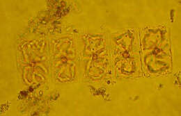
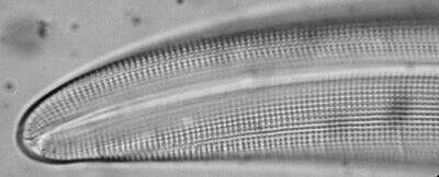
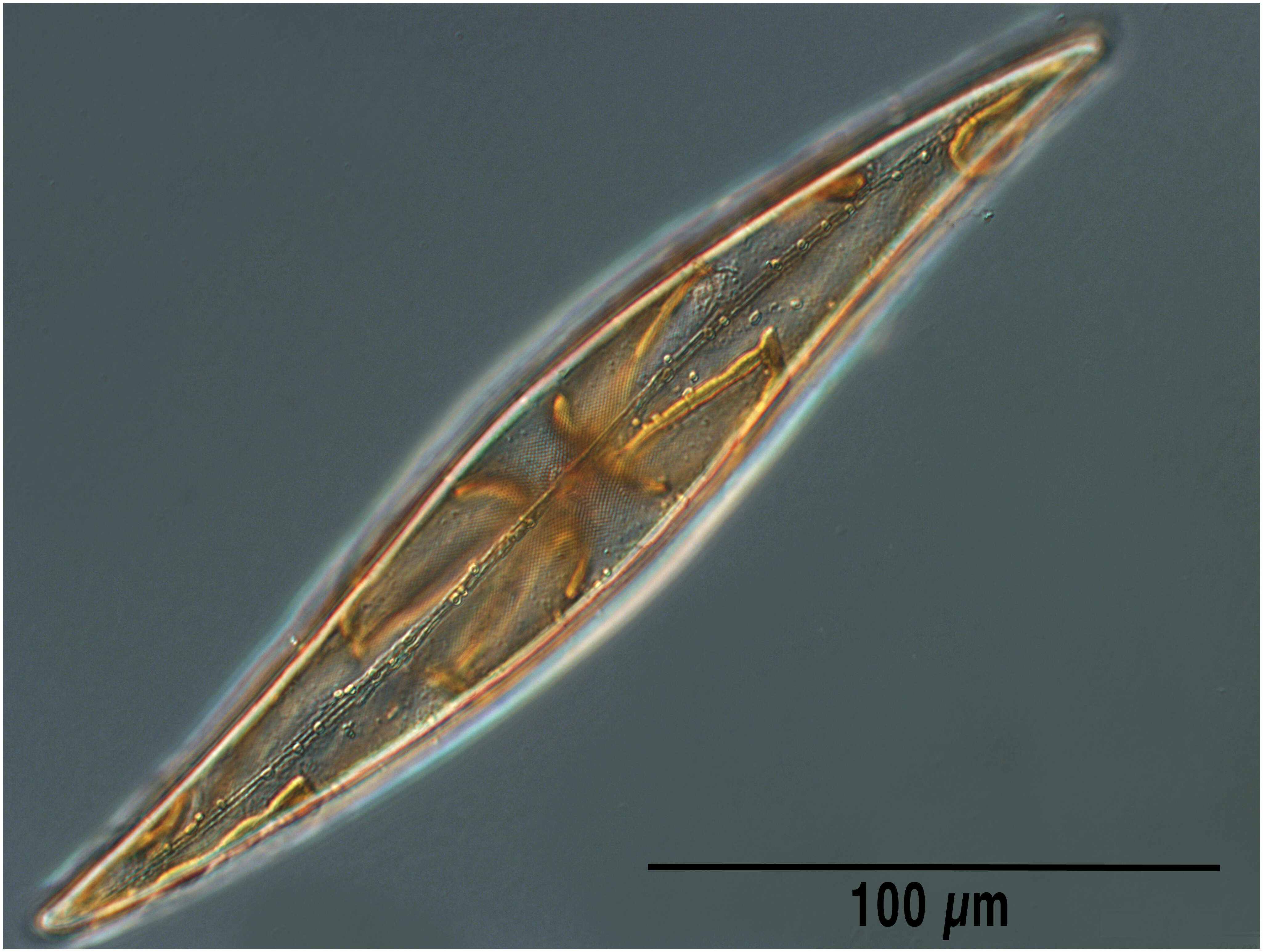
Valves are rectangular in girdle view and elliptical in valve view. Each cell has four ribbon like and folded chloroplasts, two along each side of the girdle.
-
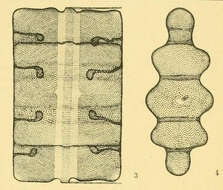
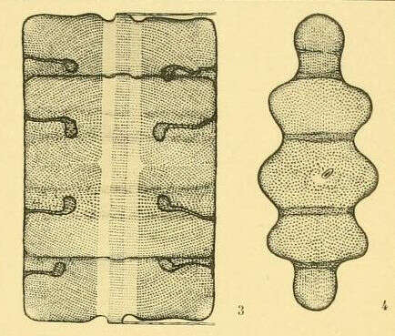
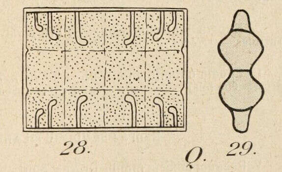
Terpsinoe musica by H. and M. PeragalloDiatomées marines de France et des districts maritimes voisins, par H. et M. Peragallo (b. 1851 and 1853, respectively). Publication info: Grez-sur-Loing,M. J. Tempère,1897-1908.Plate 90
-
-
-
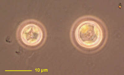
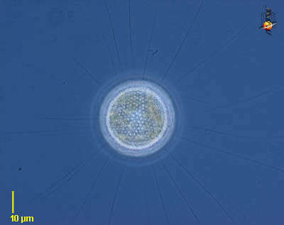
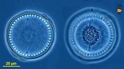
Cyclotella (sike-low-tell-a). Centric diatom, seen from valve view. Three plastid profiles are visible around the periphery of the cell. Long thin organic spines project from the cell - and are believed to have a role in flotation. The pattern of pores in the frustule is used in identification. Marine. Phase contrast.
-
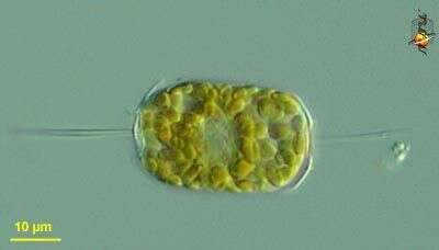
Ditylum (die-tie-lum) brightwellii. Marine centric diatom, cylindrical frustule from the ends of which is a long spine or labiate process. This contains two products of cell division, and each cell has numerous golden chloroplasts. Differential interference microscopy.
data on this strain.
-
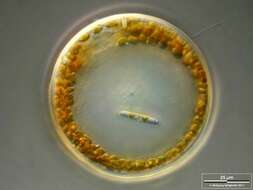
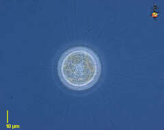
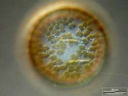
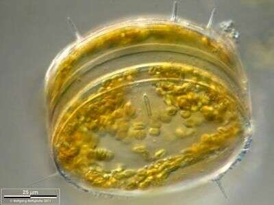
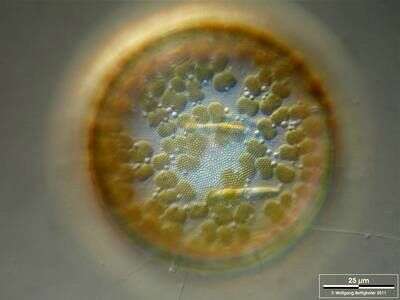
Some specimen of this centric diatom carried naviculoid ones on the valve(s). Scale bar indicates 25 µm. The image was built up using several photomicrographic frames with manual stacking technique. Sample from North Sea near Heligoland (spring diatom bloom). Images were taken using Zeiss Universal with Olympus C7070 CCD camera.
-
Image from type material.
-
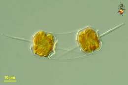
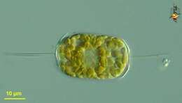
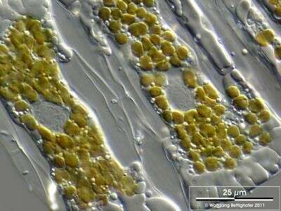
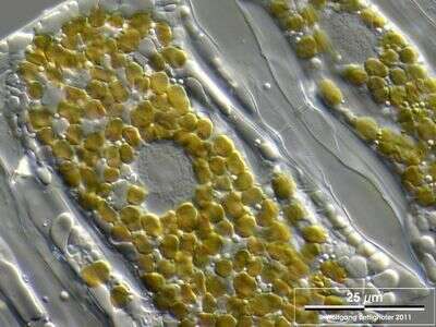
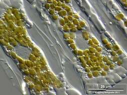
Closeup showing connection site, chloroplasts, sparkling droplets of storage matter and the nuclei with central hyalin nucleolus and the granulated karyoplasma around. Scale bar indicates 25 µm. The image was built up using several photomicrographic frames with manual stacking technique. Sample from North Sea near Heligoland (spring diatom bloom). Images were taken using Zeiss Universal with Olympus C7070 CCD camera.
-
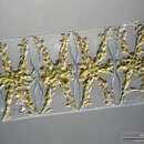
Terpsinoe musica by Henri CoupinFrom: Album général des diatomées marines, d'eau douce ou fossiles : album représentant tous les genres de diatomées et leurs principales espèces / par Henri Coupin (b. 1868).Plate 313: Seen in two directions. Fresh water. About 150 µ long.
-
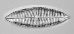
FRom the Bay of Villefranche in March 2013
-
-
O. sinensis is closely related to O. mobiliensis and O. regia but the processes are nearly parallel to the cell axis and the processes are close to the processes. The valve face between the processes is flat or concave.
-
-
Centric diatom, seen from valve view. The pattern of pores in the frustule is used in identification. Marine. Phase contrast.
-
Ditylum (die-tie-lum) brightwellii. Marine centric diatom, cylindrical frustule from the ends of which is a long spine or labiate process. Many small plastids and central nucleus. Differential interference microscopy.
data on this strain.
-
The oblique view exhibits short silicous spines, the so called occluded processes. On the lower left near the scale bar a chitinous spine is visible. Scale bar indicates 25 µm. The image was built up using several photomicrographic frames with manual stacking technique. Sample from North Sea near Heligoland (spring diatom bloom). Images were taken using Zeiss Universal with Olympus C7070 CCD camera.
-
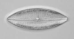
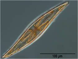
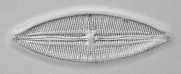
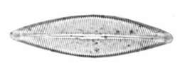
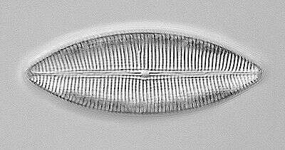
Formerly Brebissonia boeckii, material from the Netherlands.
-
Closeup showing chloroplasts, sparkling droplets of storage matter and the nuclei with central hyalin nucleolus and the granulated karyoplasma around. Scale bar indicates 25 µm. The image was built up using several photomicrographic frames with manual stacking technique. Sample from North Sea near Heligoland (spring diatom bloom). Images were taken using Zeiss Universal with Olympus C7070 CCD camera.
-
-
-
Cyclotella (sike-low-tell-a). Centric diatom, frustule only, seen from valve view, with the two frustules seen at slightly different focal planes. . The pattern of pores in the frustule is used in identification. Phase contrast.
-
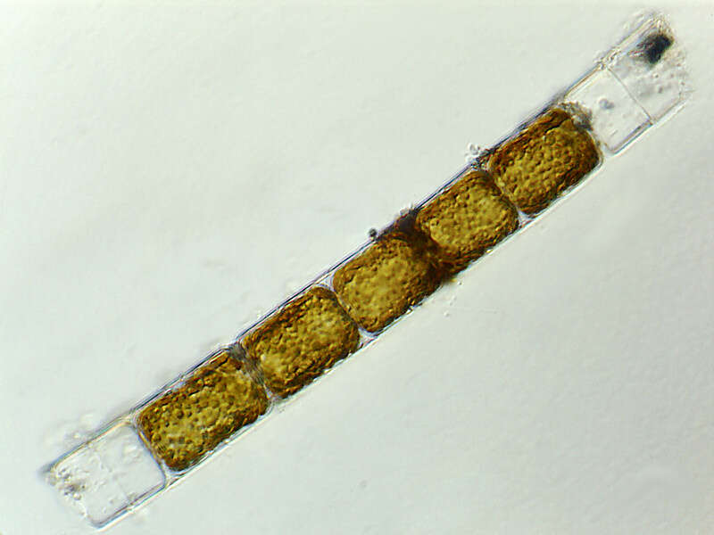
Eucampia (you-camp-ee-a) zoodiacus is a filament forming diatom (stramenochrome). Adjacent cells are attached by two interlocking apical elevations. Differential interference microscopy.
data on this strain.
-
Some specimen of this centric diatom carried naviculoid ones on the valve(s). Scale bar indicates 25 µm. The image was built up using several photomicrographic frames with manual stacking technique. Sample from North Sea near Heligoland (spring diatom bloom). Images were taken using Zeiss Universal with Olympus C7070 CCD camera.