-
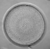
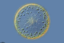
Fig 2: Coscinodiscus wailesii Light micrograph of valve face of a live cell
-
-
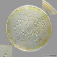
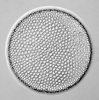
Centric diatom, seen from valve view. This is an empty frustule of a large marine species. The pattern of pores in the frustule is used in identification. Marine. Phase contrast.
-
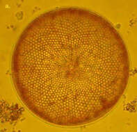
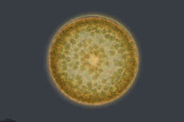
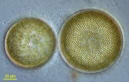
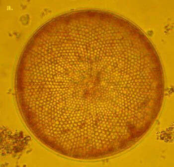
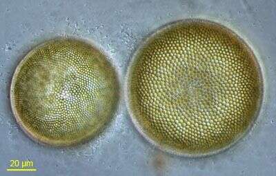
C. radiatus is one of the smaller Coscinodiscus species. It is box shaped in girdle view and the valves are very flat. The areolae form disctinct rows radiating from the valve centre. C. radiatus is a cosmopolitan species.
-
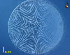
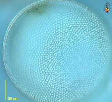
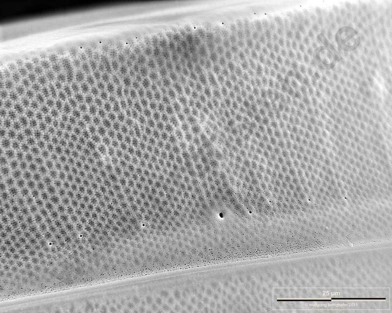
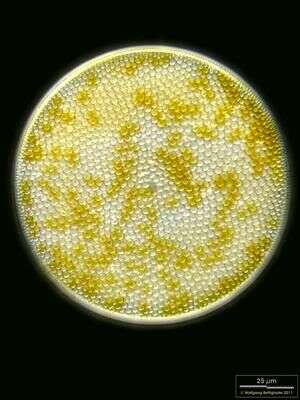
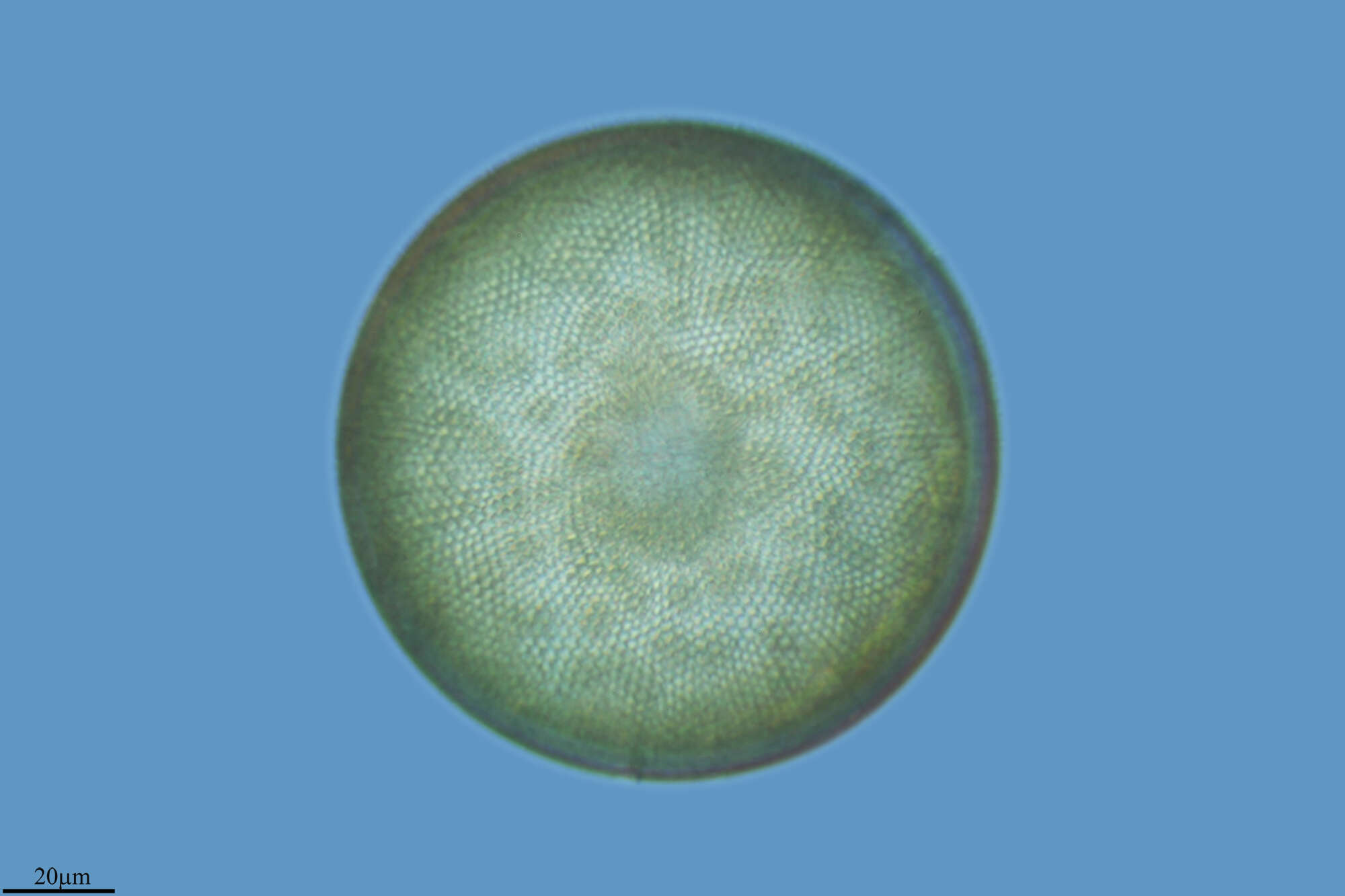
Coscinodiscus radiatus.Valvar view. Scale bar indicates 25 m. Sample from North Sea near Heligoland (spring diatom bloom). The image was built up using several photomicrographic frames with manual stacking technique. Images were taken using Zeiss Universal with Olympus C7070 CCD camera.For more look at
www.protisten.de/english/gallery_main/gallery_main.htmlFor high-resolution images please ask postmaster@protisten.de..
-
Grove, O, Galicia, Spain
-
Grove, O, Galicia, Spain
-
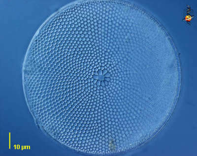
Coscinodiscus wailesii.Valvar view. Insets showing marginal ring of labiate processes (upper right) and the hyaline central area (lower left). Scale bar indicates 100 m. Sample from North Sea near Heligoland (spring diatom bloom). The image was built up using several photomicrographic frames with manual stacking and stitching technique. Images were taken using Zeiss Universal with Olympus C7070 CCD camera.For more look at
www.protisten.de/english/gallery_main/gallery_main.htmlFor high-resolution images please ask postmaster@protisten.de..
-
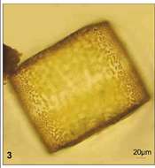
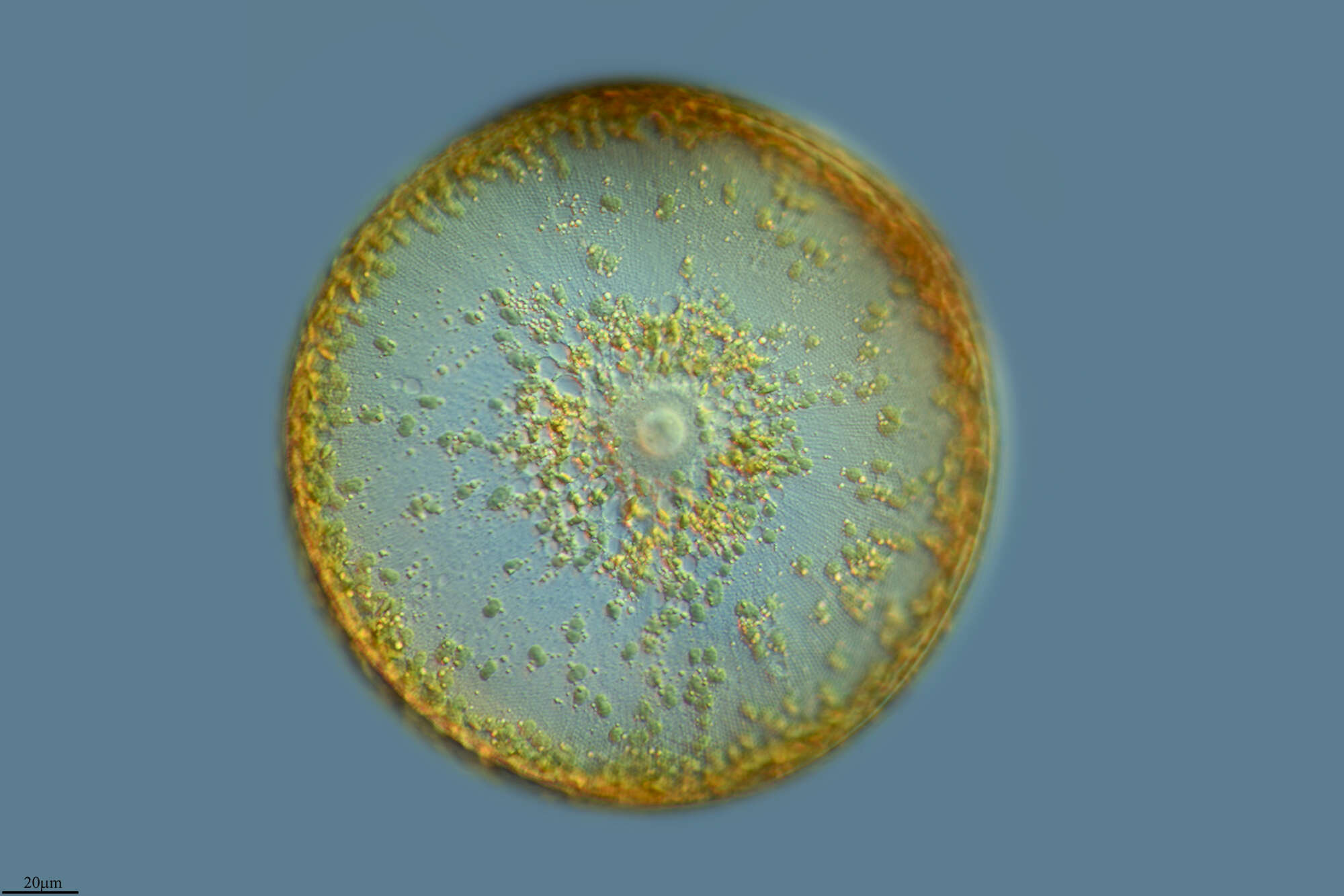
Fig 4: Coscinodiscus wailesii Light micrograph central hyaline area of a live cell
-
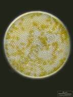
Coscinodiscus (coss-co-no-disc-us) a centric diatom, seen from valve view. This is an empty frustule of a large marine species. The pattern of pores in the frustule is used in identification. Marine. Phase contrast.
-
-
Grove, O, Galicia, Spain
-
Grove, O, Galicia, Spain
-
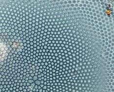
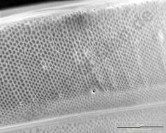
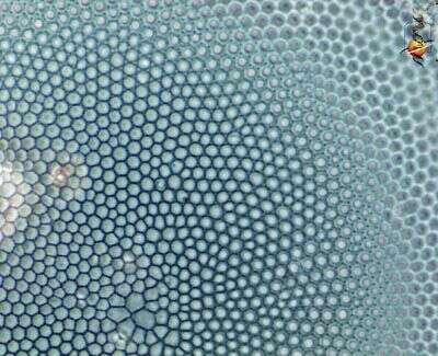
Coscinodiscus wailesii.Closeup of the lateral side of the valve. Scale bar indicates 25 m. Sample from North Sea near Heligoland (spring diatom bloom). The image was built up using several photomicrographic frames with manual stacking technique. Use of SEM equipment courtesy of Lab Dr. Karl-Heinz Schffner, Solingen, Germany. For more look at
www.protisten.de/english/gallery_main/gallery_main.htmlFor high-resolution images please ask postmaster@protisten.de.
-
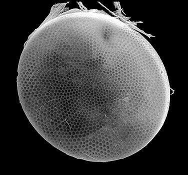
Fig 5: Scanned electron micrograph image of C.wailesii in the valve view.
-
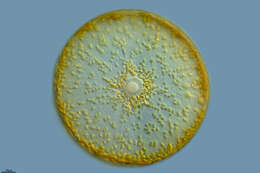
Coscinodiscus (a centric diatom), seen from valve view. This is an empty frustule of a large marine species. The pattern of pores in the frustule is used in identification. Marine. Differential interference contrast.
-
Valvar view. Scale bar indicates 25 µm. Sample from North Sea near Heligoland (spring diatom bloom). The image was built up using several photomicrographic frames with manual stacking technique. Images were taken using Zeiss Universal with Olympus C7070 CCD camera.
-
Grove, O, Galicia, Spain
-
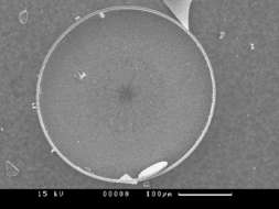
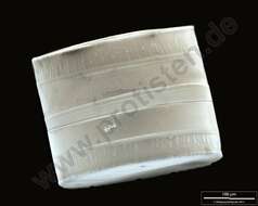
Coscinodiscus wailesii.SEM of girdle view. The ligulae which fit in the open girdle bands are weakly visible. Scale bar indicates 100 m. Sample from North Sea near Heligoland (spring diatom bloom). The image was built up using several photomicrographic frames with manual stacking technique. Use of SEM equipment courtesy of Lab Dr. Karl-Heinz Schffner, Solingen, Germany. For more look at
www.protisten.de/english/gallery_main/gallery_main.htmlFor high-resolution images please ask postmaster@protisten.de.
-
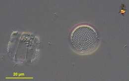
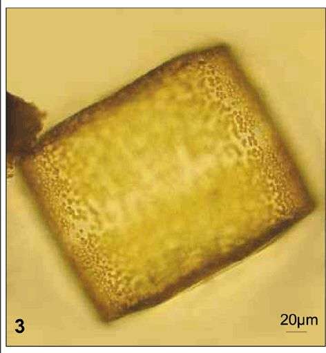
Fig 3: Coscinodiscus wailesii Light micrograph of a Lugol's preserved cell in girdle view
-
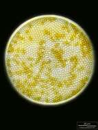
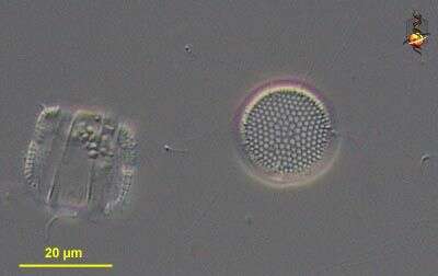
Coscinodiscus (caw-skin-owe-disk-us) a centric diatom (stramenopile), this genus is common in the marine plankton and has hundreds of species, some of which can achieve a very large size. The cell to the left is in girdle view, with the two valves visible to either end and girdle bands in the middle of the cell, the cell to the right is seen from valve (end) view. This genus has small thickenings (processes) around the margin of the valve. The species are mostly distinguished by the pattern of sculpting of the frustule. Differential interference microscopy.
data on this strain.
-
Scanning electron microscope image of valve. The organism is tentatively identified as C. radiatus. Sample taken from the water column off Martha's Vineyard, Massachusetts. Image by Charley O'Kelly and Shauna Murray.
-
Grove, O, Galicia, Spain
-
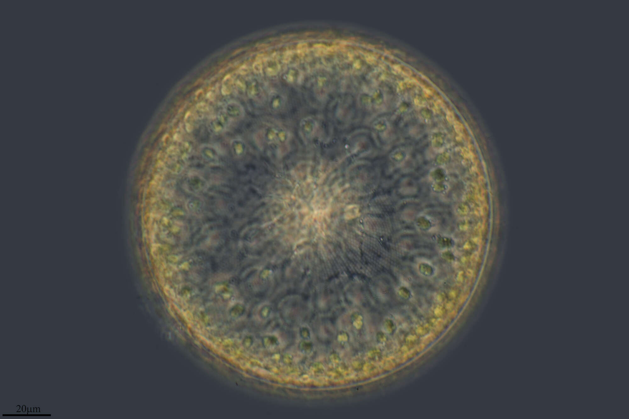
Cells of this centric diatom observed in the water column from Lake Pontchartrain, differential interference contrast optics.