-
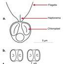
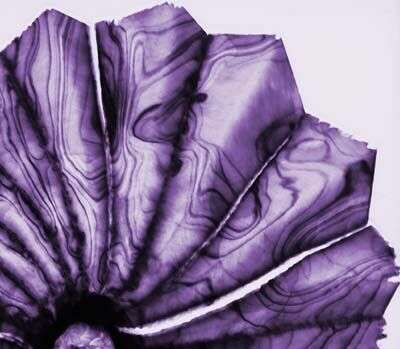
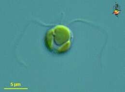
Chrysochromulina (cry-so-crumb-you-line-a) a single-celled haptophyte, with two similar flagella, a short haptonema lying between the flagella, and golden plastids. This may be C. herdlensis. Differential interference microscopy.
data on this strain.
-
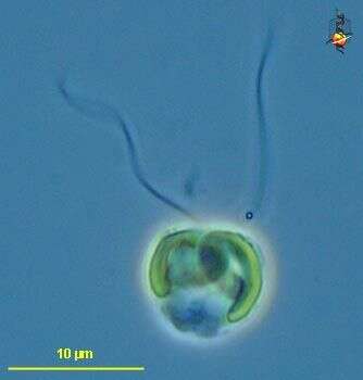
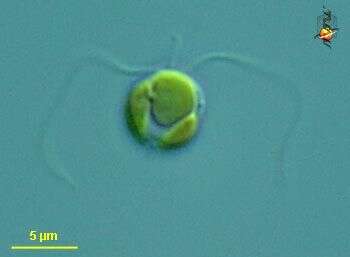
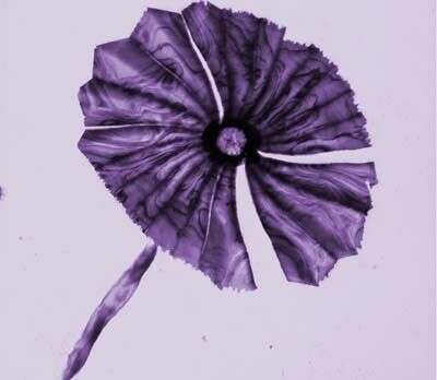
Chrysochromulina (cry-so-crumb-you-line-a) a single-celled haptophyte, with two similar flagella, a short haptonema lying between the flagella, and two golden plastids. This may be C. herdlensis. Differential interference microscopy.
data on this strain.
-
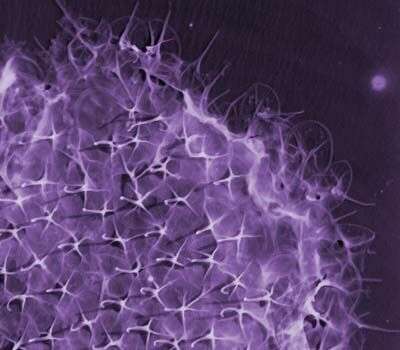
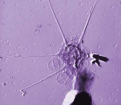
This image was made from samples taken during a scientific cruise in the Pacific. Water was filtered to concentrate the organisms that were present, then dried onto a thin sheet of plastic and then shadowed with a fine layer of metal to provide contrast. The preparation was then observed with an electron-microscope. This technique has been used to document the diversity of marine microbes, especially, protists in the oceans.
-
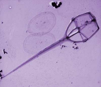
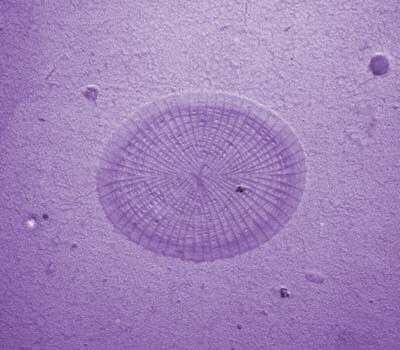
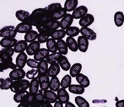
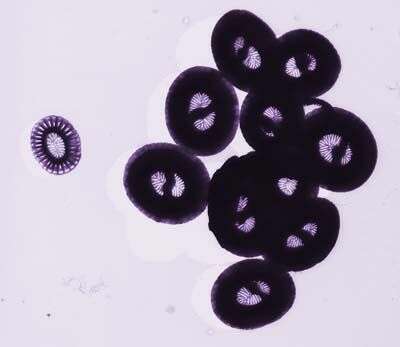
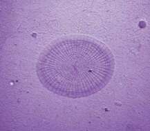
Detail of holococcoliths from the haploid life-cycle phase of S. pulchra
-
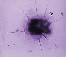
This image was made from samples taken during a scientific cruise in the Pacific. Water was filtered to concentrate the organisms that were present, then dried onto a thin sheet of plastic and then shadowed with a fine layer of metal to provide contrast. The preparation was then observed with an electron-microscope. This technique has been used to document the diversity of marine microbes, especially, protists in the oceans.
-
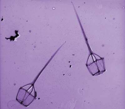
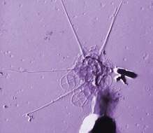
This image was made from samples taken during a scientific cruise in the Pacific. Water was filtered to concentrate the organisms that were present, then dried onto a thin sheet of plastic and then shadowed with a fine layer of metal to provide contrast. The preparation was then observed with an electron-microscope. This technique has been used to document the diversity of marine microbes, especially, protists in the oceans. Jeremy Young says ""The stem-like object at bottom left is not part of the specimen but a random piece of junk"". It does however cutify the image.
-
This image was made from samples taken during a scientific cruise in the Pacific. Water was filtered to concentrate the organisms that were present, then dried onto a thin sheet of plastic and then shadowed with a fine layer of metal to provide contrast. The preparation was then observed with an electron-microscope. This technique has been used to document the diversity of marine microbes, especially, protists in the oceans.
-
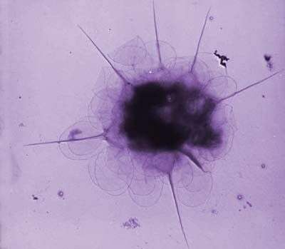
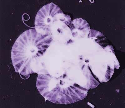
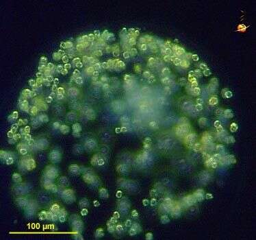
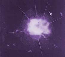
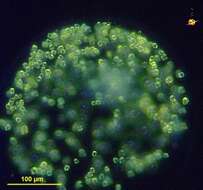
Phaeocystis (fay-owe-sis-tiss), an unusual haptophyte (prymnesiophyte) in which cells form spherical gelatinous colonies, and may cause problems with fish respiration. This medium-sized colony may be of P. globosa. Dark ground illumination.
data on this strain.
-
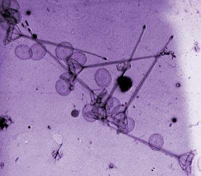
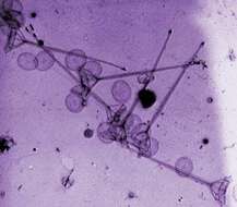
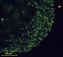
Phaeocystis (fay-owe-sis-tiss), an unusual haptophyte (prymnesiophyte) in which cells form spherical gelatinous colonies, and may cause problems with fish respiration. This is a small element of a large colony .
data on this strain.
-
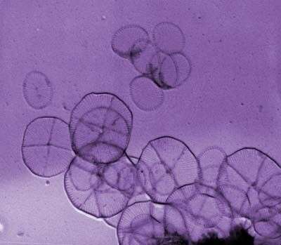
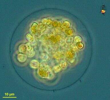
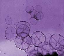
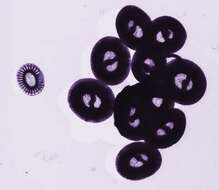
Phaeocystis (fay-owe-sis-tiss), an unusual haptophyte (prymnesiophyte) in which cells form spherical gelatinous colonies, and may cause problems with fish respiration. This small colony is probably P. globosa.
data on this strain.
-
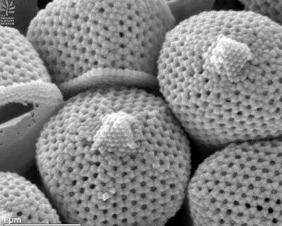
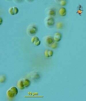
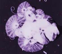
Phaeocystis (fay-owe-sis-tiss), an unusual haptophyte (prymnesiophyte) in which cells form spherical gelatinous colonies, and may cause problems with fish respiration. Detail, probably P. globosa. Differential interference microscopy.
data on this strain.