-
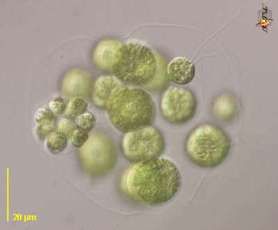
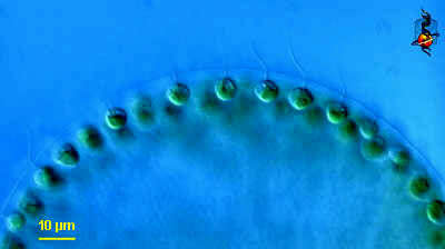
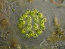
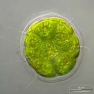
Eudorina (you-door-ine-a) is a colonial volvocid - motile green alga. In this case 16 cells are embedded in a sphere of common mucus. Each individual cell has two flagella, and a cup-shaped chloroplast - a bit like a Chlamydomonas cell. There are a number of related genera which form colonies using mucus as a common matrix, but they differ in the numbers of cells involved, how tightly packed they are, and the shape of the colony. This image illustrates the way that a daughter colony of tightly packed cells is formed within a parental colony. Phase contrast.
-
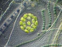
Eudorina (you-door-ine-a) is a colonial volvocid - motile green alga. In this case 16 cells are embedded in a sphere of common mucus. Each individual cell has two flagella, and a cup-shaped chloroplast - a bit like a Chlamydomonas cell. There are a number of related genera which form colonies using mucus as a common matrix, but they differ in the numbers of cells involved, how tightly packed they are, and the shape of the colony. This image illustrates the flagella very well Phase contrast.
-
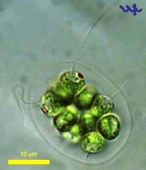
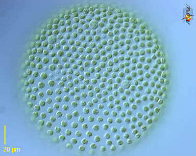
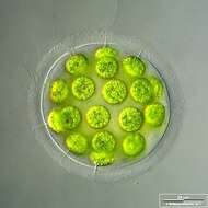
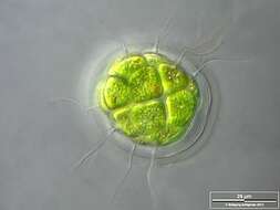
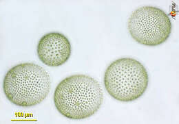
Eudorina, a motile colonial volvocid flagellate. Colonies may be cylindrical, elliptical or spherical as seen in this image. Colonies may be composed of 8 to 128 cells. This colony is composed of 16 individuals embedded in a clear gelatinous matrix. Two equal length flagella protrude through the matrix from each cell. Cells may be arranged in tiers or more randomly as seen here. One large bright green cup-shaped chloroplast. Small red stigma. Sexual and asexual reproduction occurs. From temporary rainwater pool in grass field near Boise, Idaho. Phase contrast.
-
Eudorina, a motile colonial volvocid flagellate. Colonies may be cylindrical, elliptical or spherical as seen in this image. Colonies may be composed of 8 to 128 cells. This colony is composed of 16 individuals embedded in a clear gelatinous matrix. Two equal length flagella protrude through the matrix from each cell. Cells may be arranged in tiers or more randomly as seen here. One large bright green cup-shaped chloroplast. Small red stigma. Sexual and asexual reproduction occurs. From temporary rainwater pool in grass field near Boise, Idaho. Brightfield.
-
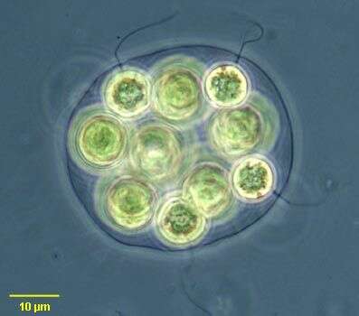
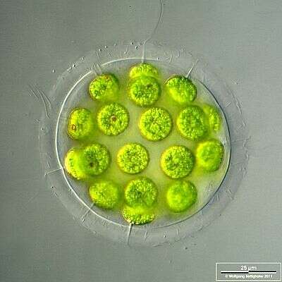
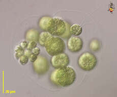
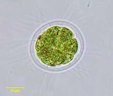
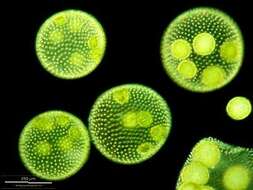
Multilayer image shows mucilaginous envelope, flagellae, structure of chloroplasts and in some cases the stigma. Scale bar indicates 25 µm. Sample from the pond Hegne Moor situated in the vicinity of Lake Constance (Bodensee, Southern Germany). Images were taken using Zeiss Universal with Olympus C7070 CCD camera.
-
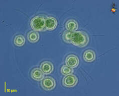
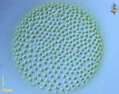
Eudorina-colony accompanied by cyanobacteria (Anabaeba) and phototrophic eubacteria Pelodictyon (small bluegreen spherules embedded in mucilage). Scale bar indicates 50 µm.Sample from the pond Hegne Moor situated in the vicinity of Lake Constance. The image was built up using several photomicrographic frames with manual stacking technique. Images were taken using Zeiss Universal with Olympus C7070 CCD camera.Image under Creative Commons License V 3.0 (CC BY-NC-SA).
-
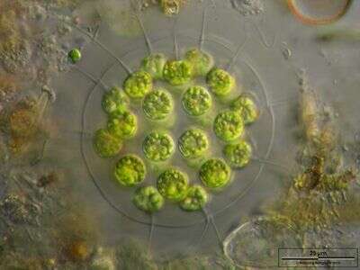
Scale bar indicates 25 µm.Sample from the pond Hegne Moor situated in the vicinity of Lake Constance. The image was built up using several photomicrographic frames with manual stacking technique. Images were taken using Zeiss Universal with Olympus C7070 CCD camera.Image under Creative Commons License V 3.0 (CC BY-NC-SA).
-
-
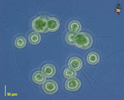
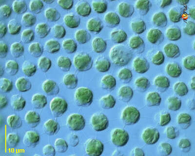
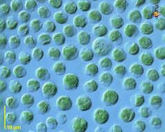
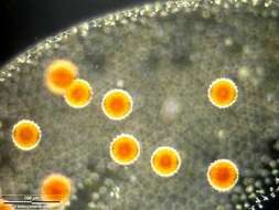
Portrait of Pandorina. Colonial flagellate with bright green chloroplasts. Usually 8-16 cells each with 2 equal-length flagella all enclosed within clear gelatinous envelope. From freshwater pond near Boise, Idaho. Brightfield.
-
Pandorina morum is rare in Lake Kinneret. This specimen was collected at the shore by the Kinneret Limnological Laboratory in March 2006.
-
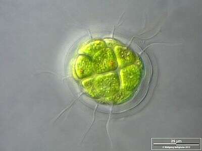
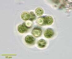
DOF image shows the layers of the mucilaginous sheath, several flagellae and stigmae of two cells. Scale bar indicates 25 µm. Sample from sphagnum pond situated in the northern alpine region of Austria near Salzburg. Images were taken using Zeiss Universal with Olympus C7070 CCD camera.
-
Scale bar indicates 25 µm.Sample from the pond Hegne Moor situated in the vicinity of Lake Constance. The image was built up using several photomicrographic frames with manual stacking technique. Images were taken using Zeiss Universal with Olympus C7070 CCD camera.Image under Creative Commons License V 3.0 (CC BY-NC-SA).
-
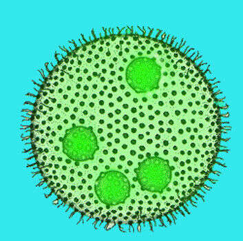
Volvox (vol-vox) is the iconic colonial volvocid - motile green alga. In this case hundred of cells are embedded in a sphere of common mucus. Each individual cell is tiny, linked to others by fine threads of cytoplasm, has two flagella, and a cup-shaped chloroplast - a bit like a Chlamydomonas cell. There are a number of related genera which form colonies using mucus as a common matrix, but they differ in the numbers of cells involved, how tightly packed they are, and the shape of the colony. Differential interference contrast.
-
Volvox (vol-vox) is the iconic colonial volvocid - motile green alga. In this case hundred of cells are embedded in a sphere of common mucus. Each individual cell is tiny, linked to others by fine threads of cytoplasm, has two flagella, and a cup-shaped chloroplast - a bit like a Chlamydomonas cell. There are a number of related genera which form colonies using mucus as a common matrix, but they differ in the numbers of cells involved, how tightly packed they are, and the shape of the colony. This is a detail of the surface of the colony, showing the flagella projecting from beyond the edge of the mucus. Differential interference contrast.
-
Volvox (vol-vox) is the iconic colonial volvocid - motile green alga. In this case hundred of cells are embedded in a sphere of common mucus. Each individual cell is tiny, linked to others by fine threads of cytoplasm, has two flagella, and a cup-shaped chloroplast - a bit like a Chlamydomonas cell. There are a number of related genera which form colonies using mucus as a common matrix, but they differ in the numbers of cells involved, how tightly packed they are, and the shape of the colony. This picture is a detail of the surface showing the chloroplasts within individual cells and showing the links among cells. Differential interference contrast.
-
Volvox (vol-vox) is the iconic colonial volvocid - motile green alga. In this case hundred of cells are embedded in a sphere of common mucus. Each individual cell is tiny, linked to others by fine threads of cytoplasm, has two flagella, and a cup-shaped chloroplast - a bit like a Chlamydomonas cell. There are a number of related genera which form colonies using mucus as a common matrix, but they differ in the numbers of cells involved, how tightly packed they are, and the shape of the colony. This picture is of several colonies, the two lower ones contain a number of smaller daughter colonies within. Differential interference contrast.
-
-
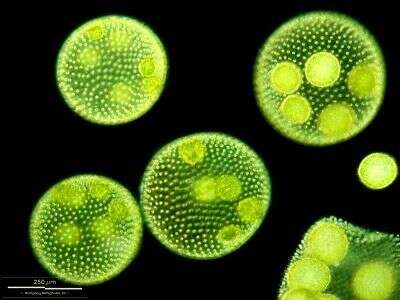
Cell colonies with daugther colonies. One parent disintegrates and let the daughters run free. Scale bar indicates 250 µm.Sample from ponds situated in the vicinity of Lake Constance (Bodensee, Southern Germany). The image was built up using several photomicrographic frames with manual stacking technique. Images were taken using Zeiss Universal with Olympus C7070 CCD camera.
-
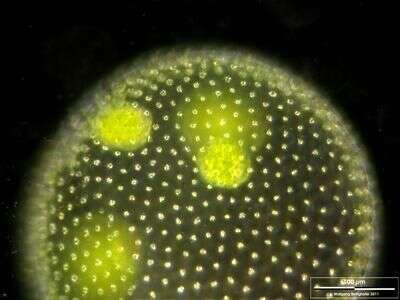
Cell colony with daugther colonies. Scale bar indicates 100 µm.Sample from ponds situated in the vicinity of Lake Constance (Bodensee, Southern Germany). The image was built up using several photomicrographic frames with manual stacking technique. Images were taken using Zeiss Universal with Olympus C7070 CCD camera.
-
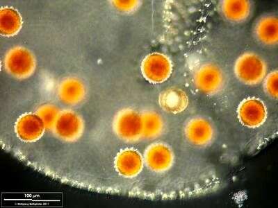
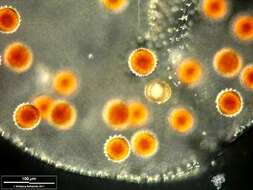
Desintegrated parent colony with zygotes. Scale bar indicates 100 µm.Sample from ponds situated in the vicinity of Lake Constance (Bodensee, Southern Germany). The image was built up using several photomicrographic frames with manual stacking technique. Images were taken using Zeiss Universal with Olympus C7070 CCD camera.
-
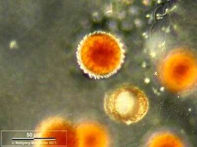
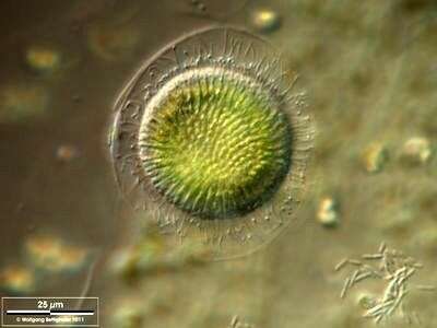
Closeup of a zygote. Mucilaginous sheath is visible. Scale bar indicates 50 µm. Sample from ponds situated in the vicinity of Lake Constance (Bodensee, Southern Germany). The image was built up using several photomicrographic frames with manual stacking technique. Images were taken using Zeiss Universal with Olympus C7070 CCD camera.
-
Desintegrated parent colony with zygotes. Scale bar indicates 100 µm.Sample from ponds situated in the vicinity of Lake Constance (Bodensee, Southern Germany). The image was built up using several photomicrographic frames with manual stacking technique. Images were taken using Zeiss Universal with Olympus C7070 CCD camera.
-
An association of Volvox microgametes (spermatozoids). Scale bar indicates 25 µm.Sample from ponds situated in the vicinity of Lake Constance (Bodensee, Southern Germany). The image was built up using several photomicrographic frames with manual stacking technique. Images were taken using Zeiss Universal with Olympus C7070 CCD camera.
-
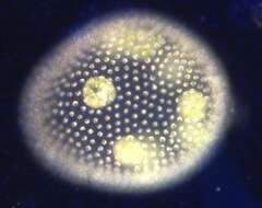
Dark ground image of colony, with daughter colonies enclosed within.