-
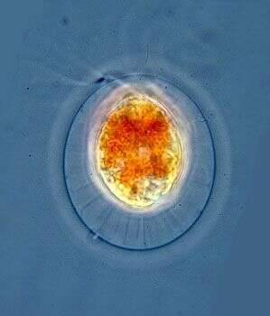
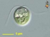
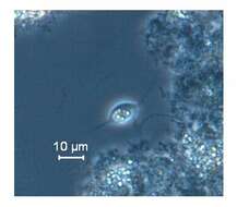
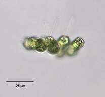
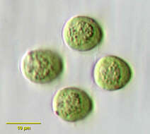
Phase contrast micrograph of living cell.
-
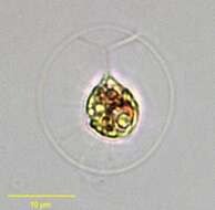
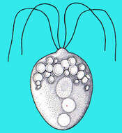
Portrait of Haematococcus pluvialis (Flotow, 1844), a widely distributed volvocid flagellate. Thin cytoplasmic strands traverse the clear mucilaginous layer to connect the protoplast to the spherical cell wall. Single large cup-shaped chloroplast. Two equal-length flagella are also seen traversing the mucilaginous layer. Reddish carotenoid pigments may obscure the stigma and chloroplast. From fish farm aquaculture pond near Boise, Idaho. Phase contrast.
-
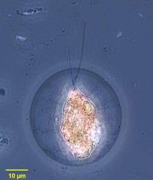
Portrait of Haematococcus pluvialis (Flotow, 1844), a widely distributed volvocid flagellate. Thin cytoplasmic strands traverse the clear mucilaginous layer to connect the protoplast to the spherical cell wall. Single large cup-shaped chloroplast. Two equal-length flagella are also seen traversing the mucilaginous layer. Reddish carotenoid pigments may obscure the stigma and chloroplast. From fish farm aquaculture pond near Boise, Idaho. Brightfield.
-
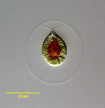
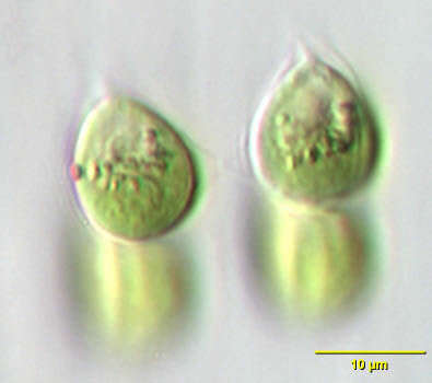
Portrait of Haematococcus pluvialis (Flotow, 1844), a widely distributed volvocid flagellate. Thin cytoplasmic strands traverse the clear mucilaginous layer to connect the protoplast to the spherical cell wall. Single large cup-shaped chloroplast. Two equal-length flagella are also seen traversing the mucilaginous layer. Reddish carotenoid pigments (concentrated in cell center here) may obscure the stigma and chloroplast. From ephemeral freshwater pool near Boise, Idaho, March, 2005. DIC.
-
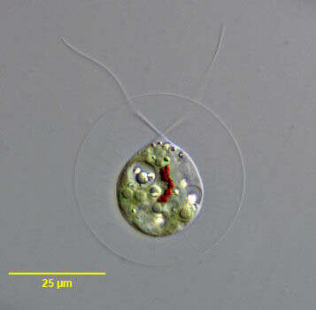
Portrait of Haematococcus pluvialis (Flotow, 1844), a widely distributed volvocid flagellate. Thin cytoplasmic strands traverse the clear mucilaginous layer to connect the protoplast to the spherical cell wall. Single large cup-shaped chloroplast. Two equal-length flagella are also seen traversing the mucilaginous layer. Reddish carotenoid pigments are concentrated in the cell center here. The inconspicuous stigma is seen at 1 o'clock near the proroplast surface. Several pyrenoids are visible here. There are multiple small contractile vacuoles. refractile cytoplasmic crystals are present in this individual. From ephemeral freshwater pool near Boise, Idaho, March, 2005. DIC.
-
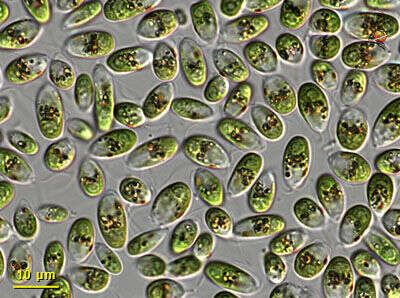
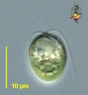
Dunaliella (done-al-ee-ella), a solitary volvocid (flagellated green algal cell). Cell surrounded by a cellulosic wall, with two similar flagella emerging from near the apex. The photosynthetic pigments are located within a cup-shaped chloroplast which has a large pyrenoid with associated polysaccharide materials located posteriorly. The nucleus is located within the cup. From marine and usually hypersaline habitats, grown commercially because of the tendency to produce large quantities of beta carotene when intensely illuminated. Differential interference contrast.
-
Dunaliella is a green alga and is a common member of the phytoplankton in salty water bodies. These cells were abundant in a collection taken at the margins of Mono lake (at Navy Beach). Each cell has a cup-shaped or bowl-shpaed chloroplast at the posterior end of the cell, aneriorly they have two equally long flagella. Differential interference contrast optics.
-
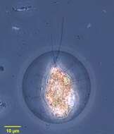
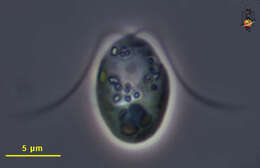
This is an un-named species of Dunaliella, a green alga and member of the phytoplankton that is often associated with hypersaline conditions. The chloroplast is bowl-shaped and fills the base of the cell. There is a yellowy eyespot to the left side of the plastid in this image. The nucleus with nucleolus lies near the front of the cell. There are two equally long flagella which beat in a breast-stroke motion. Phase contrast micrograph.
-
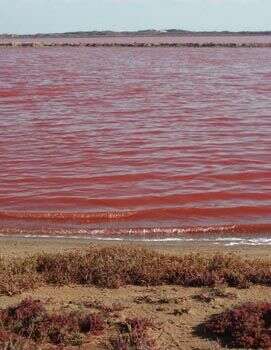
This image is of a sea-water lagoon in Western Australia. The water is red because of the very large numbers of Dunaliella salina, a green alga that is commercially harvested for carotenes as food additives and coloring agents.
-
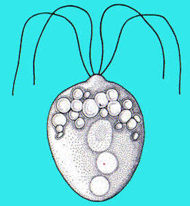
Polytomella (paul-ee-toe-mell-a) is one of a small number of green (Viridaeplantae) algal genera which lack plastids. There are four flagella inserting in a square pattern - and only two opposed flagella can be seen in this image. The flagella insert in small dimples at the anterior end of the cell. Often found in habitats rich in organic matter and low in oxygen. Phase contrast.
-
-
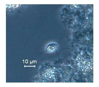
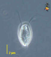
This flagellated protozoa was observed from a perchlorate treating laboratory scale anaerobic bioreactor (ORP~ -200 mV) fed with acetate as substrate. It was characterized by a pair of anterior flagella, tailing body and intracellular starch granules. Krishnakumar B and Anupama V.N. of NIIST (CSIR) are involved in the bioreactor study. The phase contrast image shows cells with intracellular starch granules and the same released to the medium.
-
-
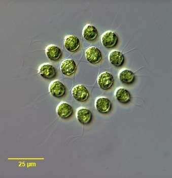
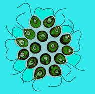
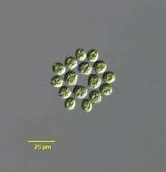
Gonium pectorale, a common colonial volvocid flagellate. Colonies usually consist of 16 cells (twelve peripheral and four central) in a plate-like arrangement of one layer. An extracellular matrix that is not easily seen connects cells. Cells have a cup-shaped chloroplast with a small anterior stigma. Lateral view of colony. From freshwater pond near Boise, Idaho. Phase contrast.
-
Gonium pectorale, a common colonial volvocid flagellate. Colonies usually consist of 16 cells (twelve peripheral and four central) in a plate-like arrangement of one layer. An extracellular matrix that is not easily seen connects cells. Cells have a cup-shaped chloroplast with a small anterior stigma. Anteroposterior view of colony. From freshwater pond near Boise, Idaho. Brightfield illumination.
-
Lateral view of Gonium pectorale (Muller), a common colonial volvocid flagellate. Colonies usually consist of 16 biflagellate cells (twelve peripheral and four central) in a plate-like arrangement of one layer. An extracellular matrix that is not easily seen connects cells. Cells have a cup-shaped chloroplast with a small anterior stigma. Lateral view of colony. Collected from a freshwater pond near Boise, Idaho. Brightfield
-
Anterior apical view of Gonium pectorale (Muller), a common colonial volvocid flagellate. Colonies usually consist of 16 biflagellate cells (twelve peripheral and four central) in a plate-like arrangement of one layer. An extracellular matrix (not easily seen without phase or differential intereference contrast techniques) connects cells. Cells have a cup-shaped chloroplast with a small anterior stigma. Collected from a freshwater pond near Boise, Idaho. DIC.
-
Anterior apical view of Gonium pectorale (Muller), a common colonial volvocid flagellate. Colonies usually consist of 16 biflagellate cells (twelve peripheral and four central) in a plate-like arrangement of one layer. An extracellular matrix that is not easily seen connects cells. Cells have a cup-shaped chloroplast with a small anterior stigma. Lateral view of colony. Collected from a freshwater pond near Boise, Idaho. DIC.
-
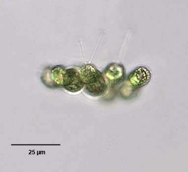
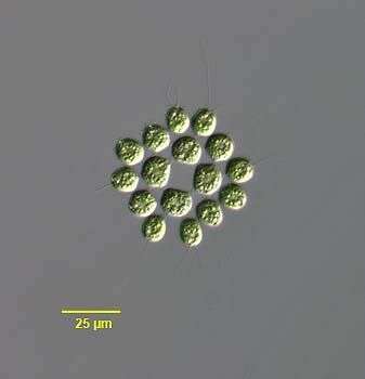
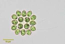
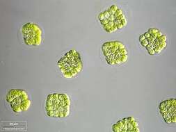
Young colonies, cell divisions are almost over, the flagellae aren't yet fully formed. Scale bar indicates 25 µm. Sample from the Lake Constance (vicinity of Bodman). The image was built up using several photomicrographic frames with manual stacking technique. Images were taken using Zeiss Universal with Olympus C7070 CCD camera.Image under Creative Commons License V 3.0 (CC BY-NC-SA).
-
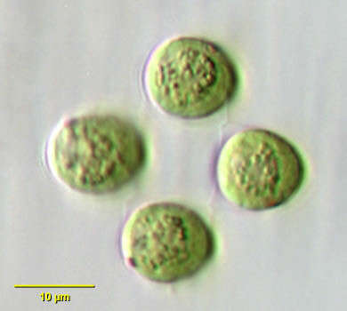
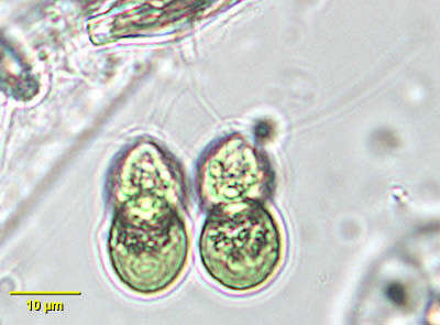
Tetrabaena socialis, a colonial volvocid flagellate. T. socialis was formerly known as Gonium sociale until the monospecific genus Tetrabaena was erected (Nozaki,H. and Itoh, M. J. Phycol. 30:353-365, 1994).The colonies are plate-shaped with four cells all oriented in the same direction. Extracellular matrix joining adjacent cells can be seen. The gelatinous envelope surrounding the whole colony is not seen in these images. The ovoid cells have two equal-length flagella, a small eyespot, large cup-shaped chloroplast and posterior round pyrenoid. From freshwater pond near Boise, Idaho. Oblique illumination.
-
Tetrabaena, a green alga (Chlorophyta) in which four cells form a colony within a delicate mucoid matrix. Each cell with two apical flagella. This image shows an eyespot in one cell.
-
Tetrabaena, a green alga (Chlorophyta) in which four cells form a colony within a delicate mucoid matrix. Each cell with two apical flagella that beat with a breast-stroke pattern.
-
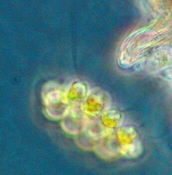
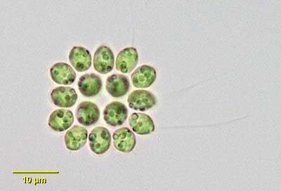
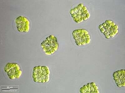
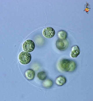
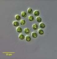
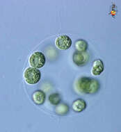
Eudorina (you-door-ine-a) is a colonial volvocid - motile green alga. In this case 16 cells are embedded in a sphere of common mucus. Each individual cell has two flagella, and a cup-shaped chloroplast - a bit like a Chlamydomonas cell. There are a number of related genera which form colonies using mucus as a common matrix, but they differ in the numbers of cells involved, how tightly packed they are, and the shape of the colony. Differential interference contrast.