-
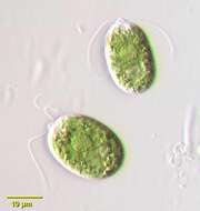
Close-up view. In the center of the cell we see the pyrenoid amidst the stellate chloroplast. The right cell shows, in addition, the nucleus and one of the two contractile vacuoles in maximum extension. The scale bar indicates 10 m. Sample from sphagnum pond Dosenmoor near Neumuenster (Schleswig-Holstein, Germany). Images were taken using Zeiss Axioplan with Canon 600D CCD camera.For permission to use of (high-resolution) images please contact postmaster@protisten.de.
-
Vinuesa, Castille and Leon, Spain
-
All Biocode files are based on field identifications to the best of the researcher’s ability at the time.
-
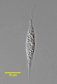
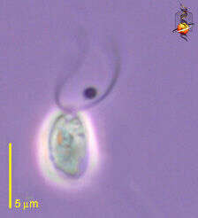
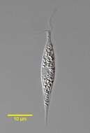
Hyalogonium klebsii (Klebs) Pascher,1927, a colorless volvocid flagellate with two equal anterior flagella. The body is elongate and fusiform. A red stigma is present on the left. Many small starch grains are seen in the cytoplasm.The nucleus is central. There are two anterior contractile vacuoles.This genus may be confused with the euglenid Cyclidiopsis but Hyalogonium is smaller, biflagellate with thinner flagella, and lacks large paramylon bodies and the anterior canal opening. From standing rainwater pool near Boise, Idaho December 2005. DIC.
-
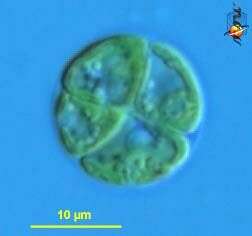
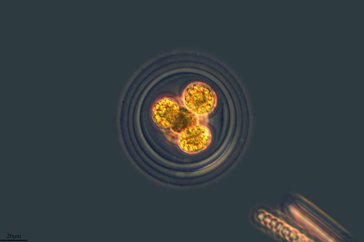
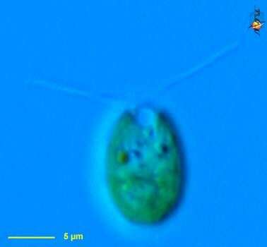
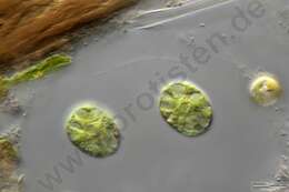
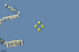
Chlamydomonas (clam-ee-doe-moan-ass), a solitary volvocid (flagellated green algal cell). Cell surrounded by a cellulosic wall, this is a division form in which four daughter cells are being produced at the same time. Animations by Rosemary Arbur of flagellar beat patterns are available
here. Differential interference contrast.
-
Phase contrast microscopy.
-
Melgar de Tera, Castille and Leon, Spain
-
Differential interference contrast
-
Rabano De Aliste, Castille and Leon, Spain
-
Galende, Castille and Leon, Spain
-
Canencia, Madrid, Spain
-
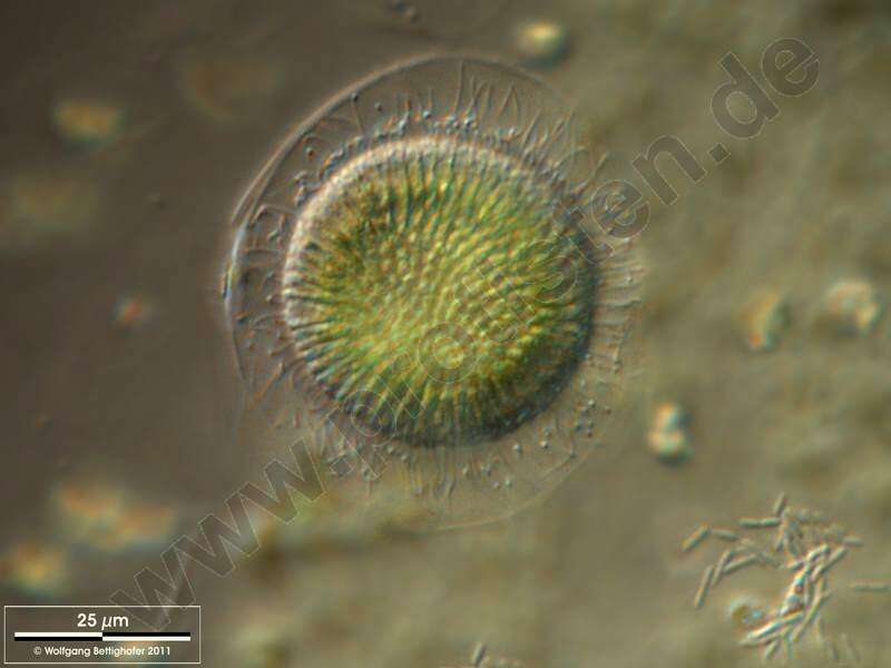
DOF image shows the layers of the mucilaginous sheath, several flagellae and stigmae of two cells. Scale bar indicates 25 m.Sample from a small wetland near Schladming (northern alpine region of Austria near Salzburg). Images were taken using Zeiss Universal with Olympus C7070 CCD camera.For permission to use of (high-resolution) images please contact postmaster@protisten.de.
-
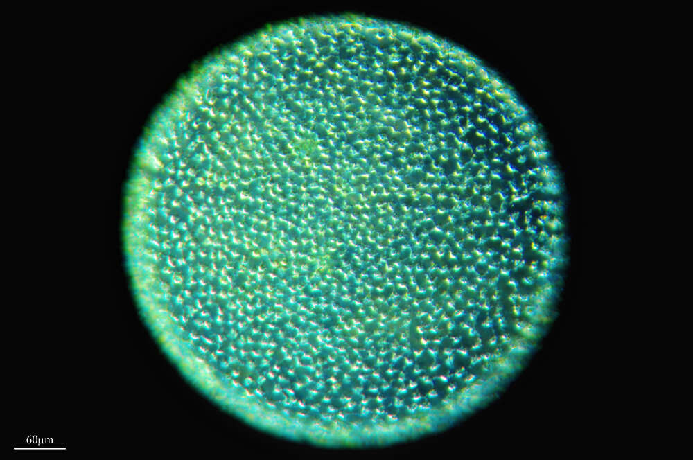
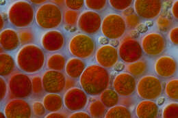
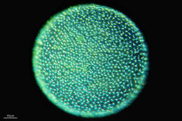
Scale bar indicates 50 m.Sample from a pond on the isle of Hiddensee (German Baltic Sea). The image was built up using several photomicrographic frames with manual stacking technique. The images were taken using Zeiss Universal with Olympus C7070 CCD camera.For permission to use of (high-resolution) images please contact postmaster@protisten.de.
-
All Biocode files are based on field identifications to the best of the researcher’s ability at the time.
-
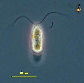
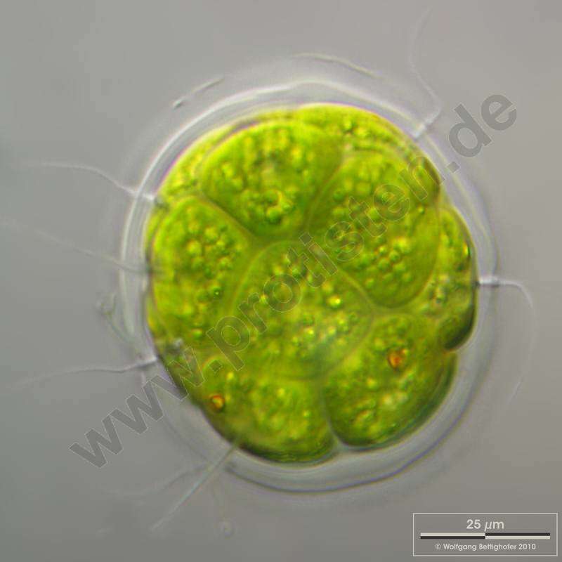
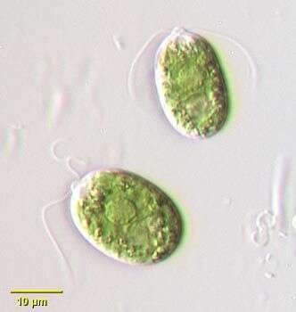
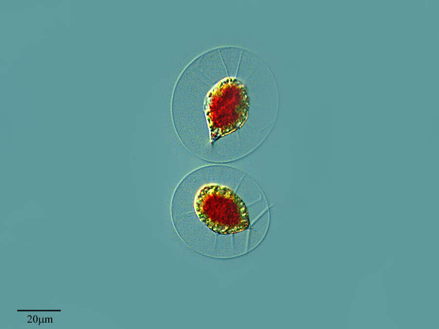
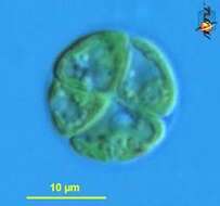
Chlamydomonas (clam-ee-doe-moan-ass), a solitary volvocid (flagellated green algal cell). Cell surrounded by a cellulosic wall, with two similar flagella emerging from near the apex. The photosynthetic pigments are located within a cup-shaped chloroplast which has a large pyrenoid with associated polysaccharide materials located posteriorly. The nucleus is located within the cup. This image shows the small red eyespot (orange colour here) and one anterior contractile vacuole. Differential interference contrast.
-
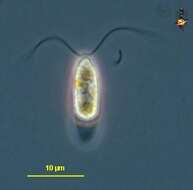
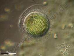
Chlamydomonas, a volvocid flagellate. The genus is very large probably with many synonymous species. Two equal length flagella emerge from a prominent anterior papilla in this species. A small contractile vacuole can be seen just posterior to the flagellar insertion site. A well-demarcated central nucleus can be seen in these images. A very small stigma is present. There is a single large cup shaped chloroplast in this species. A large pyrenoid is present in the posterior half of the cell. Some species have a gelatinous sheath although the cell wall is closely applied to the protoplast in this species. From freshwater pond near Boise, Idaho. Oblique illumination.
-
Melgar de Tera, Castilla y Len, Espaa
-
Lardero, La Rioja, Espaa
-
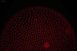
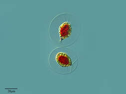
An association of Volvox microgametes (spermatozoids). Scale bar indicates 25 m. Sample from ponds situated in the vicinity of Lake Constance. The image was built up using several photomicrographic frames with manual stacking technique. Images were taken using Zeiss Universal with Olympus C7070 CCD camera.For permission to use of (high-resolution) images please contact postmaster@protisten.de.
-
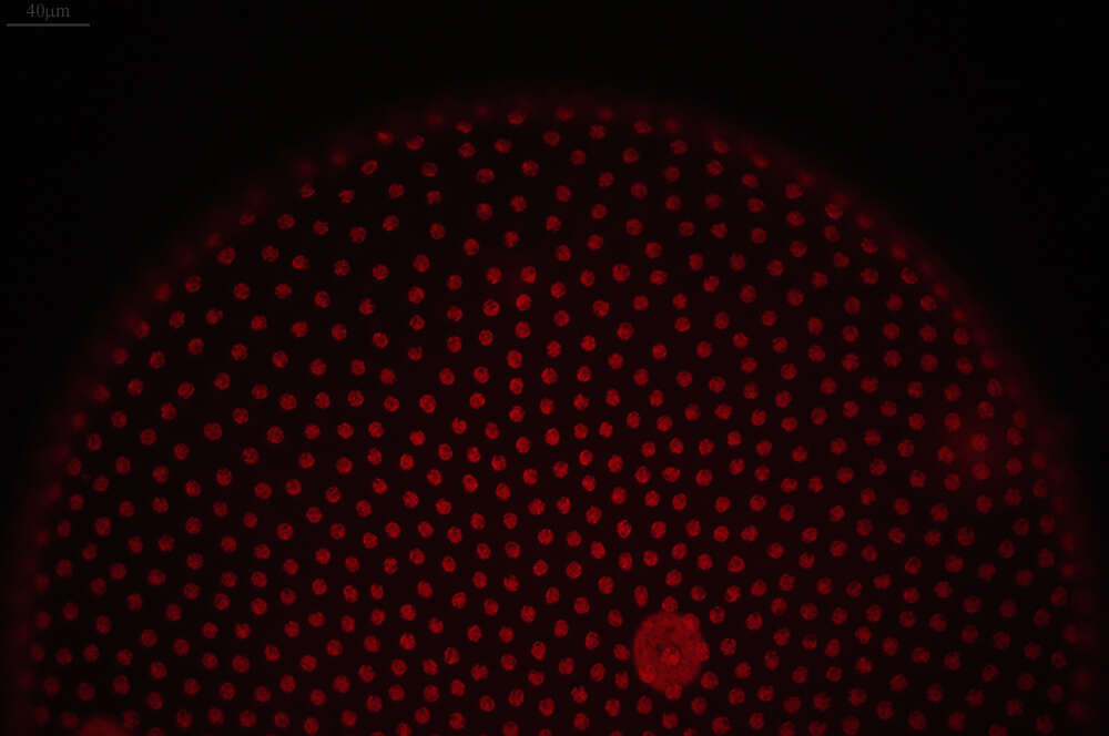
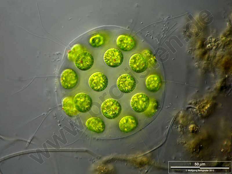
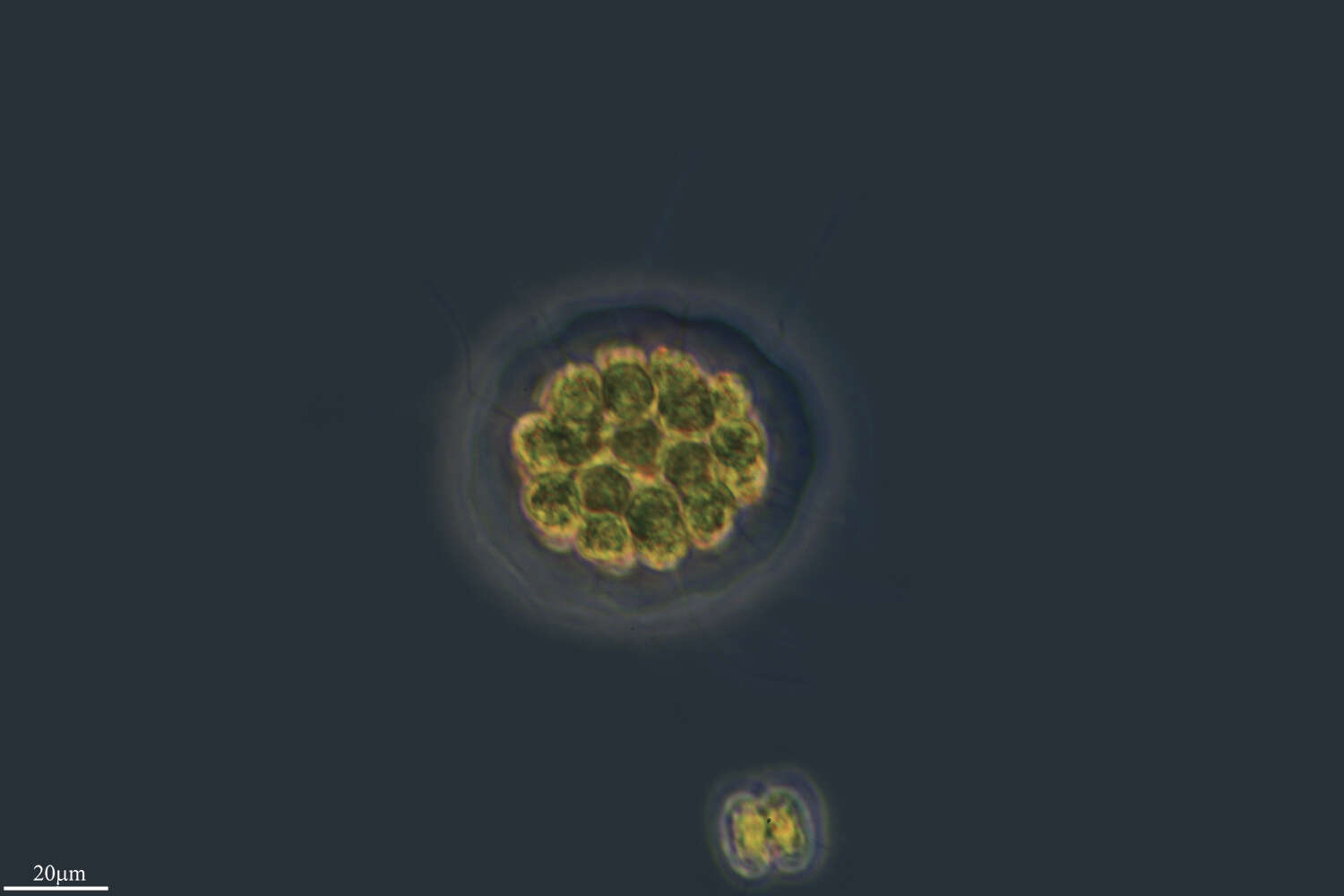
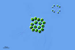
This alga consists of a coenobium. The peripheral cells have two flagella, which point out radially from the centre of the coenobium.
-
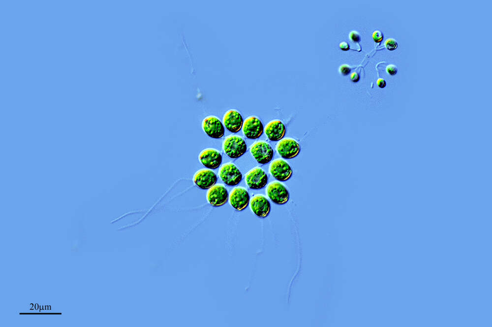
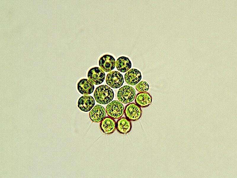
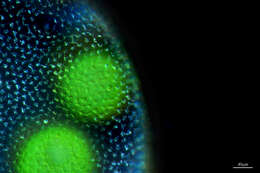
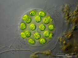
Cells producing daughter colonies. Scale bar indicates 50 m.The specimen was gathered in the wetlands of Nationalpark Unteres Odertal ( 100 km north east of Berlin). The image was built up using several photomicrographic frames with manual stacking technique. Images were taken using Zeiss Universal with Olympus C7070 CCD camera.For permission to use of (high-resolution) images please contact postmaster@protisten.de.
-
Ribadelago de Franco, Castille and Leon, Spain
-
All Biocode files are based on field identifications to the best of the researcher’s ability at the time.
-
Chlamydomonas (clam-ee-doe-moan-ass), a solitary volvocid (flagellated green algal cell). Cell surrounded by a cellulosic wall, with two similar flagella emerging from near the apex. The photosynthetic pigments are located within a cup-shpaed chloroplast which has a large pyrenoid with associated polysaccharide materials. Many taxa described (there are books on this genus). Eyespot located within plastid. Flagella beat with a breast-stroke pattern. Phase contrast.